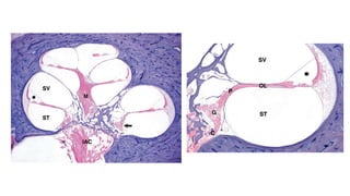
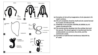
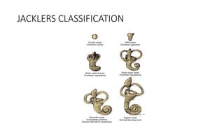
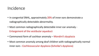
Inner Ear Malformations and
Implantation
Dr Utkal Mishra
Department of ENT-Head & Neck Surgery, AIIMS,Bhopal
RELEVANT ANATOMY
EMBRYOLOGY
JACKLERS CLASSIFICATION
Incidence
• In congenital SNHL, approximately 20% of inner ears demonstrate a
radiographically detectable abnormality.
• Most common radiographically detectable inner ear anomaly -
Enlargement of the vestibular aqueduct
• Commonest form of cochlear anomaly – Mondini’s dysplasia
• Most common anomaly among deaf children with radiographically normal
inner ears - Cochleosaccular dysplasia (Scheibe’s dysplasia)
Classification of Malformations
Malformations limited to membranous labyrinth
Complete Membranous Labyrinthine Dysplasia
Partial Membranous Labyrinthine Dysplasia
Cochleosaccular dysplasia (Scheibe)
Cochlear basal turn dysplasia (Alexander)
Malformations of Osseous & Membranous
Labyrinth
Complete Labyrinthine Aplasia (Michel’s Aplasia)
Cochlear Anomalies
Cochlear aplasia
Cochlear hypoplasia
Incomplete Partition
Common cavity
Labyrinthine anomalies
Enlargement of vestibular aqueduct
Enlargement of cochlear aqueduct
Malformations of IAC & Cochlear nerve
Cochlear nerve hypoplasia
Cochlear nerve aplasia
Narrow IAC
Classification of inner ear anomalies and their
definitions
Complete Membranous Labyrinthine
Dysplasia
• Extremely rare
• Associated with 2 syndromes
1. Jervell-Lange-Nielsen syndrome
2. Usher syndrome
Partial Membranous Labyrinthine Dysplasia
Commonest cause of congenital deafness. (90%)
Two forms
• Cochleosaccular dysplasia (Scheibe) - the organ of Corti is
partially or completely missing.
• Cochlear basal turn dysplasia (Alexander) - dysplasia is limited to
the basal turn of the cochlea. Minimally symptomatic, almost normal
hearing
Relative Incidence of Cochlear Malformations
Malformation Incidence (%)
Incomplete partition (Mondini’s dysplasia) 55
Common cavity 26
Cochlear hypoplasia 15
Cochlear aplasia 3
Complete labyrinthine aplasia (Michel’s aplasia) 1
NORMAL COCHLEA
Note that the normal cochlea appears to have only 1.5
turns on the coronal scan, as a result of the oblique angle
of section in relation to the axial scans of the modiolus.
T2-weighted magnetic
resonance image
Complete Labyrinthine Aplasia (Michel’s Aplasia)
• Exceedingly rare
• Developmental arrest occurs before the formation of otic
vesicle.
• Complete absence of inner ear.
• Associated with anencephaly and thalidomide exposure.
• No CI, ABI
Cochlear Aplasia
• Cochlea is completely absent as a result of an
arrest in the development of the cochlear bud at
the fifth week of gestation
• Radiographically, only a vestibule and SCCs are
present.
• No CI, ABI
Cochlear Hypoplasia
• Arrest during the sixth week of gestation
• Radiographically, a small bud protrudes from the
vestibule (usually 1 to 3 mm)
CI possible
• Suggested approach: Transmastoidal.
• Implant advice: Shorter electrode.
• Special care: the round window can be difficult to
localize and the facial nerve can have an aberrant course.
• Outcome - Guarded
Most Challenging
Incomplete Partition Deformity(Sennaroglu and Saatci Classification)
• Type I lacks the entire modiolus and interscalar septa
and demonstrates a cystic appearance.
• Type II has a normal basal turn but a cystic apex
• Type III has deficient modiolus and partial interscalar
septation at the cochlea’s periphery. It is X linked.
• The best way to distinguish the differences between
these entities is on MRI.
Incomplete Partition (Mondini)
• Commonest type of cochlear malformation >50%.
• Cochlea has only 1.5 turns.
• Middle and apical turns unite to form a cystic apex.
• Three components:
• Cystic cochlear apex with a normal basal turn
• Dilated vestibule
• Enlarged vestibular aqueduct.
Cochlear Implantation in IP-2
• Suggested approach: Transmastoid Posterior Tympanotomy approach.
• Implant advice: Normal Implant.
• Special care: Other than some oozing of the perilymph and special attention to
the tight closure of the cochleostomy, there is no difference from a normal
implantation.
• Outcome - Good
Common Cavity
• A deformed inner ear in which the
cochlea and vestibule are confluent,
forming an ovoid cystic space without
internal architecture, may be
explained by an arrest at the week 4
otocyst stage.
• An empty ovoid space typically longer
in its horizontal dimension is seen
radiographically.
Cochlear aplasia with vestibular dilatation (CAVD) usually has a
vestibule and semicircular canals at their usual location at the
posterolateral part of the IAC fundus.
A common cavity (CC) is an
ovoid or round structure. The
IAC usually enters the cavity at
its center. It contains cochlear
and vestibular neural
elements.
Cochlear Implantation in Common Cavity
• Suggested approach: Transmastoid labyrinthotomy
/ Subtotal Petrosectomy
• Implant advice: outer wall “uncoiled” electrode,
(The straight Cochlear Corporation K electrode
and the Med-El Combi 40+ electrode)
• Special care: often an aberrant course of the facial
nerve is encountered.
• Outcome: Poor (0 - 40%)
• The dome of the cavity is flattened and drilled until the
endosteum is exposed for a 1-mm diameter.
• The endosteum is opened using a small right-angle pick.
• If a CSF gusher is encountered, the head of the bed is
elevated and when the gusher stops the electrode is
inserted.
• The stimulable neural elements of the common cavity are
most likely lying on the outer wall of the cochlea
Labyrinthine Anomalies
• Commonest SCC anomaly detected in cochlear
anomalies – Lateral SCC
• The diameter of “the bony island” within the LSC
(yellow arrows) is a good measure for defining
malformed or not malformed.
Enlargement of the Vestibular Aqueduct
• Normal – 0.4 – 1 mm
• Enlargement of the VA is diagnosed when its diameter
exceeds 1.5 mm
• EVA is defined as an enlargement of the aqueduct
exceeding the diameter of the posterior semicircular canal
on an axial image
• Whereas the VA is well visualized on axial CT the dilated
endolymphatic sac is better seen with T2-weighted MRI
• Commonly found in Type 2 Incomplete partition or
Mondini’s.
Enlargement of the Vestibular Aqueduct
• Characterized by progressive or fluctuating
sensorineural hearing loss, beginning in
childhood.
• LVA is associated with 3 syndromes
• Pendred syndrome
• BOR syndrome
• Wardenberg Syndrome
CHARGE Syndrome
• Coloboma of the eye
• Heart defects
• Atresia of the nasal choanae
• Retardation of growth
• Genital and/or urinary abnormalities
• Ear abnormalities – LOP EAR
• Deafness – Profound SNHL
• The facial nerve is more commonly
affected in children with CHARGE
syndrome
Cochlear aperture atresia (red arrow) This phenomenon
is also referred to as “trapped cochlea” and is strongly
associated with CHARGE syndrome There appears to be
no connection between the IAC and the cochlea on CT
A cochlear nerve canal, also known as cochlear aperture, of
diameter less than 1.5 mm is most often associated with CN
hypoplasia
Cochlear Nerve Hypoplasia and Aplasia
References
THANK YOU

Inner ear malformations and Implantation

  • 1.
    Inner Ear Malformationsand Implantation Dr Utkal Mishra Department of ENT-Head & Neck Surgery, AIIMS,Bhopal
  • 2.
  • 7.
  • 9.
  • 10.
    Incidence • In congenitalSNHL, approximately 20% of inner ears demonstrate a radiographically detectable abnormality. • Most common radiographically detectable inner ear anomaly - Enlargement of the vestibular aqueduct • Commonest form of cochlear anomaly – Mondini’s dysplasia • Most common anomaly among deaf children with radiographically normal inner ears - Cochleosaccular dysplasia (Scheibe’s dysplasia)
  • 11.
    Classification of Malformations Malformationslimited to membranous labyrinth Complete Membranous Labyrinthine Dysplasia Partial Membranous Labyrinthine Dysplasia Cochleosaccular dysplasia (Scheibe) Cochlear basal turn dysplasia (Alexander) Malformations of Osseous & Membranous Labyrinth Complete Labyrinthine Aplasia (Michel’s Aplasia) Cochlear Anomalies Cochlear aplasia Cochlear hypoplasia Incomplete Partition Common cavity Labyrinthine anomalies Enlargement of vestibular aqueduct Enlargement of cochlear aqueduct Malformations of IAC & Cochlear nerve Cochlear nerve hypoplasia Cochlear nerve aplasia Narrow IAC
  • 12.
    Classification of innerear anomalies and their definitions
  • 13.
    Complete Membranous Labyrinthine Dysplasia •Extremely rare • Associated with 2 syndromes 1. Jervell-Lange-Nielsen syndrome 2. Usher syndrome
  • 14.
    Partial Membranous LabyrinthineDysplasia Commonest cause of congenital deafness. (90%) Two forms • Cochleosaccular dysplasia (Scheibe) - the organ of Corti is partially or completely missing. • Cochlear basal turn dysplasia (Alexander) - dysplasia is limited to the basal turn of the cochlea. Minimally symptomatic, almost normal hearing
  • 15.
    Relative Incidence ofCochlear Malformations Malformation Incidence (%) Incomplete partition (Mondini’s dysplasia) 55 Common cavity 26 Cochlear hypoplasia 15 Cochlear aplasia 3 Complete labyrinthine aplasia (Michel’s aplasia) 1
  • 16.
    NORMAL COCHLEA Note thatthe normal cochlea appears to have only 1.5 turns on the coronal scan, as a result of the oblique angle of section in relation to the axial scans of the modiolus. T2-weighted magnetic resonance image
  • 17.
    Complete Labyrinthine Aplasia(Michel’s Aplasia) • Exceedingly rare • Developmental arrest occurs before the formation of otic vesicle. • Complete absence of inner ear. • Associated with anencephaly and thalidomide exposure. • No CI, ABI
  • 18.
    Cochlear Aplasia • Cochleais completely absent as a result of an arrest in the development of the cochlear bud at the fifth week of gestation • Radiographically, only a vestibule and SCCs are present. • No CI, ABI
  • 19.
    Cochlear Hypoplasia • Arrestduring the sixth week of gestation • Radiographically, a small bud protrudes from the vestibule (usually 1 to 3 mm) CI possible • Suggested approach: Transmastoidal. • Implant advice: Shorter electrode. • Special care: the round window can be difficult to localize and the facial nerve can have an aberrant course. • Outcome - Guarded Most Challenging
  • 20.
    Incomplete Partition Deformity(Sennarogluand Saatci Classification) • Type I lacks the entire modiolus and interscalar septa and demonstrates a cystic appearance. • Type II has a normal basal turn but a cystic apex • Type III has deficient modiolus and partial interscalar septation at the cochlea’s periphery. It is X linked. • The best way to distinguish the differences between these entities is on MRI.
  • 21.
    Incomplete Partition (Mondini) •Commonest type of cochlear malformation >50%. • Cochlea has only 1.5 turns. • Middle and apical turns unite to form a cystic apex. • Three components: • Cystic cochlear apex with a normal basal turn • Dilated vestibule • Enlarged vestibular aqueduct.
  • 22.
    Cochlear Implantation inIP-2 • Suggested approach: Transmastoid Posterior Tympanotomy approach. • Implant advice: Normal Implant. • Special care: Other than some oozing of the perilymph and special attention to the tight closure of the cochleostomy, there is no difference from a normal implantation. • Outcome - Good
  • 23.
    Common Cavity • Adeformed inner ear in which the cochlea and vestibule are confluent, forming an ovoid cystic space without internal architecture, may be explained by an arrest at the week 4 otocyst stage. • An empty ovoid space typically longer in its horizontal dimension is seen radiographically. Cochlear aplasia with vestibular dilatation (CAVD) usually has a vestibule and semicircular canals at their usual location at the posterolateral part of the IAC fundus. A common cavity (CC) is an ovoid or round structure. The IAC usually enters the cavity at its center. It contains cochlear and vestibular neural elements.
  • 24.
    Cochlear Implantation inCommon Cavity • Suggested approach: Transmastoid labyrinthotomy / Subtotal Petrosectomy • Implant advice: outer wall “uncoiled” electrode, (The straight Cochlear Corporation K electrode and the Med-El Combi 40+ electrode) • Special care: often an aberrant course of the facial nerve is encountered. • Outcome: Poor (0 - 40%) • The dome of the cavity is flattened and drilled until the endosteum is exposed for a 1-mm diameter. • The endosteum is opened using a small right-angle pick. • If a CSF gusher is encountered, the head of the bed is elevated and when the gusher stops the electrode is inserted. • The stimulable neural elements of the common cavity are most likely lying on the outer wall of the cochlea
  • 25.
    Labyrinthine Anomalies • CommonestSCC anomaly detected in cochlear anomalies – Lateral SCC • The diameter of “the bony island” within the LSC (yellow arrows) is a good measure for defining malformed or not malformed.
  • 26.
    Enlargement of theVestibular Aqueduct • Normal – 0.4 – 1 mm • Enlargement of the VA is diagnosed when its diameter exceeds 1.5 mm • EVA is defined as an enlargement of the aqueduct exceeding the diameter of the posterior semicircular canal on an axial image • Whereas the VA is well visualized on axial CT the dilated endolymphatic sac is better seen with T2-weighted MRI • Commonly found in Type 2 Incomplete partition or Mondini’s.
  • 27.
    Enlargement of theVestibular Aqueduct • Characterized by progressive or fluctuating sensorineural hearing loss, beginning in childhood. • LVA is associated with 3 syndromes • Pendred syndrome • BOR syndrome • Wardenberg Syndrome
  • 28.
    CHARGE Syndrome • Colobomaof the eye • Heart defects • Atresia of the nasal choanae • Retardation of growth • Genital and/or urinary abnormalities • Ear abnormalities – LOP EAR • Deafness – Profound SNHL • The facial nerve is more commonly affected in children with CHARGE syndrome Cochlear aperture atresia (red arrow) This phenomenon is also referred to as “trapped cochlea” and is strongly associated with CHARGE syndrome There appears to be no connection between the IAC and the cochlea on CT A cochlear nerve canal, also known as cochlear aperture, of diameter less than 1.5 mm is most often associated with CN hypoplasia
  • 29.
  • 30.
  • 31.