Imaging modalities of the petrous bone
•Download as PPTX, PDF•
38 likes•3,227 views

This document describes various imaging modalities and techniques used to examine the petrous bone and inner ear anatomy. It outlines CT and MRI sequences that can be used and discusses what structures like the external auditory canal, middle ear, semicircular canals and cochlea appear as on different scans. It also provides examples of pathologies that can be imaged in the temporal bone like cholesteatoma, otitis media, fractures and tumors.

More Related Content
What's hot
What's hot (20)
Viewers also liked
Viewers also liked (20)
Similar to Imaging modalities of the petrous bone
Similar to Imaging modalities of the petrous bone (20)
More from Abdellah Nazeer
More from Abdellah Nazeer (20)
Imaging modalities of the petrous bone
- 1. Imaging modalities of the petrous bone. Dr/ ABD ALLAH NAZEER. MD.
- 2. Diagram for the external, middle and inner ear.
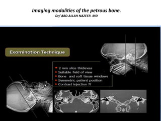
- 3. CT techniques for petrous examination
- 9. 1. External auditory canal. 2. Tympanic membrane. 3. Ossicles (3: malleus, 3': incus, 3'': stapes). 4. Prussack space. 5. Scutum. 6. Tympanic tegmen. 7. Tympanic sinus. 8. Facial nerve fossa. 9. Facial nerve. 10. Antrum of mastoid. 11. Oval window. 12. Vestibule. 13. Semicircular canals (13: anterior, 13': lateral, 13'': posterior). 14.Cochlea. 15.Internal auditory canal. 16. Vestibular aqueduct.
- 10. Axial images.
- 30. Inner ear.
- 36. One and half and basal turn of the cochlea.
- 45. Necrotizing otitis externa (NOE), also known as malignant otitis externa, is a severe invasive infection of the external auditory canal (EAC) which can spread rapidly to involve the surrounding soft tissue, adjacent neck spaces and skull base.
- 46. (A-C) Malignant otitis externa: coronal contrast-enhanced CT scan (A) shows soft tissue thickening of the EAC (arrow). Axial HRCT image (B) shows irregularity of the right TM joint (arrowhead) and mastoid (arrowhead). Axial contrast-enhanced CT scan (C) shows a temporalis abscess (arrow).
- 49. External auditory canal exostoses.
- 50. (A-C) Cholesteatoma of the EAC: non-contrast coronal CT scan (A) and axial HRCT image (B) show a hypodense lesion in the right EAC (arrow in A and arrowhead in B) invading the mastoid (stage III). 3D volume rendered image (C) shows a small, post-biopsy defect in the mastoid wall.
- 51. Squamous cell carcinoma of the external auditory canal.
- 52. The middle ear is an-air filled chamber in the petrous part of the temporal bone that is split into two parts: the tympanic cavity proper and the epitympanic recess or attic. It contains the three auditory ossicles whose purpose is to transmit sound vibrations from the tympanic membrane to the medial wall of the inner ear. Related pathology: Chronic otitis media. Cholesteatoma. Otosclerosis. Trauma. Vascular anomalies. Aberrant carotid artery. Dehiscent jugular bulb.
- 70. Diffusion weighted imaging (A) and apparent diffusion coefficient map (B) reveal restricted diffusion by a lesion situated in the right external auditory canal (arrow)
- 71. A and B, CT shows soft tissue (arrow) in the mastoid defect, external auditory canal, and epitympanum with bony erosion of the lateral semicircular canal. C and D, MR images show the extent of cholesteatoma and demonstrate a large area of hyperintensity on HASTE DWI in the mastoid defect and middle ear with T2 hypointensity (arrow, C), and mild T1 hyperintensity but no definite enhancement (arrow, D). A portion of the right lateral semicircular canal is obscured by the soft-tissue mass (C), again consistent with the fistula on CT.
- 72. DWI sequences obtained in a patient with postoperative changes. Increased diffusion signal intensity is seen in the right middle ear and mastoid defect (arrow), with cholesteatoma confirmed at surgery.
- 74. Bilateral cholesteatoma with typical restricted diffusion.
- 75. (A-D) Cholesteatoma of the EAC: coronal T1W MRI image (A) shows a hypointense lesion (arrow) in the right temporal bone. Axial STIR MRI image (B) shows a hyperintense lesion (arrow) extending into the mastoid. Diffusion-weighted (C) and axial apparent diffusion coefficient (ADC) (D) images show restricted diffusion (arrows).
- 77. Cholesterol granuloma. CT (A) demonstrates an expansive mass involving the PA; there is no evidence of bony erosion. Typically T1 WI (B) and T2 WI (D;E) show an hyperintense lesion with mass effect on the right prepontine cistern. No CE is seen (C).
- 78. Petrous apex meningocele/ Arachnoid cyst.
- 79. Mucocele.
- 85. Aberrant course of the ICA in a 25-year-old man presenting with pulsatile tinnitus. A, Enhanced axial CT image demonstrates an abnormal lateral course of the right ICA through the middle ear (white arrow). Also note dehiscence of the overlying bony plate. B, Anteroposterior projection image from the MR angiogram of the same patient demonstrates decreased caliber and lateral deviation of the aberrant ICA on the “right reversed-7 sign” (black arrow).
- 87. Temporal bone fractures are usually a sequela of blunt head injury, generally from severe trauma. Early identification of temporal bone trauma is essential to managing the injury and avoiding complications. Classification: direction Temporal bone fractures classically are described concerning the long axis of the petrous temporal bone, being classified as: Longitudinal fractures. Transverse fractures. Mixed fractures.
- 91. Right and left axial petrous temporal bone CT.
- 94. Bilateral vestibular aqueduct syndrome.
- 101. Right ear shows bony defect displayed by oblique reformation (double window view) (a). Coronal plane also shows the defect in (b). The left ear shows similar findings in (c and d).
- 104. Axial (a) and coronal (b) HRCT images of the right temporal bone in an adult patient with right-sided CHL. A hypodense demineralised plaque (arrow) is noted in the region of the fissula ante fenestram in keeping with fenestral otosclerosis
- 115. Bilateral calcification of the basal turn of cochlea, related to labyrinthine ossificans.
- 117. VIII cn schwannoma.
- 118. Endolymphatic sac tumor. Lesion shows high signal on T2 WI (A), within hyperintense foci on T1-wi B), typically located in the posterior petrous ridge. Post gadolinium T1 WI (C) reveals heterogeneous CE. Notice the involvement of the internal auditory canal. Axial CT and 3D VR reconstruction (D, E) show typical permeative bone changes of ELST; notice the involvement of internal auditory canal.
- 119. Metastasis. Ax T2 WI (A) reveals area of inhomogeneous high signal filling the right PA, post gadolinium Ax/CorT1 WI (B;C) show diffuse CE. Notice adjacent enhancing fat of the clivus and of VIII C.N (arrow in C).