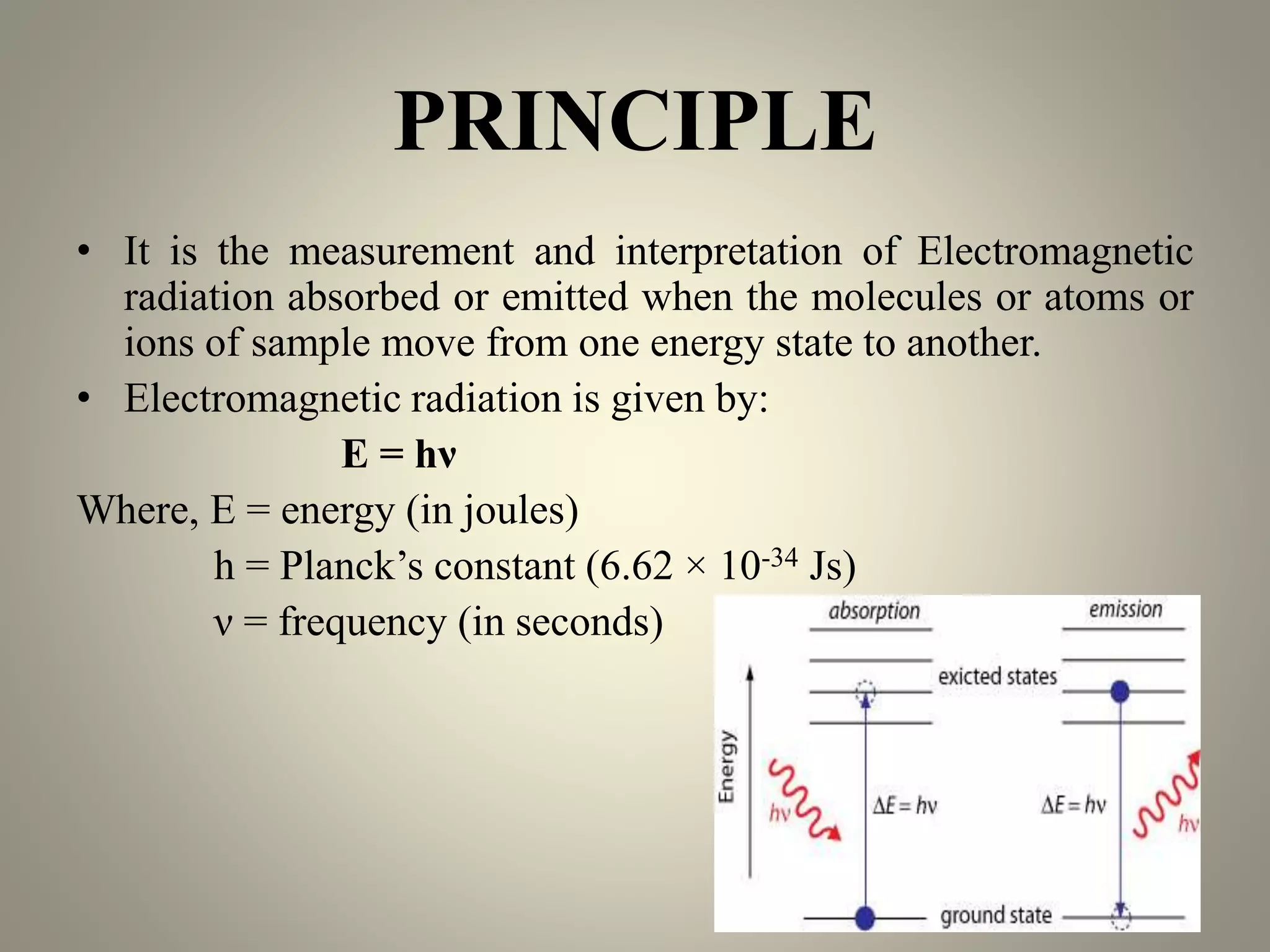
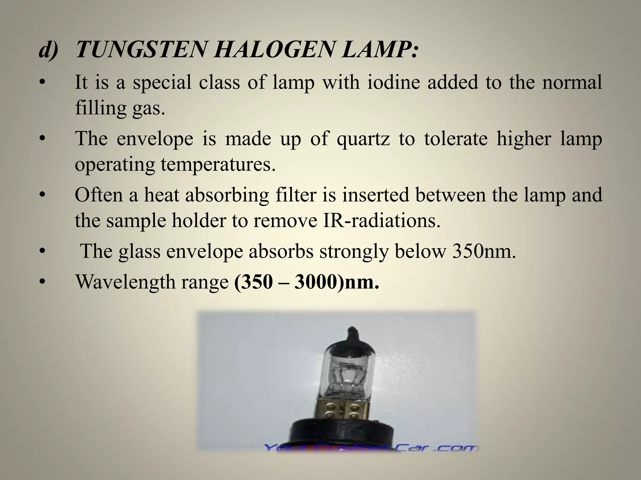
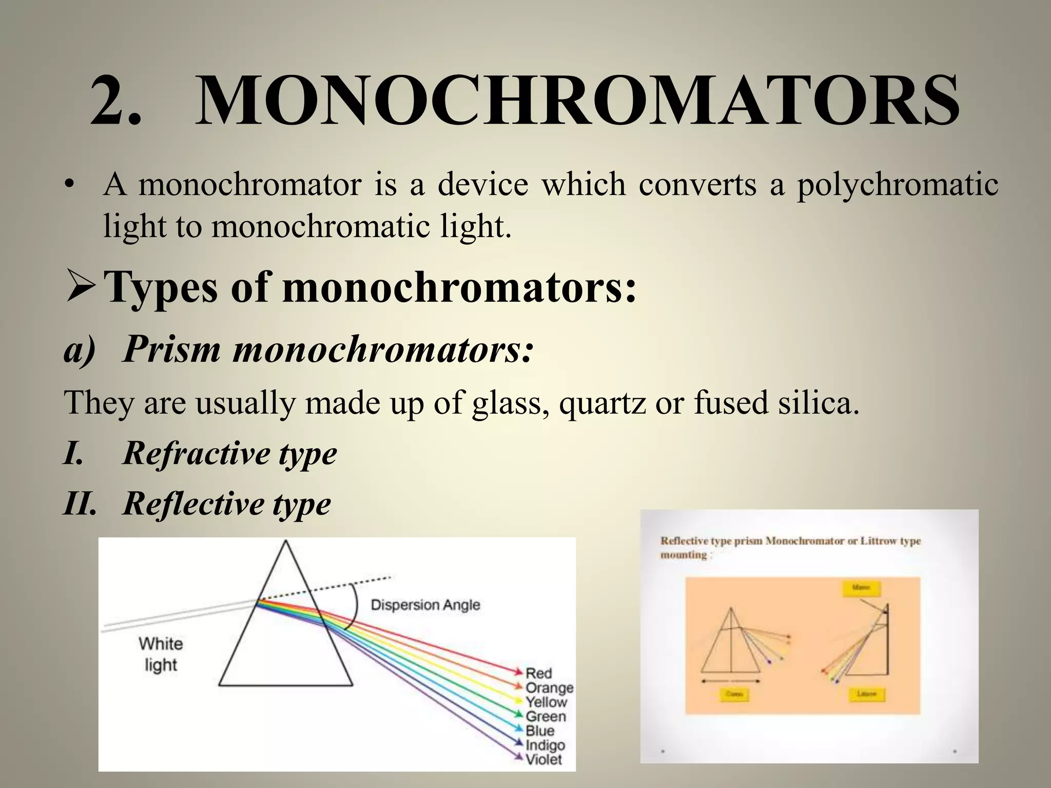
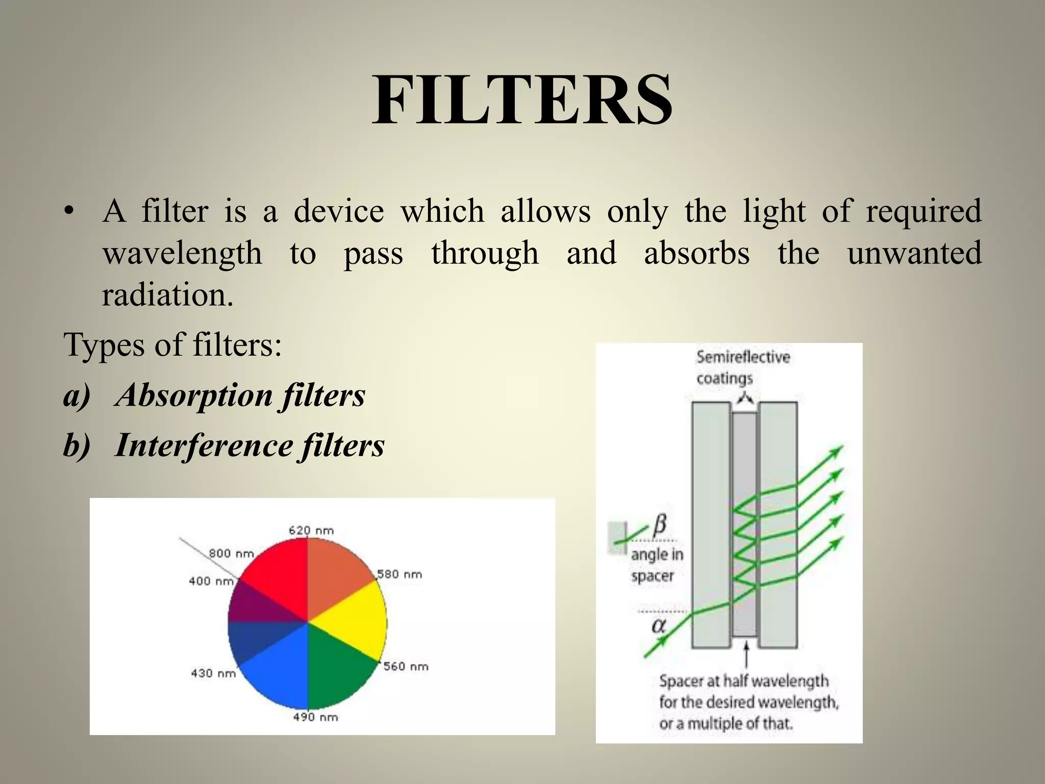
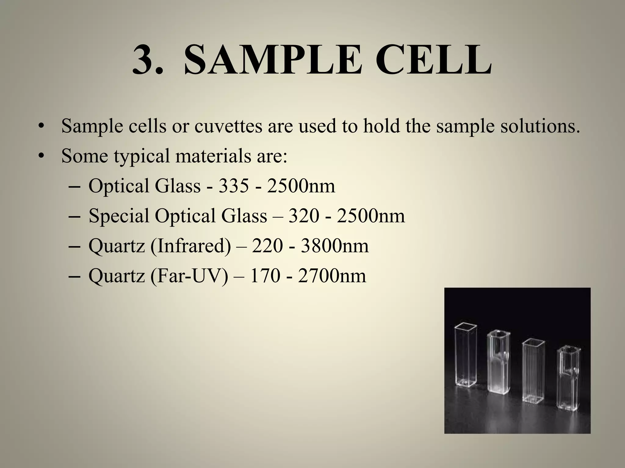
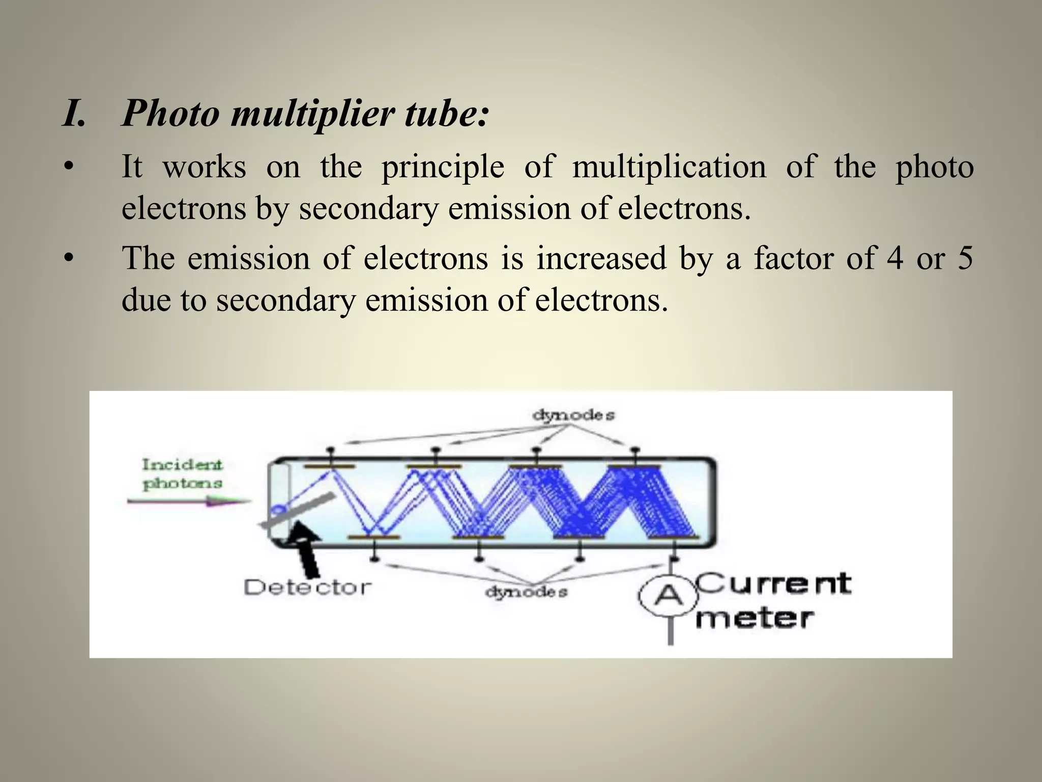
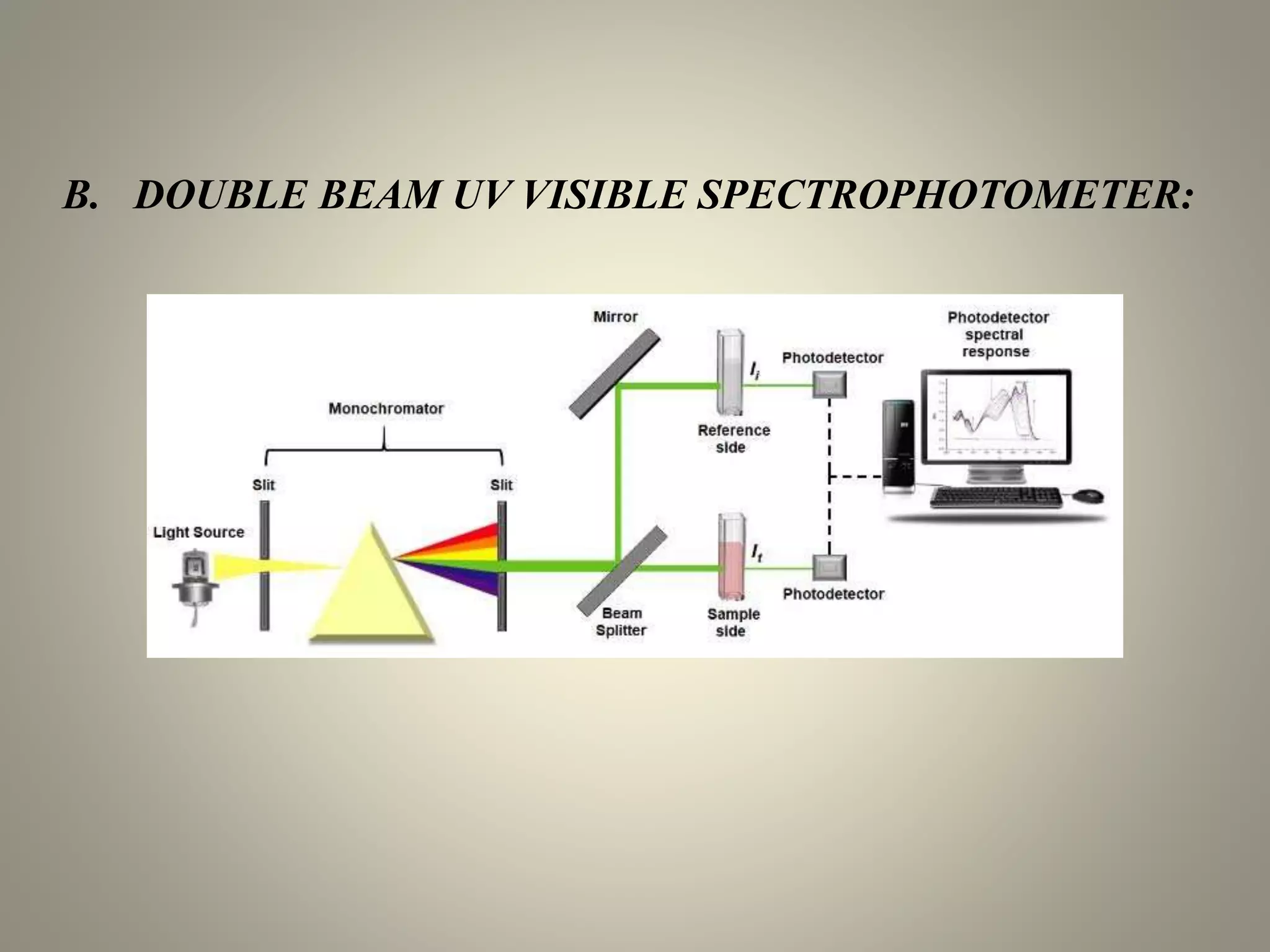
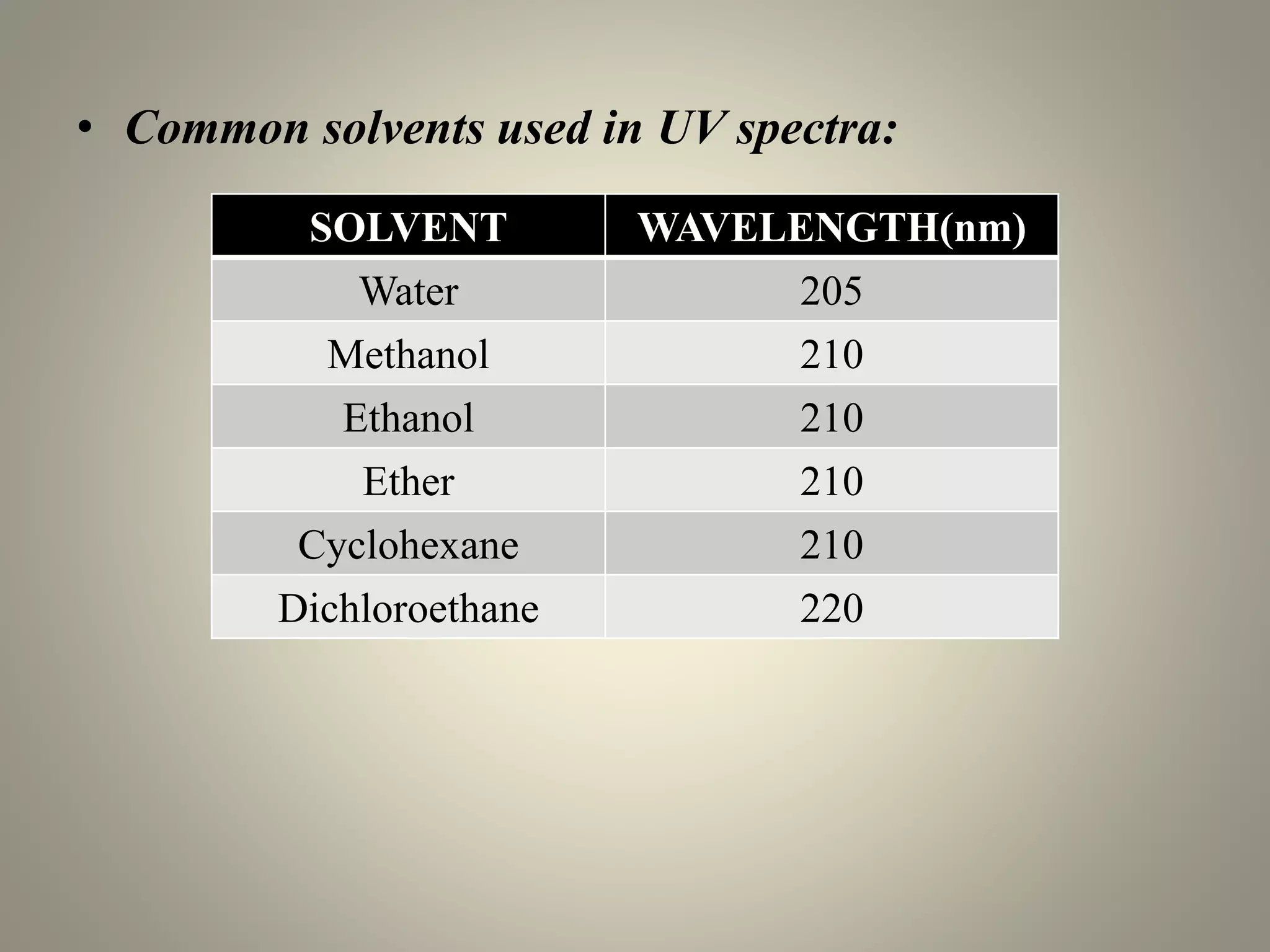
UV-visible spectroscopy is a technique that uses light in the visible and adjacent ranges. It works by measuring how much light is absorbed by a sample at each wavelength. There are several types of electronic transitions that can occur when molecules absorb this light. The amount of light absorbed follows Beer's law and is proportional to the concentration and path length of the sample. A UV-visible spectrophotometer consists of a light source, monochromator, sample holder, detector, and recording device. This technique has many applications including detection of impurities, structure elucidation, and quantitative analysis in pharmaceutical analysis.
principle, application and instrumentation of UV- visible Spectrophotometer Ayetenew Abita Desa
This Presentation powerpoint includes the principle, application, and instrumentation of UV- Visible Spectrophotometer. It covers beer-lambert low and its quantitative applications. It also includes the qualitative applications in different fields of study. Presented at Addis Ababa University, School of medicine, department of medical biochemistry.
This presentation include the detailed explanation of various parts of a UV-Visible spectrophotometer and two types of UV-Visible spectrophotometers-Single beam and Doube beam. It also include the comparison between single beam and double beam spectrophotometers.
IR SPECTROSCOPY, INTRODUCTION, PRINCIPLE, THEORY, FATE OF ABSORBED RADIATION, FERMI RESONANCE, FINGERPRINT REGION, VIBRATIONS, FACTORS AFFECTING ABSORPTION OF IR RADIATION, SAMPLING TECHNIQUES, APPLICATIONS OF IR SPECTROSCOPY.
This presentation gives you thorough knowledge about the IR Spectroscopy. This include basic principle, type of vibrations, factors influencing vibrational frequency, instrumentation and applications of IR Spectroscopy. This is the most widely used technique for identifying unknown functional group depending on the vibrational frequency.
In this slide contains principle, instrumentation, methodology, and application of gel chromatography.
Presented by: SATHEES CHANDRA (Department of pharmaceutical analysis).
RIPER, anantapur
spectrofluorometer is the instrument for recording fluorescence emission and absorption spectra When a beam of light is incident on certain substances they emit visible light or radiations. This is known as fluorescence. Fluorescence starts immediately after the absorption of light and stops as soon as the incident light is cut off. The substances showing this phenomenon are known as flourescent substances.
Uv visible spectroscopy with InstrumentationSHIVANEE VYAS
It is the branch of science that deals with the study of the interaction of matter with light.
OR
It is the branch of science that deals with the study of the interaction of electromagnetic radiation with matter.
Electromagnetic radiation is energy that is propagated through free space or through a material medium in the form of electromagnetic waves, such as radio waves, visible light, and gamma rays, etc. Electromagnetic waves consist of discrete packages of energy which are called as photons.
UV spectroscopy, Electronic transitions, law of UV, Deviations of UV, chromop...Rajesh Singh
This PowerPoint Presentation includes the principle, electronic transitions, application, chromophore, Auxochrome, Deviations and instrumentation of UV- Visible Spectrophotometer. It covers beer-lambert low and its quantitative applications. It also includes the qualitative applications in different fields of study. Presented by Rajesh Singh in GLA University Mathura.
The Principle of UV-Visible Spectroscopy is based on the absorption of ultraviolet light or visible light by chemical compounds, which results in the production of distinct spectra. Spectroscopy is based on the interaction between light and matter. When the matter absorbs the light, it undergoes excitation and de-excitation, resulting in the production of a spectrum.
When matter absorbs ultraviolet radiation, the electrons present in it undergo excitation. This causes them to jump from a ground state (an energy state with a relatively small amount of energy associated with it) to an excited state (an energy state with a relatively large amount of energy associated with it).
introduction
mechanisms of protein drug binding
binding of drugs
binding of drugs to blood components
determination of protein drug binding
factors affecting
significance
Introduction
working principle
fragmentation process
general rules for fragmentation
general modes of fragmentation
metastable ions
isotopic peaks
applications
POTENTIAL SOURCES OF ELEMENTAL IMPURITIESMehulJain143
INTRODUCTION
INDENTIFICATION OF POTENTIAL ELEMENTAL IMPURITIES
FACTORS AFFECTING
EVALUATION
RISK ASSESSMENT AND CONTROL OF ELEMENTAL IMPURITIES
GENERAL PRINCIPLES
A brief information about the SCOP protein database used in bioinformatics.
The Structural Classification of Proteins (SCOP) database is a comprehensive and authoritative resource for the structural and evolutionary relationships of proteins. It provides a detailed and curated classification of protein structures, grouping them into families, superfamilies, and folds based on their structural and sequence similarities.
This presentation explores a brief idea about the structural and functional attributes of nucleotides, the structure and function of genetic materials along with the impact of UV rays and pH upon them.
What is greenhouse gasses and how many gasses are there to affect the Earth.moosaasad1975
What are greenhouse gasses how they affect the earth and its environment what is the future of the environment and earth how the weather and the climate effects.
Richard's aventures in two entangled wonderlandsRichard Gill
Since the loophole-free Bell experiments of 2020 and the Nobel prizes in physics of 2022, critics of Bell's work have retreated to the fortress of super-determinism. Now, super-determinism is a derogatory word - it just means "determinism". Palmer, Hance and Hossenfelder argue that quantum mechanics and determinism are not incompatible, using a sophisticated mathematical construction based on a subtle thinning of allowed states and measurements in quantum mechanics, such that what is left appears to make Bell's argument fail, without altering the empirical predictions of quantum mechanics. I think however that it is a smoke screen, and the slogan "lost in math" comes to my mind. I will discuss some other recent disproofs of Bell's theorem using the language of causality based on causal graphs. Causal thinking is also central to law and justice. I will mention surprising connections to my work on serial killer nurse cases, in particular the Dutch case of Lucia de Berk and the current UK case of Lucy Letby.
This pdf is about the Schizophrenia.
For more details visit on YouTube; @SELF-EXPLANATORY;
https://www.youtube.com/channel/UCAiarMZDNhe1A3Rnpr_WkzA/videos
Thanks...!
Slide 1: Title Slide
Extrachromosomal Inheritance
Slide 2: Introduction to Extrachromosomal Inheritance
Definition: Extrachromosomal inheritance refers to the transmission of genetic material that is not found within the nucleus.
Key Components: Involves genes located in mitochondria, chloroplasts, and plasmids.
Slide 3: Mitochondrial Inheritance
Mitochondria: Organelles responsible for energy production.
Mitochondrial DNA (mtDNA): Circular DNA molecule found in mitochondria.
Inheritance Pattern: Maternally inherited, meaning it is passed from mothers to all their offspring.
Diseases: Examples include Leber’s hereditary optic neuropathy (LHON) and mitochondrial myopathy.
Slide 4: Chloroplast Inheritance
Chloroplasts: Organelles responsible for photosynthesis in plants.
Chloroplast DNA (cpDNA): Circular DNA molecule found in chloroplasts.
Inheritance Pattern: Often maternally inherited in most plants, but can vary in some species.
Examples: Variegation in plants, where leaf color patterns are determined by chloroplast DNA.
Slide 5: Plasmid Inheritance
Plasmids: Small, circular DNA molecules found in bacteria and some eukaryotes.
Features: Can carry antibiotic resistance genes and can be transferred between cells through processes like conjugation.
Significance: Important in biotechnology for gene cloning and genetic engineering.
Slide 6: Mechanisms of Extrachromosomal Inheritance
Non-Mendelian Patterns: Do not follow Mendel’s laws of inheritance.
Cytoplasmic Segregation: During cell division, organelles like mitochondria and chloroplasts are randomly distributed to daughter cells.
Heteroplasmy: Presence of more than one type of organellar genome within a cell, leading to variation in expression.
Slide 7: Examples of Extrachromosomal Inheritance
Four O’clock Plant (Mirabilis jalapa): Shows variegated leaves due to different cpDNA in leaf cells.
Petite Mutants in Yeast: Result from mutations in mitochondrial DNA affecting respiration.
Slide 8: Importance of Extrachromosomal Inheritance
Evolution: Provides insight into the evolution of eukaryotic cells.
Medicine: Understanding mitochondrial inheritance helps in diagnosing and treating mitochondrial diseases.
Agriculture: Chloroplast inheritance can be used in plant breeding and genetic modification.
Slide 9: Recent Research and Advances
Gene Editing: Techniques like CRISPR-Cas9 are being used to edit mitochondrial and chloroplast DNA.
Therapies: Development of mitochondrial replacement therapy (MRT) for preventing mitochondrial diseases.
Slide 10: Conclusion
Summary: Extrachromosomal inheritance involves the transmission of genetic material outside the nucleus and plays a crucial role in genetics, medicine, and biotechnology.
Future Directions: Continued research and technological advancements hold promise for new treatments and applications.
Slide 11: Questions and Discussion
Invite Audience: Open the floor for any questions or further discussion on the topic.
2. INTRODUCTION
• Spectroscopy is the branch of science that deals with the
study of interaction of electromagnetic radiation with matter.
• Atomic Spectroscopy:
This Spectroscopy is concerned with the interaction of
electromagnetic radiation with atoms.
• Molecular Spectroscopy:
This Spectroscopy deals with the interaction of
electromagnetic radiation with molecule.
3. Frequency (ν):
• It is defined as the number of time electrical field radiation
oscillates in one second.
• unit for frequency is Hertz (Hz).
Wavelength (λ):
• It is the distance between two nearest parts of the wave in the
same phase i.e. distance between two nearest crest or troughs.
4. PRINCIPLE
• It is the measurement and interpretation of Electromagnetic
radiation absorbed or emitted when the molecules or atoms or
ions of sample move from one energy state to another.
• Electromagnetic radiation is given by:
E = hν
Where, E = energy (in joules)
h = Planck’s constant (6.62 × 10⁻³⁴ Js)
ν = frequency (in seconds)
5. UV SPECTROSCOPY
• UV spectroscopy is concerned with the study of absorption of
UV radiation which ranges from 200nm to 400nm, colored
compounds absorb the radiation from 400nm to 800nm(visible
region).
• Colorless compounds absorb the radiation at UV region. In
both UV spectroscopy and visible spectroscopy, the valence
electrons absorb energy and there by molecules undergo
transition from ground state to excited state.
• This absorption is characteristic and depends on nature of
electrons present in the valence shell of the compound.
6. ELECTRONIC TRANSITIONS
• The absorption of light by a sample in the ultraviolet or
visible region is accompanied by a change in the
electronic state of the molecules in the sample.
• The energy supplied by the light will promote electrons
from their ground state orbital to higher energy or excited
state orbital or anti-bonding orbital.
• Any molecule has either n, π or σ or combination of these
electrons.
7. σ- σ*transitions:
• σ electron from orbital is excited to corresponding anti-
bonding orbital σ*.The energy required is large for this
transition.
• Example: Methane (CH4) has C-H bond only and can undergo
σ → σ* transition and shows absorbance maxima at 125nm.
π-π* transitions:
• π electron in a bonding orbital is excited to corresponding anti-
bonding orbital π*.
• Compounds containing multiple bonds like alkenes, alkynes,
carbonyl, nitriles, aromatic compounds, etc undergo π → π*
transitions.
• Example: Alkenes generally absorb in the region 170 to
205nm.
8. n- σ* transitions:
• Saturated compounds containing atoms with lone pair of
electrons like O, N, S and halogens are capable of n → σ*
transition.
• These transitions usually requires less energy than σ → σ*
transitions.
• The number of organic functional groups with n→ σ* peaks in
UV region is small (150 – 250nm).
n - π* transitions:
• An electron from non-bonding orbital is promoted to anti-
bonding π* orbital.
• Compounds containing double bond involving hetero atoms
(C=O, C≡N, N=O) undergo such transitions.
• n → π* transitions require minimum energy and show
absorption at longer wavelength around 300nm.
9. ELECTRONIC TRANSITION
A transition in which a bonding σ electron is excited to an anti-
bonding σ orbital is referred to as σ to σ* transition. In the same
way π to π* represents the transition of one electron of a lone pair
(non- bonding electron pair) to an anti-bonding π orbital.
σ-σ* > n-σ* > n-π* > π-π*
10. LAWS
Beer’s law:
• This law states that, “the amount of light absorbed by a
material is proportional to the concentration”.
Lambert’s law:
• This law is states that “The amount of the light absorbed is
proportional to the thickness of the absorbing material & is
independent of the intensity of the incident light”.
11. Beer-Lambert’s law
• This combined law states that the amount of light
absorbed is proportional to the Concentration of the
absorbing substance & to the thickness of the absorbing
material.
A = ε b c
A = absorbance
ε = molar absorbtivity with units of L /mol.cm
b = path length of the sample (cuvette)
c = Concentration of the compound in solution,
expressed in mol /L
13. 1. LIGTH SOURCE
Ideal Characteristics of a Light Source:
a. It should be stable and should not show fluctuation.
b. It should provide light of sufficient intensity.
c. It should be economical.
d. It should emit a continuous spectrum.
e. It should be simple in construction and operation.
14. TYPES OF LIGHT SOURCE
a. Hydrogen Discharge Lamp
b. Deuterium Lamp
c. Xenon Arc Lamp
d. Tungsten Halogen Lamp
a) HYDROGEN DISCHARGE LAMP:
In Hydrogen discharge lamp pair of electrodes is enclosed in
a glass tube filled with hydrogen gas. When current is passed
through these electrodes maintained at high voltage, discharge
of electrons occurs which excites hydrogen molecules which
in turn cause emission of UV radiation.
15. b) DEUTERIUM LAMP:
It is similar to Hydrogen discharge lamp but instead of
Hydrogen gas, Deuterium gas is used. It provides radiation in
the range (185 - 380nm). The spectroscopic technique is not
useful below 200nm since oxygen absorbs strongly at
185nm.The region below 200nm is called vacuum UV-
region.
16. c) XENON ARC LAMP:
• In this xenon gas is stored under pressure. The UV- light
produced by this lamp is of a greater intensity compared to
hydrogen discharge lamp.
• Since the lamp operates at a high voltage, it becomes very
hot during operations and hence needs thermal insulation.
• Emission of visible radiation also occurs along with the UV-
radiation.
• Wavelength range (200 – 1000)nm.
17. d) TUNGSTEN HALOGEN LAMP:
• It is a special class of lamp with iodine added to the normal
filling gas.
• The envelope is made up of quartz to tolerate higher lamp
operating temperatures.
• Often a heat absorbing filter is inserted between the lamp and
the sample holder to remove IR-radiations.
• The glass envelope absorbs strongly below 350nm.
• Wavelength range (350 – 3000)nm.
18. 2. MONOCHROMATORS
• A monochromator is a device which converts a polychromatic
light to monochromatic light.
Types of monochromators:
a) Prism monochromators:
They are usually made up of glass, quartz or fused silica.
I. Refractive type
II. Reflective type
19. b) Grating monochromators:
• Gratings are made up of glass, quartz or alkyl halides like
KBr and NaBr. Back surface of the gratings are coated with
aluminium to make them reflective.
• Two types of gratings
I. Diffraction gratings
II. Transmission gratings
I. Diffraction gratings:
It works on the mechanism of reinforcement(strengthening).
The incident rays are reinforced with those reflected,
resulting in radiation whose wavelength is expressed by
equation.
20. λ= d(sini ± sinr)
n
Where,
n = order number (0,1,2,3)
λ = wavelength of the resultant
radiation
d = grating spacing
i = angle of incidence
r = angle of reflection
21. II. Transmission gratings:
In this type of grating the refracted rays produce
reinforcement. When the transmitted radiations reinforce
with the refracted radiations, a resultant radiation is obtained
whose wavelength is given by the equation.
λ= d(sinø)
n
λ= wavelength of the resultant radiation
d= grating spacing
Ø= angle of diffraction
n= order number(0,1,2,3)
22. FILTERS
• A filter is a device which allows only the light of required
wavelength to pass through and absorbs the unwanted
radiation.
Types of filters:
a) Absorption filters
b) Interference filters
23. 3. SAMPLE CELL
• Sample cells or cuvettes are used to hold the sample solutions.
• Some typical materials are:
– Optical Glass - 335 - 2500nm
– Special Optical Glass – 320 - 2500nm
– Quartz (Infrared) – 220 - 3800nm
– Quartz (Far-UV) – 170 - 2700nm
24. 4. DETECTORS
• Detectors are the devices which convert light energy into
electrical signals that are displayed on the read out device.
Sample absorbs a part of radiation and the remaining is
transmitted. The transmitted radiation falls on the detector
which determines the intensity of the radiation.
• Types of detectors:
I. Photo multiplier tube
II. Photovoltaic cell
III. Photo tubes
25. I. Photo multiplier tube:
• It works on the principle of multiplication of the photo
electrons by secondary emission of electrons.
• The emission of electrons is increased by a factor of 4 or 5
due to secondary emission of electrons.
26. II. Photo voltaic cell:
• When light rays falls on the selenium layer electrons are
generated and taken by the photocathode. Electrons get
accumulated which results in the generation of electric
current. The current flow causes deflection in the
galvanometer which gives the measure of the intensity of the
radiation.
27. III. Phototubes:
• When lights falls on the photocathode electrons are produced
that travel towards the collector anode and generate current.
• The amount of current generated is directly proportional to
the intensity of light.
30. APPLICATIONS
• Detection of Impurities- UV absorption spectroscopy is one
of the best methods for determination of impurities in organic
molecules.
• Structure elucidation of organic compounds- UV
spectroscopy is useful in the structure elucidation of organic
molecules, the presence or absence of unsaturation, the
presence of hetero atoms.
• Quantitative analysis- UV absorption spectroscopy can be
used for the quantitative determination of compounds that
absorb UV radiation.
• Qualitative analysis- UV absorption spectroscopy can
characterize those types of compounds which absorbs UV
radiation. Identification is done by comparing the absorption
spectrum with the spectra of known compounds.
31. Detection of Impurities
• UV absorption spectroscopy is one of the best methods for
determination of impurities in organic molecules. Additional
peaks can be observed due to impurities in the sample and it
can be compared with that of standard raw material. By also
measuring the absorbance at specific wavelength, the
impurities can be detected.
• Benzene appears as a common impurity in cyclohexane. Its
presence can be easily detected by its absorption at 255nm.
32. CHOICE OF SOLVETS
• A solvent is a liquid that dissolves another solid, liquid or
gaseous solute resulting in a solution at specified temperature.
• Solvents can be broadly classified into two categories:
i) Polar
ii) Non polar
• A drug may show varied spectrum at particular wavelength in
one particular condition but shall absorb partially at the same
wavelength in another conditions.
• The changes in the spectrum are due to
1. Nature of solvent
2. Nature of absorption band
3. Nature of solute
33. 1. Nature of solvent: most commonly used solvent is 95%
ethanol. It is best solvent as:
• It is cheap.
• Has good dissolving power.
• Does not absorbs radiations above 210nm.
In choosing a solvent, consideration must be given not only to
its transparency, but also to its possible effects on absorbing
system.
Benzene, chloroform, carbon tetrachloride can’t be used
because they absorb in the range of 240-280nm.
It should not itself absorb radiations in the region under
investigation.
It should be less polar so that it has minimum interaction with
the solute molecules.
34. • Common solvents used in UV spectra:
SOLVENT WAVELENGTH(nm)
Water 205
Methanol 210
Ethanol 210
Ether 210
Cyclohexane 210
Dichloroethane 220
35. EFFECT OF SOLVENT
• A solvent exerts a profound influence on the quality and shape
of spectrum.
• The absorption spectrum of pharmaceutical substance depends
practically upon the solvent that has to been employed to
solubilize the substance.
• A drug may absorb a maximum radiation energy at particular
wavelength in one solvent but shall absorb partially at the
same wavelength in another solvent.
• Example: Acetone in n-hexane λmax at 279nm.
Acetone in water λmax at 264nm.
36. DIFFERENCE
SPECTROSCOPY
• The selectivity and accuracy of spectrophotometric analysis of
sample containing absorbing interference may be markedly
Improved by the technique of difference spectrophotometry.
Advantages:
• The selectivity and accuracy of spectrophotometric analysis of
samples containing absorbing interferents may be markedly
improved by the technique of difference spectrophotometry.
• A substance whose spectrum is unaffected by changes of pH
can be determined by difference spectrophotometric
procedures.
37. DERIVATIVE
SPECTROSCOPY
• Another simplest method for an increasing a selectivity is
derivatization of spectra. this operation allows to remove
spectral interferences and as a consequence leads to increase
selectivity of assay.
• It involves the conversion of a normal spectrum to it’s first,
second or higher derivative spectrum.
• The normal absorption spectrum is referred to as the
fundamental zero order or D0 spectrum.
• The first derivative D1 spectrum is a plot of the rate of change
of absorbance with wavelength against wavelength dA/dʎ.
38. Multi -component analysis:
• Derivative spectrophotometry(DS) has been mainly used in
pharmaceutical analysis for assaying of a main ingredient in a
presence of others components or its degradation product.
• Calculation of some physico-chemical constants, e.g. reaction,
complexation or binding constants.
• The main disadvantage of derivative spectrophotometry is its
poor reproducibility.