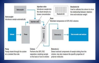
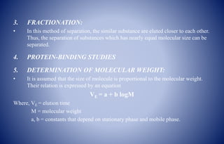
In this slide contains principle, instrumentation, methodology, and application of gel chromatography.
Presented by: SATHEES CHANDRA (Department of pharmaceutical analysis).
RIPER, anantapur
In this slide contains principle, instrumentation, methodology, and application of gel chromatography.
Presented by: SATHEES CHANDRA (Department of pharmaceutical analysis).
RIPER, anantapur
ION EXCHANGE CHROMATOGRAPHY
ByM.Vharshini
B.Sc. Bio Medical Science
Sri Ramachandra University
ION EXCHANGE CHROMATOGRAPHY
Ion-exchange chromatography is a process that allows the separation of ions and polar molecules based on their affinity to the ion exchanger.
It can be used for almost any kind of charged molecule including large proteins, small nucleotides and amino acids.
Cations or Anions can be separated using this method.
PRINCIPLE
It is based on the reversible electrostatic interaction of ions with the separation matrix (i.e.)
The separation occurs by reversible exchange of ions between the ions present in the solution and those present in the ion exchange resin.
CLASSIFICATION OF RESINS
According to the chemical nature they classified as-
1. Strong cation exchange resin
2. Weak cation exchange resin
3. Strong anion exchange resin
4. Weak anion exchange resin
According to the Source they can -
Natural resins : Cation - Zeolytes, Clay
Anion - Dolomite
Synthetic resins: Inorganic & Organic resins
◘Organic resins are polymeric resin matrix.
The resin composed of –
Polystyrene (sites for exchangeable functional groups)
Divinyl benzene(Cross linking agent)-offers stability.
Ion exchange resin should have following requirements
»It must be chemically stable.
»It should be insoluble in common solvents.
» It should have a sufficient degree of cross linking.
»The swollen resin must be denser than water.
»It must contain sufficient no. of ion exchange groups.
Physical properties of ion exchange resins
Cross linking:
It affects swelling & strength & solubility
Swelling:
When resin swells, polymer chain spreads apart
Polar solvents → swelling
Non-polar solvents → contraction
Swelling also affected electrolyte concentration.
Particle size and porosity
Increase in surface area & decrease in particle size will increase the rate of ion exchange.
Regeneration
Cation exchange resin are regenerated by treatment with acid, then washing with water.
Anion exchange resin are regenerated by treatment with NaOH, then washing with water until neutral.
EXPERIMENTAL SETUP OF ION EXCHANGE CHROMATOGRAPHY
Metrohm 850 Ion chromatography system
Instrumentation of ion exchange chromatography
PRACTICAL REQUIREMENTS
1.Column
» glass, stainless steel or polymers
2.Packing the column
» Wet packing method:
A slurry is prepared of the eluent with the stationary phase powder and then carefully poured into the column. Care must be taken to avoid air bubbles.
3.Application of the sample
After packing, sample is added to the top of the stationary phase, use syringe or pipette.
This layer is usually topped with a small layer of sand or with cotton or glass wool to protect the shape of the organic layer from the velocity of newly added eluent.
4.Mobile phase
Acids, alkalis, buffers…
6.Stationary phase
The ionic
In this slide contains principle of IR spectroscopy and sampling techniques.
Presented by: R.Banuteja (Department of pharmaceutical analysis).
RIPER, anantpur.
Liposomes-Classification, methods of preparation and application Vijay Hemmadi
liposome preparation and application
A liposome is a tiny bubble (vesicle), made out of the same material as a cell membrane. Liposomes can be filled with drugs, and used to deliver drugs for cancer and other diseases. Membranes are usually made of phospholipids, which are molecules that have a head group and a tail group
Gel chromatography, Introduction, Theory, Instrumentation, Applications .pptxVandana Devesh Sharma
Affinity chromatography- Content-Introduction
Theory
Instrumentation
Applications
Gel chromatography is a type of partition chromatography used for separating different sized molecules.
Gel chromatography is also called Gel permeation chromatography or gel filtration or gel exclusion, size exclusion, molecular- sieve chromatography.
The separation is based on the analyte molecular sizes since the gel behaves like a molecular sieve.
In size exclusion chromatography, the stationary phase is a porous matrix made up of compounds like
cross-linked polystyrene, cross-like dextrans, polyacrylamide gels, agarose gels, etc.
The gel structure being used contains pores of different diameters upto maximum size.
1.The test molecules are washed through a gel column and molecules larger than the largest pores in the gel are excluded from the gel structure.
2. Smaller molecules penetrate the gel and the extent of penetration depends on the molecular size----- This delay their movement through the column
This technique is used for the separation of proteins, polysaccharides, enzymes, and synthetic polymers. Instrumentation- A. Stationary phase- It is composed of semi-permeable, porous polymer gel beads with a well-defined range of pore sizes. eg. Dextran, Agarose, Acrylamide. 2. sample size and concentration- sample is applied in small volume (1-5% of the total bed volume).3. Column parameters- use long column, ratio of column diameter to column length (1:20 to :100). The method or steps used for gel preparation. 4. Choice of eluent/mobile phase- Buffers Ex- Phosphate buffer pH 7, NaCl solution, Ammonium acetate (CH3COO-NH4+ ), Ammonium bicarbonate (NH₄HCO₃) ethylenediamine acetate. 5. Effect of Flow rate- maintain with the help of pump. Elution carried out with buffer at optimal flow rate (Eg- 0.25-5ml/min) to give maximum resolution with optimal separation time.6. Separation of components from the sample-
Separation of component from mixture is achieved with the help of column. The retention volume (VR).7. Detection- Using UV absorption detectors. A graph of Elution Volume (ml) Vs Molecular weight. 7. Detection- Using UV absorption detectors. A graph of Elution Volume (ml) Vs Molecular weight. For calibration of the gel in column – Calibrators - (Proteins of known molecular weight. Procedure for gel filtration technique-1. Preparation of column- 2. Washing of the column- 3. Loading of the sample-4. Elution using mobile phase (buffers)5. Detection of compounds . Applications
Download and play it my friends it contain VIDEO
The technique of ion exchange chromatography is based upon the interaction between charged solute molecules and oppositely charged moieties covalently linked to chromatographic matrix.
The reasons for its widespread success is its applicability, high resolving power, high capacity and simplicity of the technique.
Separation in ion exchange chromatography depends upon the reversible adsorption of charged solute molecules to immobilized ion exchange groups of opposite charge. Most experiments are performed by following : Video For Understanding Play It
ION EXCHANGE CHROMATOGRAPHY
ByM.Vharshini
B.Sc. Bio Medical Science
Sri Ramachandra University
ION EXCHANGE CHROMATOGRAPHY
Ion-exchange chromatography is a process that allows the separation of ions and polar molecules based on their affinity to the ion exchanger.
It can be used for almost any kind of charged molecule including large proteins, small nucleotides and amino acids.
Cations or Anions can be separated using this method.
PRINCIPLE
It is based on the reversible electrostatic interaction of ions with the separation matrix (i.e.)
The separation occurs by reversible exchange of ions between the ions present in the solution and those present in the ion exchange resin.
CLASSIFICATION OF RESINS
According to the chemical nature they classified as-
1. Strong cation exchange resin
2. Weak cation exchange resin
3. Strong anion exchange resin
4. Weak anion exchange resin
According to the Source they can -
Natural resins : Cation - Zeolytes, Clay
Anion - Dolomite
Synthetic resins: Inorganic & Organic resins
◘Organic resins are polymeric resin matrix.
The resin composed of –
Polystyrene (sites for exchangeable functional groups)
Divinyl benzene(Cross linking agent)-offers stability.
Ion exchange resin should have following requirements
»It must be chemically stable.
»It should be insoluble in common solvents.
» It should have a sufficient degree of cross linking.
»The swollen resin must be denser than water.
»It must contain sufficient no. of ion exchange groups.
Physical properties of ion exchange resins
Cross linking:
It affects swelling & strength & solubility
Swelling:
When resin swells, polymer chain spreads apart
Polar solvents → swelling
Non-polar solvents → contraction
Swelling also affected electrolyte concentration.
Particle size and porosity
Increase in surface area & decrease in particle size will increase the rate of ion exchange.
Regeneration
Cation exchange resin are regenerated by treatment with acid, then washing with water.
Anion exchange resin are regenerated by treatment with NaOH, then washing with water until neutral.
EXPERIMENTAL SETUP OF ION EXCHANGE CHROMATOGRAPHY
Metrohm 850 Ion chromatography system
Instrumentation of ion exchange chromatography
PRACTICAL REQUIREMENTS
1.Column
» glass, stainless steel or polymers
2.Packing the column
» Wet packing method:
A slurry is prepared of the eluent with the stationary phase powder and then carefully poured into the column. Care must be taken to avoid air bubbles.
3.Application of the sample
After packing, sample is added to the top of the stationary phase, use syringe or pipette.
This layer is usually topped with a small layer of sand or with cotton or glass wool to protect the shape of the organic layer from the velocity of newly added eluent.
4.Mobile phase
Acids, alkalis, buffers…
6.Stationary phase
The ionic
In this slide contains principle of IR spectroscopy and sampling techniques.
Presented by: R.Banuteja (Department of pharmaceutical analysis).
RIPER, anantpur.
Liposomes-Classification, methods of preparation and application Vijay Hemmadi
liposome preparation and application
A liposome is a tiny bubble (vesicle), made out of the same material as a cell membrane. Liposomes can be filled with drugs, and used to deliver drugs for cancer and other diseases. Membranes are usually made of phospholipids, which are molecules that have a head group and a tail group
Gel chromatography, Introduction, Theory, Instrumentation, Applications .pptxVandana Devesh Sharma
Affinity chromatography- Content-Introduction
Theory
Instrumentation
Applications
Gel chromatography is a type of partition chromatography used for separating different sized molecules.
Gel chromatography is also called Gel permeation chromatography or gel filtration or gel exclusion, size exclusion, molecular- sieve chromatography.
The separation is based on the analyte molecular sizes since the gel behaves like a molecular sieve.
In size exclusion chromatography, the stationary phase is a porous matrix made up of compounds like
cross-linked polystyrene, cross-like dextrans, polyacrylamide gels, agarose gels, etc.
The gel structure being used contains pores of different diameters upto maximum size.
1.The test molecules are washed through a gel column and molecules larger than the largest pores in the gel are excluded from the gel structure.
2. Smaller molecules penetrate the gel and the extent of penetration depends on the molecular size----- This delay their movement through the column
This technique is used for the separation of proteins, polysaccharides, enzymes, and synthetic polymers. Instrumentation- A. Stationary phase- It is composed of semi-permeable, porous polymer gel beads with a well-defined range of pore sizes. eg. Dextran, Agarose, Acrylamide. 2. sample size and concentration- sample is applied in small volume (1-5% of the total bed volume).3. Column parameters- use long column, ratio of column diameter to column length (1:20 to :100). The method or steps used for gel preparation. 4. Choice of eluent/mobile phase- Buffers Ex- Phosphate buffer pH 7, NaCl solution, Ammonium acetate (CH3COO-NH4+ ), Ammonium bicarbonate (NH₄HCO₃) ethylenediamine acetate. 5. Effect of Flow rate- maintain with the help of pump. Elution carried out with buffer at optimal flow rate (Eg- 0.25-5ml/min) to give maximum resolution with optimal separation time.6. Separation of components from the sample-
Separation of component from mixture is achieved with the help of column. The retention volume (VR).7. Detection- Using UV absorption detectors. A graph of Elution Volume (ml) Vs Molecular weight. 7. Detection- Using UV absorption detectors. A graph of Elution Volume (ml) Vs Molecular weight. For calibration of the gel in column – Calibrators - (Proteins of known molecular weight. Procedure for gel filtration technique-1. Preparation of column- 2. Washing of the column- 3. Loading of the sample-4. Elution using mobile phase (buffers)5. Detection of compounds . Applications
Download and play it my friends it contain VIDEO
The technique of ion exchange chromatography is based upon the interaction between charged solute molecules and oppositely charged moieties covalently linked to chromatographic matrix.
The reasons for its widespread success is its applicability, high resolving power, high capacity and simplicity of the technique.
Separation in ion exchange chromatography depends upon the reversible adsorption of charged solute molecules to immobilized ion exchange groups of opposite charge. Most experiments are performed by following : Video For Understanding Play It
Product polishing techniques in Downstream ProcessingErin Davis
This is a presentation based on gel permeation chromatography and dialysis.This mainly deals with the basic principle behind these techniques.and its working.The major components,advantages,disadvantages,applications are also mentioned in the same.Besides these the pictoric representation helps to understand the concept clearly.
This will be helpful to learn downstream processing techniques.
GEL CHROMATOGRAPHY
GEL CHROMATOGRAPHY B.PHARM
GEL CHROMATOGRAPHY M.PHARM
SIZE EXCLUSION CHROMATOGRPHY
GEL CHROMATOGRPHY PPT
GEL CHROMATOGRAPHY SLIDESHARE
introduction
mechanisms of protein drug binding
binding of drugs
binding of drugs to blood components
determination of protein drug binding
factors affecting
significance
Introduction
working principle
fragmentation process
general rules for fragmentation
general modes of fragmentation
metastable ions
isotopic peaks
applications
POTENTIAL SOURCES OF ELEMENTAL IMPURITIESMehulJain143
INTRODUCTION
INDENTIFICATION OF POTENTIAL ELEMENTAL IMPURITIES
FACTORS AFFECTING
EVALUATION
RISK ASSESSMENT AND CONTROL OF ELEMENTAL IMPURITIES
GENERAL PRINCIPLES
Seminar of U.V. Spectroscopy by SAMIR PANDASAMIR PANDA
Spectroscopy is a branch of science dealing the study of interaction of electromagnetic radiation with matter.
Ultraviolet-visible spectroscopy refers to absorption spectroscopy or reflect spectroscopy in the UV-VIS spectral region.
Ultraviolet-visible spectroscopy is an analytical method that can measure the amount of light received by the analyte.
Earliest Galaxies in the JADES Origins Field: Luminosity Function and Cosmic ...Sérgio Sacani
We characterize the earliest galaxy population in the JADES Origins Field (JOF), the deepest
imaging field observed with JWST. We make use of the ancillary Hubble optical images (5 filters
spanning 0.4−0.9µm) and novel JWST images with 14 filters spanning 0.8−5µm, including 7 mediumband filters, and reaching total exposure times of up to 46 hours per filter. We combine all our data
at > 2.3µm to construct an ultradeep image, reaching as deep as ≈ 31.4 AB mag in the stack and
30.3-31.0 AB mag (5σ, r = 0.1” circular aperture) in individual filters. We measure photometric
redshifts and use robust selection criteria to identify a sample of eight galaxy candidates at redshifts
z = 11.5 − 15. These objects show compact half-light radii of R1/2 ∼ 50 − 200pc, stellar masses of
M⋆ ∼ 107−108M⊙, and star-formation rates of SFR ∼ 0.1−1 M⊙ yr−1
. Our search finds no candidates
at 15 < z < 20, placing upper limits at these redshifts. We develop a forward modeling approach to
infer the properties of the evolving luminosity function without binning in redshift or luminosity that
marginalizes over the photometric redshift uncertainty of our candidate galaxies and incorporates the
impact of non-detections. We find a z = 12 luminosity function in good agreement with prior results,
and that the luminosity function normalization and UV luminosity density decline by a factor of ∼ 2.5
from z = 12 to z = 14. We discuss the possible implications of our results in the context of theoretical
models for evolution of the dark matter halo mass function.
Slide 1: Title Slide
Extrachromosomal Inheritance
Slide 2: Introduction to Extrachromosomal Inheritance
Definition: Extrachromosomal inheritance refers to the transmission of genetic material that is not found within the nucleus.
Key Components: Involves genes located in mitochondria, chloroplasts, and plasmids.
Slide 3: Mitochondrial Inheritance
Mitochondria: Organelles responsible for energy production.
Mitochondrial DNA (mtDNA): Circular DNA molecule found in mitochondria.
Inheritance Pattern: Maternally inherited, meaning it is passed from mothers to all their offspring.
Diseases: Examples include Leber’s hereditary optic neuropathy (LHON) and mitochondrial myopathy.
Slide 4: Chloroplast Inheritance
Chloroplasts: Organelles responsible for photosynthesis in plants.
Chloroplast DNA (cpDNA): Circular DNA molecule found in chloroplasts.
Inheritance Pattern: Often maternally inherited in most plants, but can vary in some species.
Examples: Variegation in plants, where leaf color patterns are determined by chloroplast DNA.
Slide 5: Plasmid Inheritance
Plasmids: Small, circular DNA molecules found in bacteria and some eukaryotes.
Features: Can carry antibiotic resistance genes and can be transferred between cells through processes like conjugation.
Significance: Important in biotechnology for gene cloning and genetic engineering.
Slide 6: Mechanisms of Extrachromosomal Inheritance
Non-Mendelian Patterns: Do not follow Mendel’s laws of inheritance.
Cytoplasmic Segregation: During cell division, organelles like mitochondria and chloroplasts are randomly distributed to daughter cells.
Heteroplasmy: Presence of more than one type of organellar genome within a cell, leading to variation in expression.
Slide 7: Examples of Extrachromosomal Inheritance
Four O’clock Plant (Mirabilis jalapa): Shows variegated leaves due to different cpDNA in leaf cells.
Petite Mutants in Yeast: Result from mutations in mitochondrial DNA affecting respiration.
Slide 8: Importance of Extrachromosomal Inheritance
Evolution: Provides insight into the evolution of eukaryotic cells.
Medicine: Understanding mitochondrial inheritance helps in diagnosing and treating mitochondrial diseases.
Agriculture: Chloroplast inheritance can be used in plant breeding and genetic modification.
Slide 9: Recent Research and Advances
Gene Editing: Techniques like CRISPR-Cas9 are being used to edit mitochondrial and chloroplast DNA.
Therapies: Development of mitochondrial replacement therapy (MRT) for preventing mitochondrial diseases.
Slide 10: Conclusion
Summary: Extrachromosomal inheritance involves the transmission of genetic material outside the nucleus and plays a crucial role in genetics, medicine, and biotechnology.
Future Directions: Continued research and technological advancements hold promise for new treatments and applications.
Slide 11: Questions and Discussion
Invite Audience: Open the floor for any questions or further discussion on the topic.
(May 29th, 2024) Advancements in Intravital Microscopy- Insights for Preclini...Scintica Instrumentation
Intravital microscopy (IVM) is a powerful tool utilized to study cellular behavior over time and space in vivo. Much of our understanding of cell biology has been accomplished using various in vitro and ex vivo methods; however, these studies do not necessarily reflect the natural dynamics of biological processes. Unlike traditional cell culture or fixed tissue imaging, IVM allows for the ultra-fast high-resolution imaging of cellular processes over time and space and were studied in its natural environment. Real-time visualization of biological processes in the context of an intact organism helps maintain physiological relevance and provide insights into the progression of disease, response to treatments or developmental processes.
In this webinar we give an overview of advanced applications of the IVM system in preclinical research. IVIM technology is a provider of all-in-one intravital microscopy systems and solutions optimized for in vivo imaging of live animal models at sub-micron resolution. The system’s unique features and user-friendly software enables researchers to probe fast dynamic biological processes such as immune cell tracking, cell-cell interaction as well as vascularization and tumor metastasis with exceptional detail. This webinar will also give an overview of IVM being utilized in drug development, offering a view into the intricate interaction between drugs/nanoparticles and tissues in vivo and allows for the evaluation of therapeutic intervention in a variety of tissues and organs. This interdisciplinary collaboration continues to drive the advancements of novel therapeutic strategies.
This pdf is about the Schizophrenia.
For more details visit on YouTube; @SELF-EXPLANATORY;
https://www.youtube.com/channel/UCAiarMZDNhe1A3Rnpr_WkzA/videos
Thanks...!
Nutraceutical market, scope and growth: Herbal drug technologyLokesh Patil
As consumer awareness of health and wellness rises, the nutraceutical market—which includes goods like functional meals, drinks, and dietary supplements that provide health advantages beyond basic nutrition—is growing significantly. As healthcare expenses rise, the population ages, and people want natural and preventative health solutions more and more, this industry is increasing quickly. Further driving market expansion are product formulation innovations and the use of cutting-edge technology for customized nutrition. With its worldwide reach, the nutraceutical industry is expected to keep growing and provide significant chances for research and investment in a number of categories, including vitamins, minerals, probiotics, and herbal supplements.
A brief information about the SCOP protein database used in bioinformatics.
The Structural Classification of Proteins (SCOP) database is a comprehensive and authoritative resource for the structural and evolutionary relationships of proteins. It provides a detailed and curated classification of protein structures, grouping them into families, superfamilies, and folds based on their structural and sequence similarities.
Professional air quality monitoring systems provide immediate, on-site data for analysis, compliance, and decision-making.
Monitor common gases, weather parameters, particulates.
2. INTRODUCTION
DEFINITION:
Gel Chromatography (known as gel permeation, molecular sieving or size
exclusion chromatography) is a chromatographic technique in which the separation of
components based on the difference of molecular weight or size, and is one of the
effective methods used to isolate and analyze the bio-macromolecular substances.
The stationary phase consists of beads containing pores that span a relatively narrow
size range. When the gel is packed into a column and percolated with a solvent, it
permits the large molecular weight components to pass faster without penetration of the
pores (totally excluded).
Smaller molecules spend more time inside the beads and therefore is eluted later
(after a larger volume of mobile phase has passed through the column).
3. • When an organic solvent is used as a mobile phase, then it is tend to called as gel
permeation chromatography.
• When an aqueous solution is used to transport the sample through the column, the
technique is known as gel filtration chromatography.
In 1954, the scientist Mould and Sunge showed that the separation of uncharged
substances can be performed based on the size of molecules.
However the systematic use of this principle was introduced by Porth and Foblin in
1954 who separated the molecules of biological origin using polysaccharide gel
(Sephadex).
Stationary phase: Polysaccharide gel (cross-linked to form pores)
Mobile phase: Liquid solvent
Sample: Molecules of biological origin (different sizes)
4. ADVANTAGES:
• Short analysis time.
• Well defined separation.
• Narrow bands and good sensitivity.
• There is no sample loss.
• Small amount of mobile phase required.
• The flow rate can be set.
DISADVANTAGES:
• Limited number of peaks that can be resolved within the short time scale.
• Filtrations must be performed before using the instrument to prevent dust and other
particulates from ruining the columns and interfering with the detectors.
• The molecular masses of most of the chains will be too close for the separation to
show anything more than broad peaks.
5. PRINCIPLE
• The stationary phase in this method is an open network of polymers which are
cross-linked to each other to form the pores of consistent size. The degree of cross-
linking of polymer mainly governs the pore size.
• When the mobile phase containing mixture of solutes of various sizes is passed
through the column, the molecules which are too large to enter the pores are
“excluded” completely and get eluted with the mobile phase.
• The molecules which are smaller in size diffuse in and out of the pores, thus the
path travelled by them through the column is quite longer and they are eluted later.
• The molecules which are of intermediate size cannot diffuse into the earlier pores as
the space is already occupied by the smaller molecules hence they get separated
quite later along the length of column.
• Thus, the components of mixture get eluted from the column in the order of their
relative molecular sizes.
6. • The one with largest molecular mass or size is eluted first, followed by elution of
intermediate sized molecules. The smaller size molecules are eluted in the last order
of elution.
Large size > Intermediate size > Smallest size
7. THEORY OF SEPARATION
A column is made up of swollen gel particles and the solvent used to swell the gel in a
suitable tubular container.
An equation is given below:
Vt = V0 + Vi + Vm
Where, Vt = the total volume of the column (which can be measured),
V0 = the volume of liquid outside the gel matrix (known also void or dead volume),
Vi = the volume of liquid inside the matrix,
Vm = the volume of the gel matrix
8. TYPES OF GELS USED
• The gels used as molecular sieves are cross linked polymers.
• They are uncharged and inert i.e., don’t bind or react with the materials being
analyzed.
• Three types of gels are used:
1. Dextran (Sephadex)
2. Agarose gel
3. Acrylamide gels (synthetic gel)
Nature of Gel:
• Chemically inert
• Mechanically stable
• Ideal porous structure
• Wide pore size give low resolution
• Uniform particle size
9. 1. DEXTRAN (SEPHADEX):
• Dextran is a homopolysaccharide of glucose residues.
• It is prepared with various degrees of cross-linking to control pore size.
• It is bought as dry beads, the beads swell when water is added.
• 1-6 polymer of glucose is prepared by microbial fermentation of sucrose (glucose +
fructose).
• The resulting glucose provides the required α 1-6 glucosan polymer called dextran.
• The resulting dextran is treated with epichlorohydrin to give several types of cross-
linked dextran (sephadex).
• It is mainly used for separation of small peptides and globular proteins with small to
average molecular mass.
• Sephadex is obtained in different degrees depending on the pore size.
• High percentage of epichlorohydrin give high degree of cross linking (small pore
size).
• Lower percentage produce sephadex with large pore size.
10. Characters of Sephadex:
• Highly stable gels.
• Stable at pH 2-12.
• Their particles are free from ions.
• Insoluble in water and organic solvent.
• They swell in water and other hydrophilic solvent.
• They require bactericidal such as Hg acetate.
2. AGAROSE GEL:
• Obtained from agar and composed of alternating units of 1,3 linked β-D-gal and 1,4
linked 3,6-anhydro-α, L-galactose.
• This was subjected to epichlorohydrin to give sepharose.
Characters:
• It dissolves in H2O at 50ºC and on cooling form gel.
• Insoluble below 40ºC.
• Freezing destroys the gel.
11. 3. ACRYLAMIDE GELS (SYNTHETIC GEL):
• It is not dextran polymer.
• It is polymerized acrylamide or methylene-bis-acrylamide.
• The pore size is determined by the degree of cross-linking. The separation
properties of polyacrylamide gels are mainly
• The same as those of dextrans.
• They are sold as bio-gel P. They are available in wide range of pore sizes.
According to the swelling process, the gels are two types:
1. Soft gels (Xerogel i.e., gel only on swelling):
Example: Polyacrylamide gels, dextran or agarose (used for separation of proteins in
aqueous mobile phase).
2. Semirigid or rigid gels (aerogel i.e., gel in air):
• Polystyrene gels (separation of non-polar polymers in non-polar solvents).
• Porous glass gels (separation of polar systems).
12. COMPONENTS
• Stationary Phase
• The Mobile Phase
• The Columns
• The Pump
• Detectors
1. STATIONARY PHASE:
• Composed of semi-permeable, porous polymer gel beads with well defined range of
pore sizes.
Properties of gel beads:
• Chemically inert.
• Mechanically stable.
• Has ideal and homogeneous porous structure (wide pore size give low resolution).
• Uniform particle and pore size.
• The pore size of the gel must be carefully controlled.
13. Examples of gel:
• Dextran(Sephadex) gel: An α 1-6-polymer of glucose natural gel.
• Agarose gel: A 1,3 linked β-D-galactose and 1,4 linked 3,6-anhydro-α, L-galactose
natural gel.
• Acrylamide gel: A polymerized acrylamide, a synthetic gel.
2. THE MOBILE PHASE:
• Composed of a liquid used to dissolve the bio-molecules to make the mobile phase
permitting high detection response and wet the packing surface.
MATERIAL SOLVENT
Synthetic elastomers (polybutadiene,
polyisoprene)
Toluene
PS, PVC, Styrene-Butadiene Rubber, Epoxy resins Tetrahydrofuran (THF)
Polyolefins Tri-chloro-benzene
Polyurethane Di-methylformamide (DMF)
Proteins, polysaccharides Water / Buffers
14. 3. COLUMNS:
Commercially Available Columns include
• Analytical column- 7.5–8mm diameters.
• Preparative columns-22–25mm.
• Usual column lengths-25, 30, 50, and
60 cm.
• Narrow bore columns- 2–3mm diameter
have been introduced.
4. THE PUMP:
• These are either syringe pumps or
reciprocating pumps with a highly
constant flow rate.
17. SEPARATION PROCEDURE
1. PREPARATION OF COLUMN FOR GEL FILTRATION:
• Swelling of the gel: Some resin come in a powder form. These must be sonicated
first in the eluent or the desired buffer to swell.
• Packing the column: Make a slurry of gel + buffer and pour it into column which
is one third filled with the buffer.
• Washing the resin: After packing, pass several column volumes of the buffer
through the column to remove any air bubbles and to test the column homogeneity.
2. LOADING THE SAMPLE ONTO THE COLUMN:
• The sample must enter the resin in the form of solution using a syringe.
3. ELUTING THE SAMPLE AND DETECTION OF COMPONENTS:
• Fractions are collected as the sample elutes from the column.
18. APPLICATIONS
Gel chromatography is mainly used for the separation of sugars, polysaccharides,
proteins, lipids, polymers and other materials.
1. PURIFICATION:
• This technique is used for purification of biological molecules.
• Different proteins, enzymes, hormones, antibodies, polysaccharides have been
separated and purified by using appropriate gels.
• Low molecular weight dextrans can be separated from corn syrup oil.
2. DESALTING:
• This method of desalting is faster and more efficient than dialysis.
• Examples of desalting process include separation of monosaccharides from
polysaccharides and separation of amino acids from proteins.
19. 3. FRACTIONATION:
• In this method of separation, the similar substance are eluted closer to each other.
Thus, the separation of substances which has nearly equal molecular size can be
separated.
4. PROTEIN-BINDING STUDIES
5. DETERMINATION OF MOLECULAR WEIGHT:
• It is assumed that the size of molecule is proportional to the molecular weight.
Their relation is expressed by an equation
VE = a + b logM
Where, VE = elution time
M = molecular weight
a, b = constants that depend on stationary phase and mobile phase.