
Dr. Reshmi G R
Division – Pteropsida
Class - Leptosporangiopsida
Order – Filicales
Family – Polypodiaceae
Genus – Adiantum
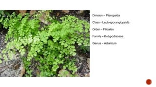
 Adiantum is commonly known as ‘Maiden hair fern’ or ‘Walking fern’ because of the shiny black rachis of
the leaves.
 It is one of the most widely distributed genera of the family growing luxuriantly in both tropical and sub
topical regions of the world.
 There are nearly 200 species.
 Nayar (1961) has investigated the morphology of 24 Indian species of Adiantum.
 Some of the common Indian species are – A. capillus-veneris, A. pedatum, A. incisum, A. caudatum, A.
venustum, A. lunulatum, A. edgowrthii etc.
 It grows ubiquitously wherever nature offers a moist, shaded locality.
 Species of Adiantum are commonly cultivated in green houses because of their attractive foliage.
Morphology of the plant:
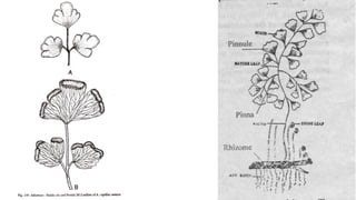
 The sporophyte is differentiated into rhizome, roots and leaves
 Rhizome
 The rhizome is a perennial, subterranean dichotomously branched structure and is creeping in A.
capillus-veneris or may be erect as in A. caudatum.
 It is covered with persistent leaf bases and hairy outgrowths called ramenta.
 The sporophytic plant body consists of an underground rhizome from which are produced leaves and
roots.
 The rhizome is covered with chaffy scales (Paleae).
 It may be erect (A. caudatum), semi erect (A. pedatum), or creeping (A. capillus-veneris).
 Studies of Nicholas (1985) in Adiantum trapeziforme indicate that the erect rhizome of the young
sporophyte quickly transforms itself into creeping.
 The rhizome may be hard or soft and brown in colour.
 The chaffy scales that cover the rhizome are of various shapes and sizes.
 Root
 The roots are adventitious and arise from the rhizome.
 The roots are stiff and black in colour.
 Occasionally they may be branched.
 Leaf
 The leaves are also called fronds and are pinnately compound (unipinnate- A. caudatum, bipinnate- A.
capillus- veneris) the young leaves are circinately coiled.
 The petiole is long, black and shiny.
 The venation is free and dichotomous in all the species.
 The vein spread in a fan-like manner in the lamina.
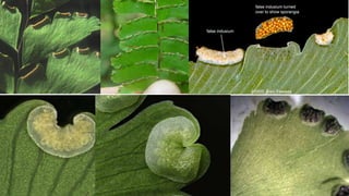
 The leaves bear marginal sori which are covered by a false indusium.
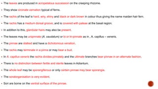
 The leaves are produced in acropetalous succession on the creeping rhizome.
 They show circinate vernation typical of ferns.
 The rachis of the leaf is hard, wiry, shiny and black or dark brown in colour thus giving the name maiden hair fern.
 The rachis has a medium dorsal groove, and is covered with paleae at the basal region.
 In addition to this, glandular hairs may also be present.
 The leaves may be unipinnate (A. xaudatum) or bi or tri-pinnate as in , A. capillus – veneris.
 The pinnae are stalked and have a dichotomous venation.
 The rachis may terminate in a pinna or may bear a bud.
 In A. capillus veneris the rachis divides pinnately and the ultimate branches bear pinnae in an alternate fashion.
 There is no distinction between fertile and sterile leaves in Adiantum.
 The whole leaf may be sporangiferous or only certain pinnae may bear sporangia.
 The soralorganisation is very evident.
 Sori are borne on the ventral surface of the pinnae.
Frond and whole plant of Adiantum
species.
A. A. capillus-veneris B. A. lunulatum C.
A. peruvianum D. A. venustum
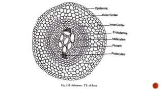
Root
 A transection shows a very prominent piliferous layer, a two zoned cortex and
the central protostele.
 The root is differentiated into epidermis, cortex and central vascular cylinder.
 The epidermis is the outermost layer and bears unicellular root hairs.
 The cortex is divided into outer wide parenchymatous and inner narrow
sclerenchymatous layer.
 Surrounding the stele is a conspicuous endodermis with prominent casparian
thickenings.
 External to phloem is a single layered pericycle.
 The stele is simple and possesses a central core of xylem in diarch condition
with phloem on either side of it.
Rhizome
 The rhizome in transverse section shows a single layered epidermis covered by cuticle.
 Some epidermal cells bear multicellular hairs.
 The Epidermis is followed by two to three layered hypodermis made up of sclerenchyma tissue.
 A parenchymatous ground tissue is present.
 The young rhizomes have amphiphloic siphonostele.
 The older rhizomes have solenostele or dictyostele
 A transection reveals the usual three zones epidermis, cortex and stele.
 The outline of the section would be wavy.
 Epidermis is single layered and the cells may be thin walled or thick walled.
 There is a cuticle external to the epidermis.
 Cortex lies internal to the epidermis.
 It may be wholly parenchymatous (A. rubellum), or it may have sclerenchyma and
parenchyma.
 In A. pectinatum, scattered masses of sclerenchyma are found embedded in the
parenchymatous ground tissue.
 In A. caudatum, sclerenchyma constitutes the hypodermal region.
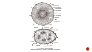
 The central vascular cylinder exhibits great variety.
 In A. capillus veneris, it is a dictyostele consisting of a ring of meristeles.
 In the young condition the stele may be a solenostele.
 In A. rubellum the stele is a typical amphiphloic solenostele, with characteristic
features such as outer endodermis, outer pericycle, outer phloem, xylem, inner
phloem, inner pericycle and inner endodermis lining the parenchymatous pith.
Petiole
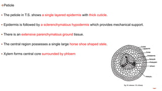
 The petiole in T.S. shows a single layered epidermis with thick cuticle.
 Epidermis is followed by a sclerenchymatous hypodermis which provides mechanical support.
 There is an extensive parenchymatous ground tissue.
 The central region possesses a single large horse shoe shaped stele.
 Xylem forms central core surrounded by phloem
TS Rachis of Adiantum species. A. A. capillus-
veneris B. A. lunulatum C. A. peruvianum; D. A.
venustum cer: ceratenchyma; cu: cuticle; end:
endodermis; gt: ground tissue; hyp: hypodermis; ph:
phloem; ste: stele; xy: xylem.
Pinnule
 The Pinnule shows upper and lower epidermis.
 The cells contain chloroplasts.
 Stomata are confined to lower epidermis.
 The mesophyll is not differentiated into palisade and spongy parenchyma.
 The vascular bundle is surrounded by sclerenchymatous bundle sheath.
 The lamina shows the two epidermal layers upper and lower the mesophyll is generally
undifferentiated.
 It is highly reduced in A. capillus – veneris, A. pedatum, etc., having only two layers of
cells.
 In A. pedatum, in some regions the mesophyll is totally absent and at such places the
two epidermal layers are closely appressed to each other.
 The mesophyll (when present) as well as the epidermal layers are chlorophyllous.
 The epidermal cells over the veins are thick walled.
 The stomata are scattered throughout the surface of the leaf.
 Paleae or ramenta may be borne even on the epidermis of the lamina.
 Vegetative propagation is brought about by buds produced at the leaf tips.
 The buds enter the ground when the leaf bends and touches the soil.
 There they develop into a new individual.
 This, in turn may repeat the process leading to the walking Habit.
 Walking habit is seen in A. caudatum.
 Adiantum is homosporous.
 The reproduction takes place by the production of spores.
 The spores are produced in sporangia.
 A group of sporangia forms sori.
 The sori on distal end of the pinnae but the reflex margins of the pinna form a protective membranous
structure called false indusium.
 The development of sporangium is of leptosporangiate type.
 The sorus does not show any definite sequence hence fall under mixed type.
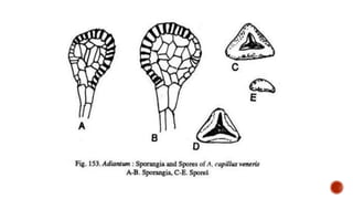
 A mature sporangium bears a multicellular stalk and a spherical or elliptical single layered structure
called capsule.
 The capsule contains haploid spores.
 The wall of the capsule is differentiated into thick walled annulus and thin walled stomium.
 On maturity the sporangium bursts and spores are released.
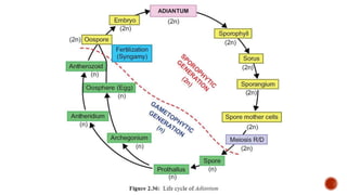
 The spores germinate and undergo repeated division to produce a prothallus.
 The prothallus is flat, green and heart shaped.
 It is monoecious and represents the gametophytic phase.
 Sex organs called antheridia and archegonia develop on the prothallus.
 Antheridia release multiflagellate antherozoids which swim in water and reach the egg of the
archegonium to accomplish fertilization.
 The fertilization results in zygote(2n) and it represents the first cell of sporophytic generation.
 The zygote develops into embryo which further differentiates into sporophyte.
 Thus Adiantum shows alternation of generation
 No distinction into fertile and sterile leaves.
 The sori are born at the distal end of the pinnae.
 But the sori are not exactly marginal.
 They are borne a little behind the tip of the veins.
 The sorus bearing margin of the leaf in curls and forms the false inducium.
 In some cases sporangia may develop at the distal ends of the veins (A. phillippense).
 In the sori paraphyses may be present in between the sporangia as in A. rubellum, A. tenerum, etc.
 The sorus is of the mixed type.
 The development is similar to what is seen in Pteris.
 A mature sporangium has a stalk made up of three rows of cells.
 The stalk terminates in a globose or biconvex capsule. The wall is single layered.
 There is an obliquely vertical annulus of 12-24 cells long.
 The annulus is separated from the stalk by two or three cells.
 The stomium also is separated from both the stalk, and the annulus.
 The rest of the sporangial wall is composed of a few large cells.
 The sporangium dehisces transversely liberating the spores.
 All the spores are of the same type.
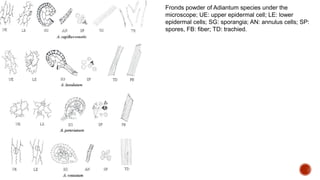
Fronds powder of Adiantum species under the
microscope; UE: upper epidermal cell; LE: lower
epidermal cells; SG: sporangia; AN: annulus cells; SP:
spores, FB: fiber; TD: trachied.
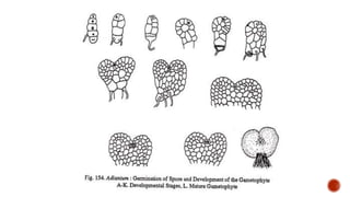
Structure and germination of the spores:
 Spores are tetrahedral in shape.
 The wall is two layered.
 Exine is thick and smooth and has a brownish tinge.
 On falling upon a suitable substratum the spore germinates.
 The first sign of germination is the rupturing of exine and the protruding out of the germ tube.
 The germ tube undergoes several transverse divisions to form a short filament.
 The lowest cell forms a lateral rhizoid.
 The terminal cell becomes an apical cell with three cutting faces.
 By the division of the apical cell, a spatulate pro-thallus is formed first.
The mature pro-thallus is cordate, photosynthetic, dorsiventrally flattened
and aerial.
The growing point is situated in the apical notch.
All the cells in the pro-thallus are parenchymatous.
The pro-thallus is one celled thick towards the margins but many celled
thick towards the centre.
In some species collenchyma may be found at the corners.
Rhizoids are produced from the ventral surface.
 The prothalli are monoecious.
 Antheridia are found in between the rhizoids towards the ventral surface.
 Arechegonia are found near the growing point towards the ventral surface.
 Structure and development of sex organs is same as in Pteris.
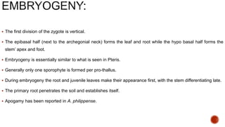
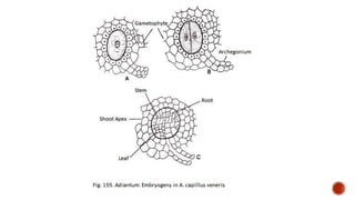
 The first division of the zygote is vertical.
 The epibasal half (next to the archegonial neck) forms the leaf and root while the hypo basal half forms the
stem’ apex and foot.
 Embryogeny is essentially similar to what is seen in Pteris.
 Generally only one sporophyte is formed per pro-thallus.
 During embryogeny the root and juvenile leaves make their appearance first, with the stem differentiating late.
 The primary root penetrates the soil and establishes itself.
 Apogamy has been reported in A. philippense.
Adiantum .pptx
Adiantum .pptx
Adiantum .pptx