Downloaded 337 times


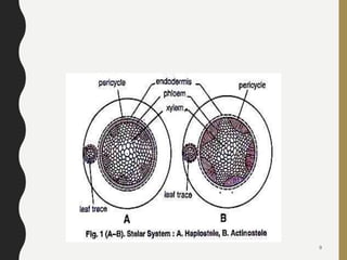

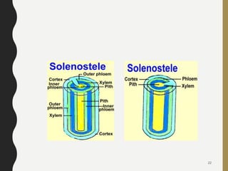
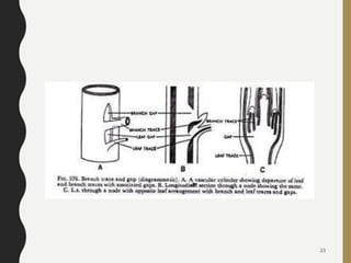
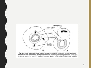
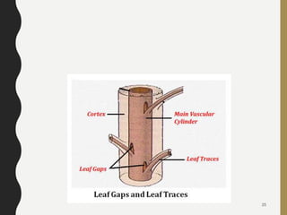
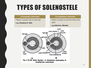
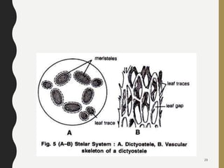


The document discusses the stelar system in plants, describing the fundamental components and types of steles such as protostele, siphonostele, and solenostele, along with their subcategories. It elaborates on the evolutionary theories of steles and includes specific examples of each type. Overall, the paper emphasizes the importance of the stele as a central vascular structure in higher plants.