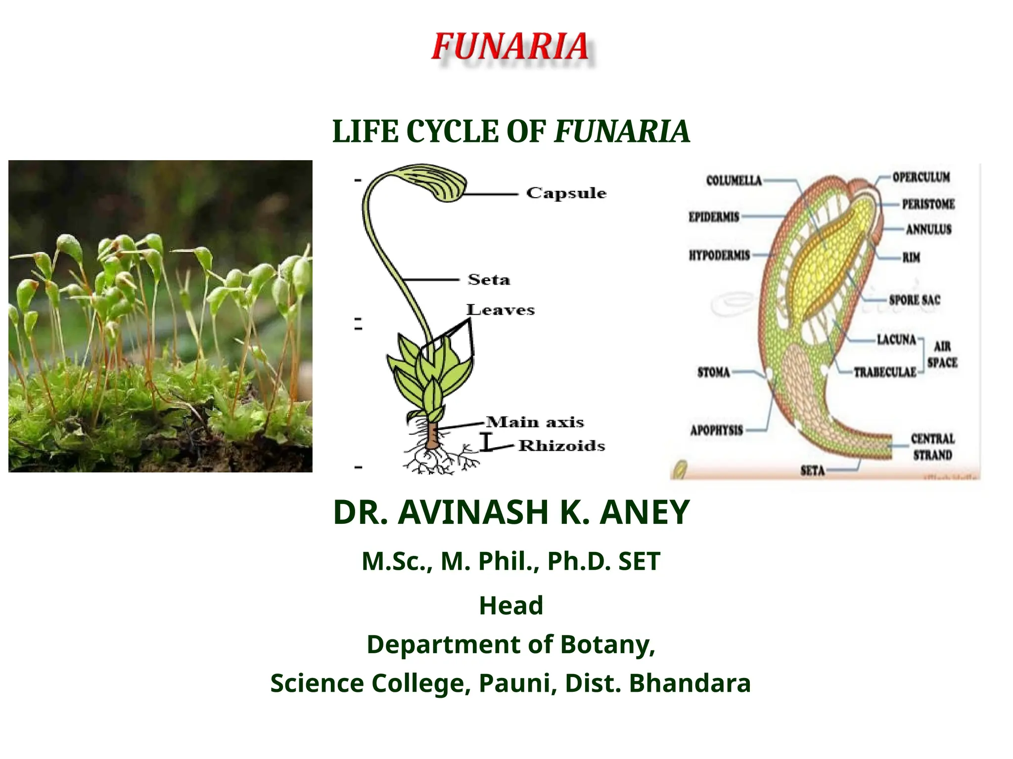
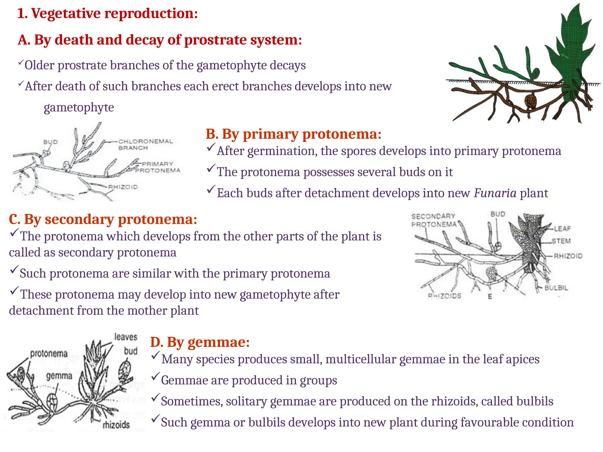
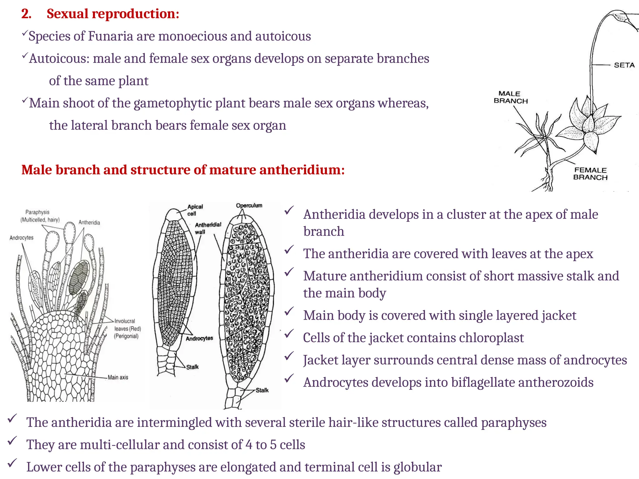
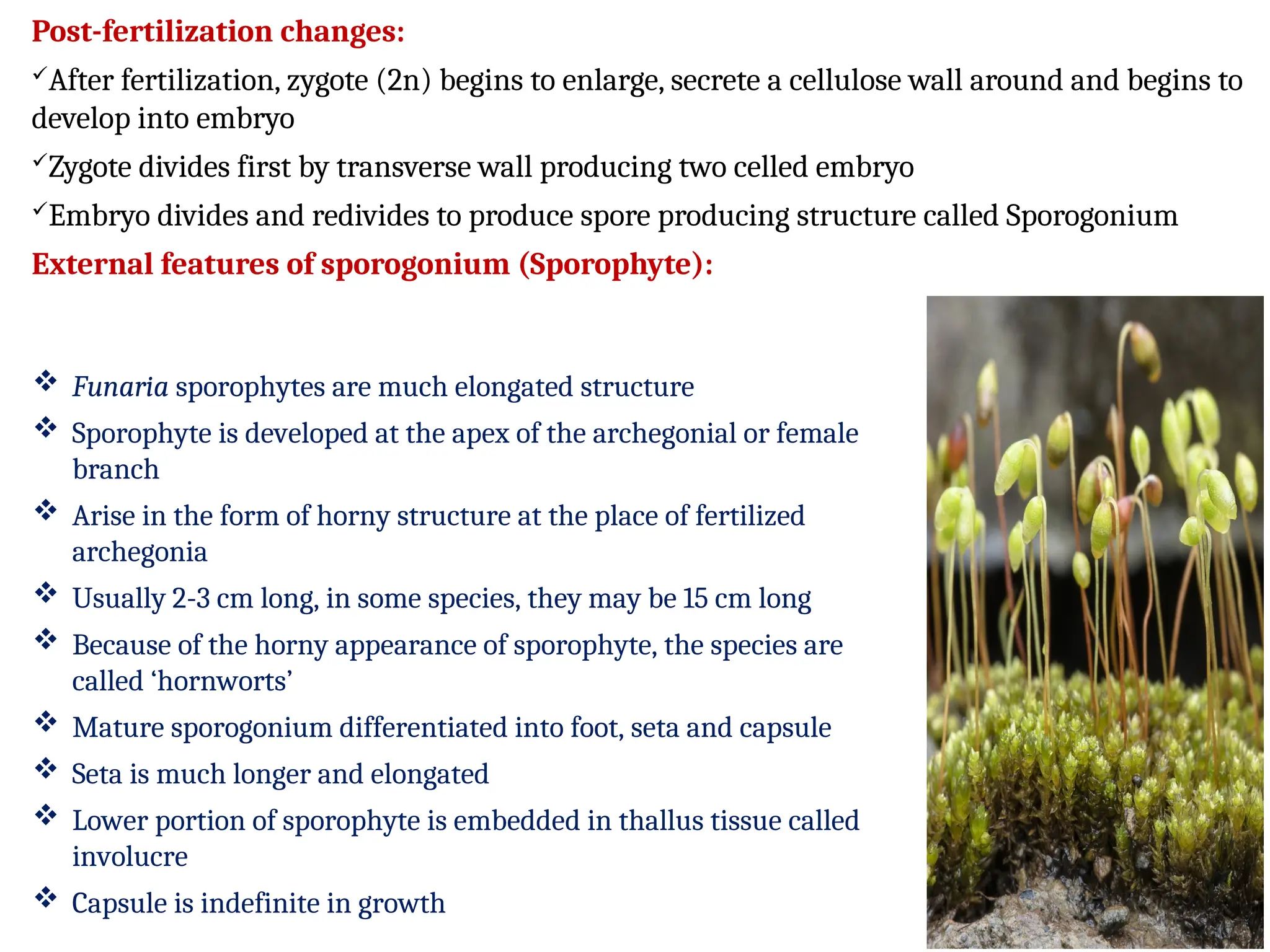
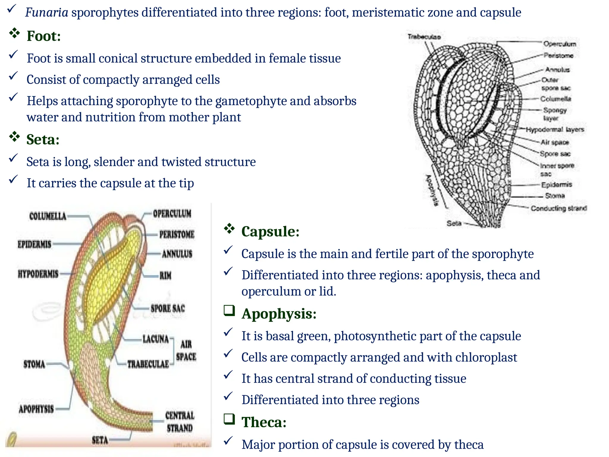
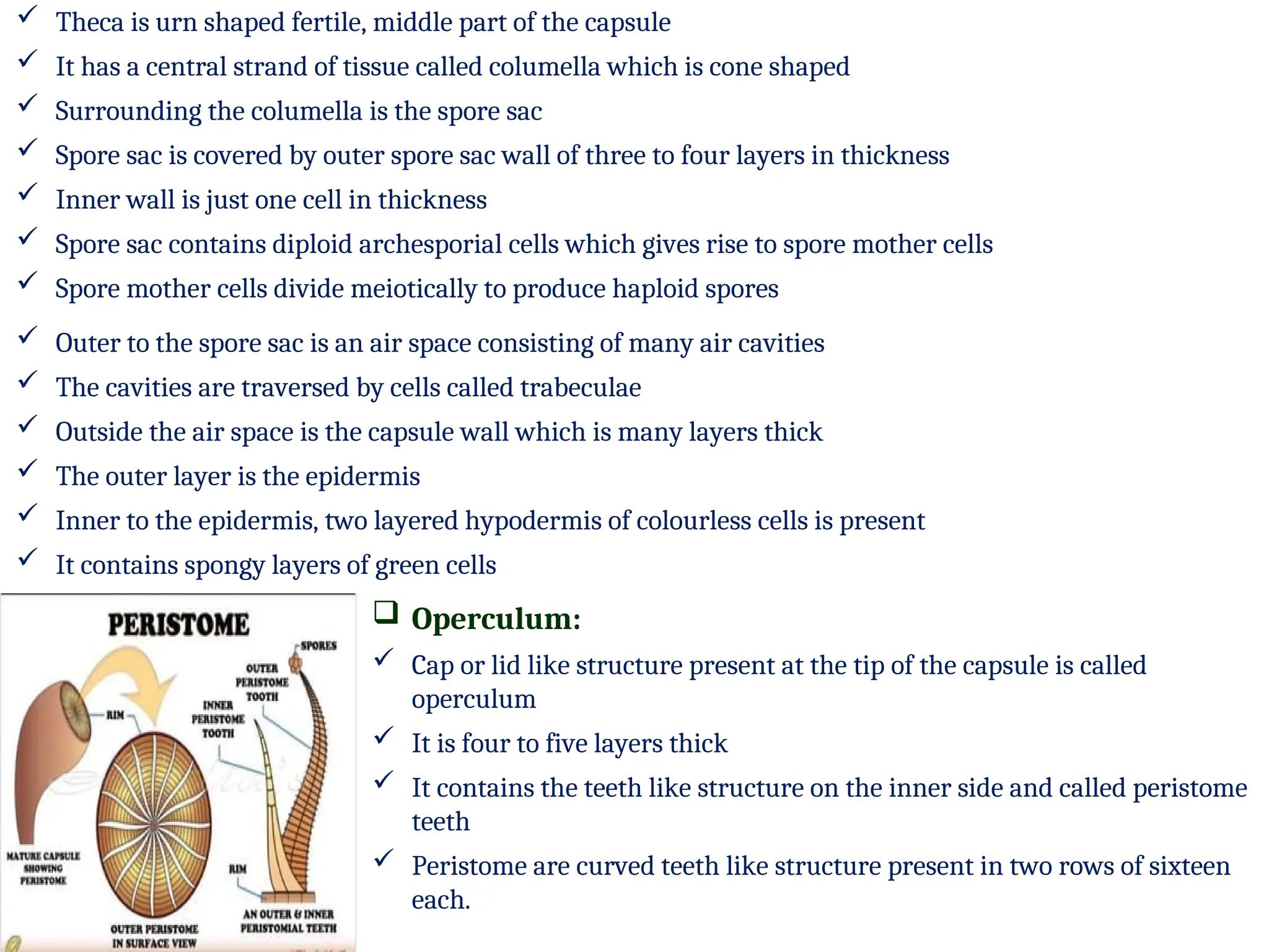
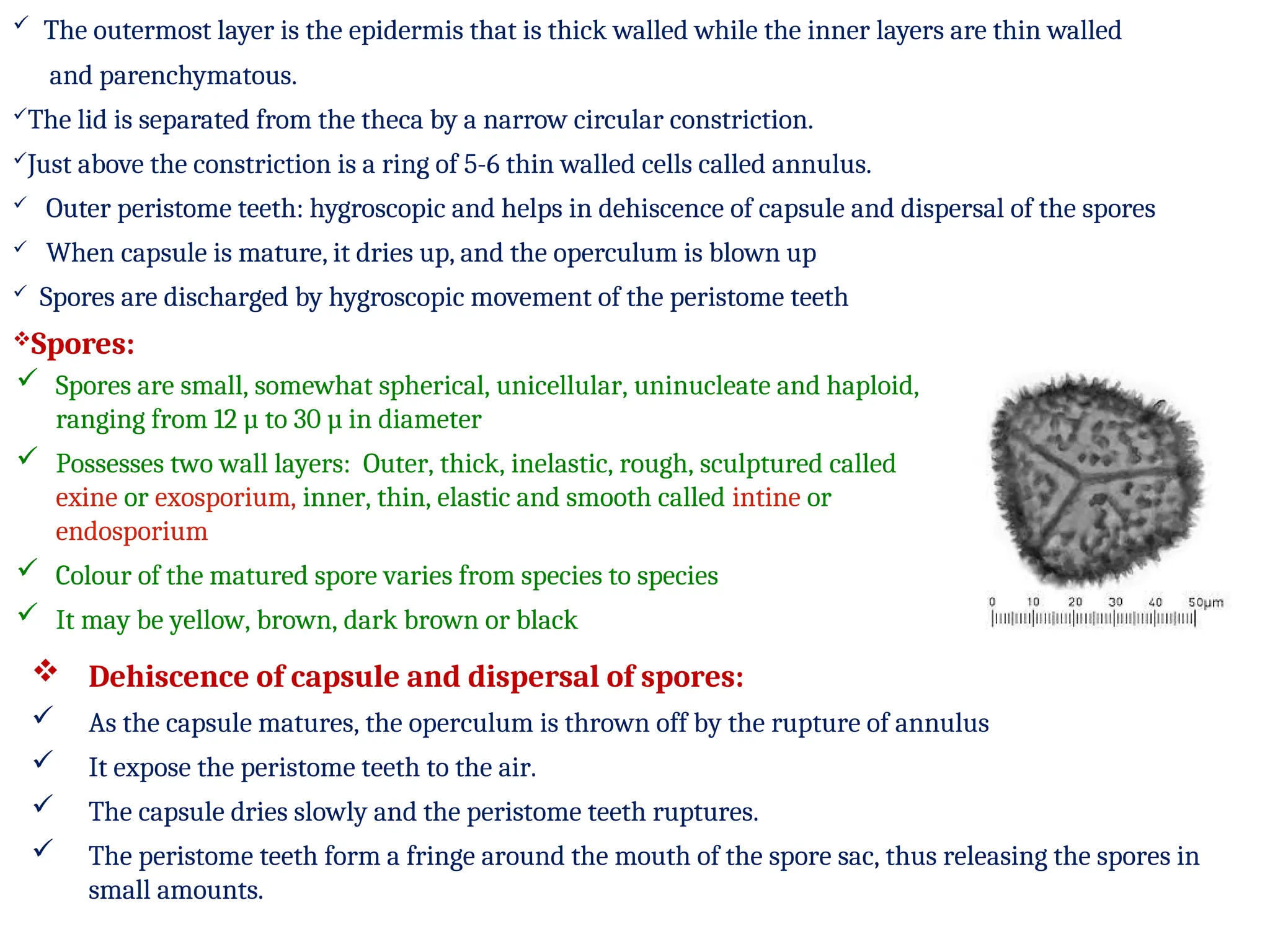
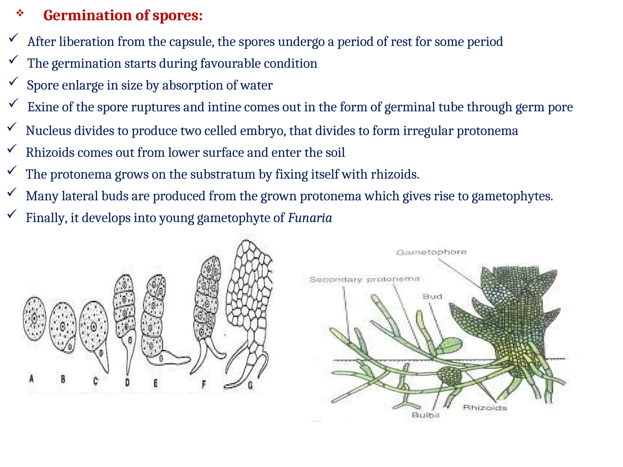
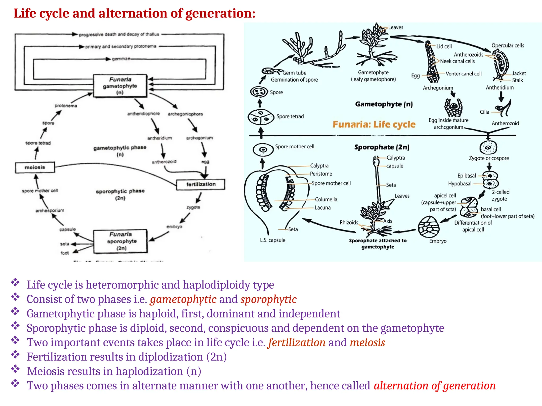
LIFE CYCLE OF FUNARIA
DR. AVINASH K. ANEY
M.Sc., M. Phil., Ph.D. SET
Head
Department of Botany,
Science College, Pauni, Dist. Bhandara
Occurrence and distribution:
Cosmopolitan in distribution and includes about 117 species
15 species reported from India
The species F. hygrometrica is best known and found throughout the world
Mosses grows luxuriantly in humus soil, sometimes they also occur on the rocks and damp
walls
Green protonema appear in the recently ploughed soil
Some mosses are epiphytic in habitat and frow upon trunks of the trees
Classification and systematic position:
Division: Bryophyta
Class: Bryopsida (Musci)
Sub-class: Bryidae
Cohert: Eubriidae
Order: Funariales
Family: Funariaceae
Genus: Funaria
External features/External morphology:
Plant body is small, green, erect gametophyte, easily collected from damp places
Arise from the alga-like filamentous protonema
Differentiated into rhizoids, stem and leaves
It bears sex organs at the apex
Rhizoids are branched, multicellular and thread-like
Septa of the rhizoids are oblique
Young rhizoids are whitish in colour and later turns brown or pink
Rhizoids absorbs water and nutrients from soil
Leaves are small, ovate, bright green and spirally arranged on the stem
Each leaf with distinct mid-rib.
 Upper leaves are somewhat larger in size and crowded at the apex
 Lower leaves are smaller and scattered on the stem
 Stem is erect, upright, green and with monopodial branching
 Internal structure:
 V.S. Leaf:
 Internally, the leaf shows distinct mid-rib and wings
 Mid-rib is several celled in thickness
 Wings are single celled in thickness
 There is a central strand in the center
 The cells of the wings contain chloroplast
 The chloroplast continuously divide and re-divide
 T.S. Stem:
 Internally, stem consists of three parts
 Epidermis:
 It is single layered
 It is outermost layer consisting of thin-walled compactly arranged cells
with chloroplasts
 This layer is photosynthetic in function
 Cuticle and stomata are absent on epidermis
 Cortex:
 Cortex is multilayered and consist of thin walled parenchymatous cells.
 Central strand:
 Consist of somewhat thick walled, compactly arranged cells
 Cells are vertically elongated.
 Central cylinder acts as conducting tissue helps in water conduction.
REPRODUCTION IN FUNARIA
Vegetative Reproduction Sexual Reproduction
By Bulbils
By Gemmae
By primary protonema
By secondary
protonema
Antheridia Archegonia
1. Vegetative reproduction:
A. By death and decay of prostrate system:
Older prostrate branches of the gametophyte decays
After death of such branches each erect branches develops into new
gametophyte
B. By primary protonema:
After germination, the spores develops into primary protonema
The protonema possesses several buds on it
Each buds after detachment develops into new Funaria plant
C. By secondary protonema:
The protonema which develops from the other parts of the plant is
called as secondary protonema
Such protonema are similar with the primary protonema
These protonema may develop into new gametophyte after
detachment from the mother plant
D. By gemmae:
Many species produces small, multicellular gemmae in the leaf apices
Gemmae are produced in groups
Sometimes, solitary gemmae are produced on the rhizoids, called bulbils
Such gemma or bulbils develops into new plant during favourable condition
2. Sexual reproduction:
Species of Funaria are monoecious and autoicous
Autoicous: male and female sex organs develops on separate branches
of the same plant
Main shoot of the gametophytic plant bears male sex organs whereas,
the lateral branch bears female sex organ
Male branch and structure of mature antheridium:
 The antheridia are intermingled with several sterile hair-like structures called paraphyses
 They are multi-cellular and consist of 4 to 5 cells
 Lower cells of the paraphyses are elongated and terminal cell is globular
 Antheridia develops in a cluster at the apex of male
branch
 The antheridia are covered with leaves at the apex
 Mature antheridium consist of short massive stalk and
the main body
 Main body is covered with single layered jacket
 Cells of the jacket contains chloroplast
 Jacket layer surrounds central dense mass of androcytes
 Androcytes develops into biflagellate antherozoids
Female branch and structure of mature archegonium:
Fertilization:
Act of union of haploid male gametes (n) with haploid female gamete (n) is called fertilization
Water is very essential for the act of fertilization
Pre-fertilization changes:
Matured antheridia opens due to water and biflagellate antherozoids liberate
Chemotactic antherozoids swim on the film of water and reaches the archegonia
Prior to fertilization, cover cells detached from archegonium and neck canal become gelatinized due to
disintegration of all NCCs and VCC
Many antherozoids enter the archegonium, travel through neck and but one lucky antherozoid
penetrate the egg and fertilization is affected to produce diploid (2n) zygote
Each
 Archegonia are developed in a cluster on the lateral female branches
 Mature archegonium is much elongated flask-shaped structure
 It has massive and elongated stalk.
 Differentiated into lower broader venter and upper elongated neck
 Neck region is covered by single layered jacket, whereas, it is two layered in
the venter region
 Venter contains lower egg (n) cell and upper VCC
 Elongated neck contains six or more neck canal cells (NCCS)
 Tip of the neck is covered by four cover cells arranged in two tiers
 Cluster of archegonia covered with sterile leaves called perichaetial leave
Post-fertilization changes:
After fertilization, zygote (2n) begins to enlarge, secrete a cellulose wall around and begins to
develop into embryo
Zygote divides first by transverse wall producing two celled embryo
Embryo divides and redivides to produce spore producing structure called Sporogonium
External features of sporogonium (Sporophyte):
 Funaria sporophytes are much elongated structure
 Sporophyte is developed at the apex of the archegonial or female
branch
 Arise in the form of horny structure at the place of fertilized
archegonia
 Usually 2-3 cm long, in some species, they may be 15 cm long
 Because of the horny appearance of sporophyte, the species are
called ‘hornworts’
 Mature sporogonium differentiated into foot, seta and capsule
 Seta is much longer and elongated
 Lower portion of sporophyte is embedded in thallus tissue called
involucre
 Capsule is indefinite in growth
 Funaria sporophytes differentiated into three regions: foot, meristematic zone and capsule
 Capsule:
 Capsule is the main and fertile part of the sporophyte
 Differentiated into three regions: apophysis, theca and
operculum or lid.
 Apophysis:
 It is basal green, photosynthetic part of the capsule
 Cells are compactly arranged and with chloroplast
 It has central strand of conducting tissue
 Differentiated into three regions
 Theca:
 Major portion of capsule is covered by theca
 Foot:
 Foot is small conical structure embedded in female tissue
 Consist of compactly arranged cells
 Helps attaching sporophyte to the gametophyte and absorbs
water and nutrition from mother plant
 Seta:
 Seta is long, slender and twisted structure
 It carries the capsule at the tip
 Theca is urn shaped fertile, middle part of the capsule
 It has a central strand of tissue called columella which is cone shaped
 Surrounding the columella is the spore sac
 Spore sac is covered by outer spore sac wall of three to four layers in thickness
 Inner wall is just one cell in thickness
 Spore sac contains diploid archesporial cells which gives rise to spore mother cells
 Spore mother cells divide meiotically to produce haploid spores
 Operculum:
 Cap or lid like structure present at the tip of the capsule is called
operculum
 It is four to five layers thick
 It contains the teeth like structure on the inner side and called peristome
teeth
 Peristome are curved teeth like structure present in two rows of sixteen
each.
 Outer to the spore sac is an air space consisting of many air cavities
 The cavities are traversed by cells called trabeculae
 Outside the air space is the capsule wall which is many layers thick
 The outer layer is the epidermis
 Inner to the epidermis, two layered hypodermis of colourless cells is present
 It contains spongy layers of green cells
 The outermost layer is the epidermis that is thick walled while the inner layers are thin walled
and parenchymatous.
The lid is separated from the theca by a narrow circular constriction.
Just above the constriction is a ring of 5-6 thin walled cells called annulus.
 Outer peristome teeth: hygroscopic and helps in dehiscence of capsule and dispersal of the spores
 When capsule is mature, it dries up, and the operculum is blown up
 Spores are discharged by hygroscopic movement of the peristome teeth
Spores:
 Spores are small, somewhat spherical, unicellular, uninucleate and haploid,
ranging from 12 µ to 30 µ in diameter
 Possesses two wall layers: Outer, thick, inelastic, rough, sculptured called
exine or exosporium, inner, thin, elastic and smooth called intine or
endosporium
 Colour of the matured spore varies from species to species
 It may be yellow, brown, dark brown or black
 Dehiscence of capsule and dispersal of spores:
 As the capsule matures, the operculum is thrown off by the rupture of annulus
 It expose the peristome teeth to the air.
 The capsule dries slowly and the peristome teeth ruptures.
 The peristome teeth form a fringe around the mouth of the spore sac, thus releasing the spores in
small amounts.
 Germination of spores:
 After liberation from the capsule, the spores undergo a period of rest for some period
 The germination starts during favourable condition
 Spore enlarge in size by absorption of water
 Exine of the spore ruptures and intine comes out in the form of germinal tube through germ pore
 Nucleus divides to produce two celled embryo, that divides to form irregular protonema
 Rhizoids comes out from lower surface and enter the soil
 The protonema grows on the substratum by fixing itself with rhizoids.
 Many lateral buds are produced from the grown protonema which gives rise to gametophytes.
 Finally, it develops into young gametophyte of Funaria
Life cycle and alternation of generation:
 Life cycle is heteromorphic and haplodiploidy type
 Consist of two phases i.e. gametophytic and sporophytic
 Gametophytic phase is haploid, first, dominant and independent
 Sporophytic phase is diploid, second, conspicuous and dependent on the gametophyte
 Two important events takes place in life cycle i.e. fertilization and meiosis
 Fertilization results in diplodization (2n)
 Meiosis results in haplodization (n)
 Two phases comes in alternate manner with one another, hence called alternation of generation
Go Out and Thank a Tree!

Life cycle of Funaria, member of class Bryopsida.ppt

  • 1.
    LIFE CYCLE OFFUNARIA DR. AVINASH K. ANEY M.Sc., M. Phil., Ph.D. SET Head Department of Botany, Science College, Pauni, Dist. Bhandara
  • 2.
    Occurrence and distribution: Cosmopolitanin distribution and includes about 117 species 15 species reported from India The species F. hygrometrica is best known and found throughout the world Mosses grows luxuriantly in humus soil, sometimes they also occur on the rocks and damp walls Green protonema appear in the recently ploughed soil Some mosses are epiphytic in habitat and frow upon trunks of the trees Classification and systematic position: Division: Bryophyta Class: Bryopsida (Musci) Sub-class: Bryidae Cohert: Eubriidae Order: Funariales Family: Funariaceae Genus: Funaria
  • 3.
    External features/External morphology: Plantbody is small, green, erect gametophyte, easily collected from damp places Arise from the alga-like filamentous protonema Differentiated into rhizoids, stem and leaves It bears sex organs at the apex Rhizoids are branched, multicellular and thread-like Septa of the rhizoids are oblique Young rhizoids are whitish in colour and later turns brown or pink Rhizoids absorbs water and nutrients from soil Leaves are small, ovate, bright green and spirally arranged on the stem Each leaf with distinct mid-rib.
  • 4.
     Upper leavesare somewhat larger in size and crowded at the apex  Lower leaves are smaller and scattered on the stem  Stem is erect, upright, green and with monopodial branching  Internal structure:  V.S. Leaf:  Internally, the leaf shows distinct mid-rib and wings  Mid-rib is several celled in thickness  Wings are single celled in thickness  There is a central strand in the center  The cells of the wings contain chloroplast  The chloroplast continuously divide and re-divide  T.S. Stem:  Internally, stem consists of three parts  Epidermis:  It is single layered  It is outermost layer consisting of thin-walled compactly arranged cells with chloroplasts  This layer is photosynthetic in function  Cuticle and stomata are absent on epidermis  Cortex:  Cortex is multilayered and consist of thin walled parenchymatous cells.  Central strand:  Consist of somewhat thick walled, compactly arranged cells  Cells are vertically elongated.  Central cylinder acts as conducting tissue helps in water conduction.
  • 5.
    REPRODUCTION IN FUNARIA VegetativeReproduction Sexual Reproduction By Bulbils By Gemmae By primary protonema By secondary protonema Antheridia Archegonia
  • 6.
    1. Vegetative reproduction: A.By death and decay of prostrate system: Older prostrate branches of the gametophyte decays After death of such branches each erect branches develops into new gametophyte B. By primary protonema: After germination, the spores develops into primary protonema The protonema possesses several buds on it Each buds after detachment develops into new Funaria plant C. By secondary protonema: The protonema which develops from the other parts of the plant is called as secondary protonema Such protonema are similar with the primary protonema These protonema may develop into new gametophyte after detachment from the mother plant D. By gemmae: Many species produces small, multicellular gemmae in the leaf apices Gemmae are produced in groups Sometimes, solitary gemmae are produced on the rhizoids, called bulbils Such gemma or bulbils develops into new plant during favourable condition
  • 7.
    2. Sexual reproduction: Speciesof Funaria are monoecious and autoicous Autoicous: male and female sex organs develops on separate branches of the same plant Main shoot of the gametophytic plant bears male sex organs whereas, the lateral branch bears female sex organ Male branch and structure of mature antheridium:  The antheridia are intermingled with several sterile hair-like structures called paraphyses  They are multi-cellular and consist of 4 to 5 cells  Lower cells of the paraphyses are elongated and terminal cell is globular  Antheridia develops in a cluster at the apex of male branch  The antheridia are covered with leaves at the apex  Mature antheridium consist of short massive stalk and the main body  Main body is covered with single layered jacket  Cells of the jacket contains chloroplast  Jacket layer surrounds central dense mass of androcytes  Androcytes develops into biflagellate antherozoids
  • 8.
    Female branch andstructure of mature archegonium: Fertilization: Act of union of haploid male gametes (n) with haploid female gamete (n) is called fertilization Water is very essential for the act of fertilization Pre-fertilization changes: Matured antheridia opens due to water and biflagellate antherozoids liberate Chemotactic antherozoids swim on the film of water and reaches the archegonia Prior to fertilization, cover cells detached from archegonium and neck canal become gelatinized due to disintegration of all NCCs and VCC Many antherozoids enter the archegonium, travel through neck and but one lucky antherozoid penetrate the egg and fertilization is affected to produce diploid (2n) zygote Each  Archegonia are developed in a cluster on the lateral female branches  Mature archegonium is much elongated flask-shaped structure  It has massive and elongated stalk.  Differentiated into lower broader venter and upper elongated neck  Neck region is covered by single layered jacket, whereas, it is two layered in the venter region  Venter contains lower egg (n) cell and upper VCC  Elongated neck contains six or more neck canal cells (NCCS)  Tip of the neck is covered by four cover cells arranged in two tiers  Cluster of archegonia covered with sterile leaves called perichaetial leave
  • 9.
    Post-fertilization changes: After fertilization,zygote (2n) begins to enlarge, secrete a cellulose wall around and begins to develop into embryo Zygote divides first by transverse wall producing two celled embryo Embryo divides and redivides to produce spore producing structure called Sporogonium External features of sporogonium (Sporophyte):  Funaria sporophytes are much elongated structure  Sporophyte is developed at the apex of the archegonial or female branch  Arise in the form of horny structure at the place of fertilized archegonia  Usually 2-3 cm long, in some species, they may be 15 cm long  Because of the horny appearance of sporophyte, the species are called ‘hornworts’  Mature sporogonium differentiated into foot, seta and capsule  Seta is much longer and elongated  Lower portion of sporophyte is embedded in thallus tissue called involucre  Capsule is indefinite in growth
  • 10.
     Funaria sporophytesdifferentiated into three regions: foot, meristematic zone and capsule  Capsule:  Capsule is the main and fertile part of the sporophyte  Differentiated into three regions: apophysis, theca and operculum or lid.  Apophysis:  It is basal green, photosynthetic part of the capsule  Cells are compactly arranged and with chloroplast  It has central strand of conducting tissue  Differentiated into three regions  Theca:  Major portion of capsule is covered by theca  Foot:  Foot is small conical structure embedded in female tissue  Consist of compactly arranged cells  Helps attaching sporophyte to the gametophyte and absorbs water and nutrition from mother plant  Seta:  Seta is long, slender and twisted structure  It carries the capsule at the tip
  • 11.
     Theca isurn shaped fertile, middle part of the capsule  It has a central strand of tissue called columella which is cone shaped  Surrounding the columella is the spore sac  Spore sac is covered by outer spore sac wall of three to four layers in thickness  Inner wall is just one cell in thickness  Spore sac contains diploid archesporial cells which gives rise to spore mother cells  Spore mother cells divide meiotically to produce haploid spores  Operculum:  Cap or lid like structure present at the tip of the capsule is called operculum  It is four to five layers thick  It contains the teeth like structure on the inner side and called peristome teeth  Peristome are curved teeth like structure present in two rows of sixteen each.  Outer to the spore sac is an air space consisting of many air cavities  The cavities are traversed by cells called trabeculae  Outside the air space is the capsule wall which is many layers thick  The outer layer is the epidermis  Inner to the epidermis, two layered hypodermis of colourless cells is present  It contains spongy layers of green cells
  • 12.
     The outermostlayer is the epidermis that is thick walled while the inner layers are thin walled and parenchymatous. The lid is separated from the theca by a narrow circular constriction. Just above the constriction is a ring of 5-6 thin walled cells called annulus.  Outer peristome teeth: hygroscopic and helps in dehiscence of capsule and dispersal of the spores  When capsule is mature, it dries up, and the operculum is blown up  Spores are discharged by hygroscopic movement of the peristome teeth Spores:  Spores are small, somewhat spherical, unicellular, uninucleate and haploid, ranging from 12 µ to 30 µ in diameter  Possesses two wall layers: Outer, thick, inelastic, rough, sculptured called exine or exosporium, inner, thin, elastic and smooth called intine or endosporium  Colour of the matured spore varies from species to species  It may be yellow, brown, dark brown or black  Dehiscence of capsule and dispersal of spores:  As the capsule matures, the operculum is thrown off by the rupture of annulus  It expose the peristome teeth to the air.  The capsule dries slowly and the peristome teeth ruptures.  The peristome teeth form a fringe around the mouth of the spore sac, thus releasing the spores in small amounts.
  • 13.
     Germination ofspores:  After liberation from the capsule, the spores undergo a period of rest for some period  The germination starts during favourable condition  Spore enlarge in size by absorption of water  Exine of the spore ruptures and intine comes out in the form of germinal tube through germ pore  Nucleus divides to produce two celled embryo, that divides to form irregular protonema  Rhizoids comes out from lower surface and enter the soil  The protonema grows on the substratum by fixing itself with rhizoids.  Many lateral buds are produced from the grown protonema which gives rise to gametophytes.  Finally, it develops into young gametophyte of Funaria
  • 14.
    Life cycle andalternation of generation:  Life cycle is heteromorphic and haplodiploidy type  Consist of two phases i.e. gametophytic and sporophytic  Gametophytic phase is haploid, first, dominant and independent  Sporophytic phase is diploid, second, conspicuous and dependent on the gametophyte  Two important events takes place in life cycle i.e. fertilization and meiosis  Fertilization results in diplodization (2n)  Meiosis results in haplodization (n)  Two phases comes in alternate manner with one another, hence called alternation of generation
  • 15.
    Go Out andThank a Tree!