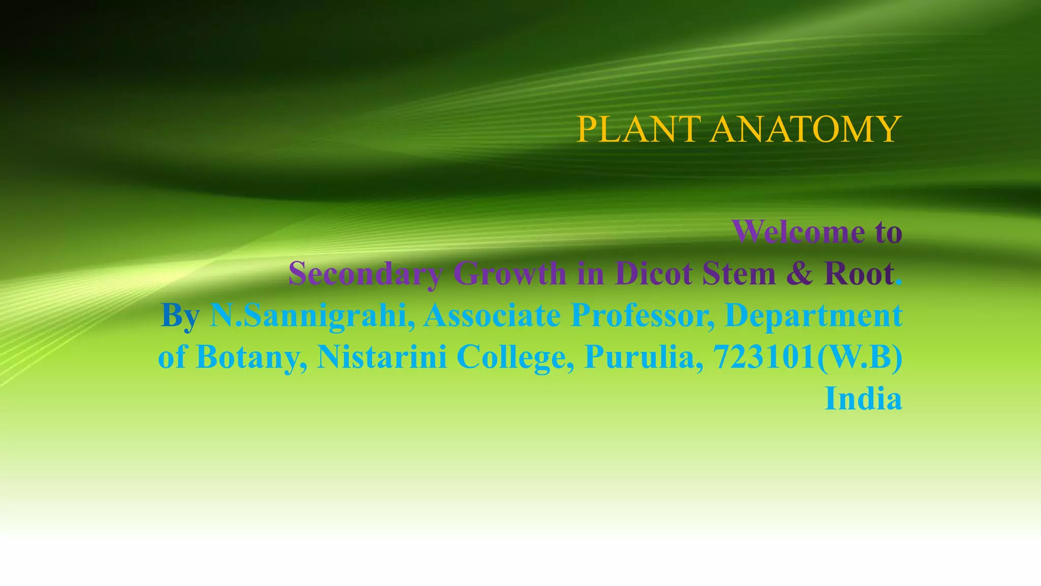
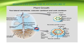
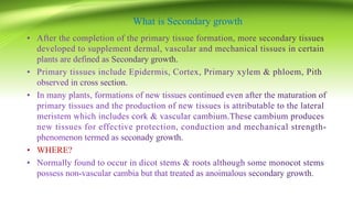
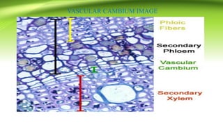
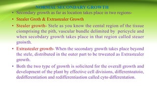
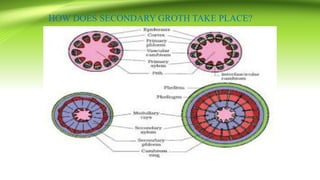
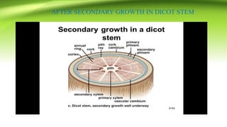
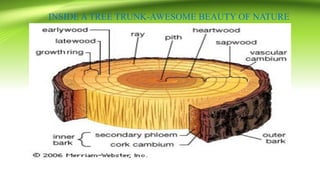
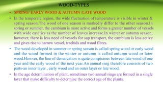
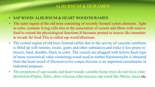
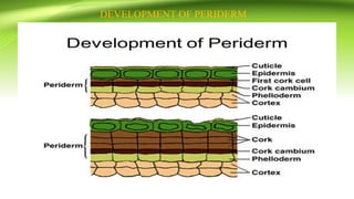
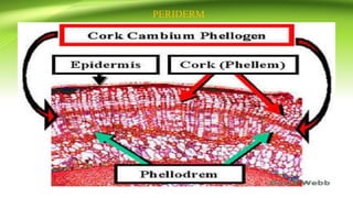
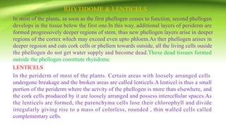
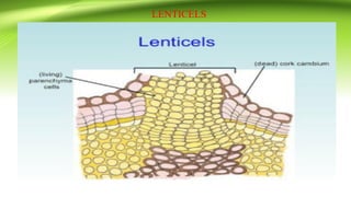
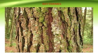
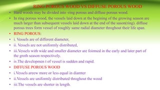
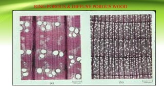
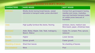
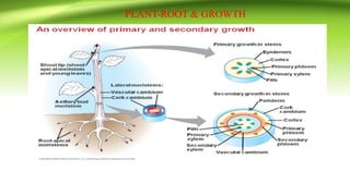
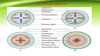
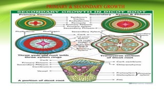
Vascular Cambium & Seasonal activity & its Role in Stem & RootFatima Ramay
Vascular Cambium & Seasonal activity & its Role in Stem & Root:
The vascular cambium (pl. cambia or cambiums) is a lateral meristem in the vascular tissue of plants.
The vascular cambium is a cylindrical layer of cambium that runs through the stem of a plant that undergoes secondary growth.
In Dicots:
The vascular cambium is in dicot stems and roots, located between the xylem and the phloem in the stem and root of a vascular plant, and is the source of both the secondary xylem growth (inwards, towards the pith) and the secondary phloem growth (outwards).
In Monocots:
Monocot stems, such as corn, palms and bamboos, do not have a vascular cambium and do not exhibit secondary growth by the production of concentric annual rings. They cannot increase in girth by adding lateral layers of cells as in conifers and woody dicots.
Cambium of some plants remains active for the entire period of their life, i.e., cambial cells divide and resulting cells mature to form xylem and phloem elements.
This type of seasonal activity usually found in the plants present in the tropical regions, and not all plants show cambial activity.
Percentage of ringless trees in the rain forests of;India : 75%Amazon : 43%Malaysia : 15%
In regions with definite seasonal climate; seasonal activity of cambium ceased with onset of unfavorable conditions; In Autumn, it enters the dormant state and lasts for the end of summer; In Spring, cambium again becomes active.
Duration of cambial activity is also affected by day-length, e.g., In Robinia pseudoacacia, cambium is dormant under short-day condition.
The cambium cells formed in circular in cross section from the beginning onwards.
The cambial ring is partially primary (fascicular cambium) and partially secondary (interfascicular cambium).
Periderm originates from the cortical cells (extra stelar in origin).
In Dicot stem, for mechanical support xylem is with comparatively smaller vessels, greater fibers and less parenchyma.
More amount of cork is produces for protection.
Lenticels on periderm are very prominent.
The cambial ring formed is wavy in the beginning and later becomes circular.
The cambium ring is completely secondary in origin.
Periderm originates from the pericycle (intra stelar in origin).
In Dicot root, xylem is with big thin walled vessels with few fibers and more parenchyma.
Less amount of cork is produced as root is underground.
Lenticels on periderm are not very prominent.
Gnetum: A Powerpoint Presentation on Gymnospemsshivduraigaran
The Gymnosperms are a group of seed-producing plants (spermatophytes) that includes conifers (Pinophyta), cycads, Ginkgo, and gnetophytes. The term "gymnosperm" comes from the Greek composite word γυμνόσπερμος (γυμνός gymnos, "naked" and σπέρμα sperma, "seed"), meaning "naked seeds". The name is based on the unenclosed condition of their seeds (called ovules in their unfertilized state). The non-encased condition of their seeds stands in contrast to the seeds and ovules of flowering plants (angiosperms), which are enclosed within an ovary. Gymnosperm seeds develop either on the surface of scales or leaves, which are often modified to form cones, or solitary as in Yew, Torreya, Ginkgo.
The gymnosperms and angiosperms together compose the spermatophytes or seed plants. The gymnosperms are divided into six phyla. Organisms that belong to the Cycadophyta, Ginkgophyta, Gnetophyta, and Pinophyta (also known as Coniferophyta) phyla are still in existence while those in the Pteridospermales and Cordaitales phyla are now extinct.
By far the largest group of living gymnosperms are the conifers (pines, cypresses, and relatives), followed by cycads, gnetophytes (Gnetum, Ephedra and Welwitschia), and Ginkgo biloba (a single living species). Roots in some genera have fungal association with roots in the form of micorrhiza(Pinus), while in some others(Cycas) small specialised roots called coralloid roots are associated with nitrogen fixing cyanobacteria.
Gnetum is a genus of gymnosperms, the sole genus in the family Gnetaceae and order Gnetales. They are tropical evergreen trees, shrubs and lianas. Unlike other gymnosperms, they possess vessel elements in the xylem. Some species have been proposed to have been the first plants to be insect-pollinated as their fossils occur in association with extinct pollinating scorpion flies. Molecular phylogenies based on nuclear and plastid sequences from most of the species indicate hybridization among some of the Southeast Asian species. Fossil-calibrated molecular-clocks suggest that the Gnetum lineages now found in Africa, South America and Southeast Asia are the result of ancient long-distance dispersal across seawater
The Shoot apex is also known as the terminal bud of plants that grows from 0.1-1.0 mm and consists of the apical meristem, developing leaves and the immediate surrounding leaf primordial. The shoot apex is present in both dicot and monocot plants.
This PPT contains the application of Plant Anatomy in the field of Pharmacognosy & Plant systemics with number of examples to explore the beauty of this subject .
Structure, Development & Function of PeridermFatima Ramay
A group of secondary tissues forming a protective layer which replaces the epidermis of many plant stems, roots, and other parts.
Although periderm may develop in leaves and fruits, its main function is to protects stems and roots.
The periderm consists of three different layers:
Phelloderm
Phellogen (cork cambium)
Phellem (cork)
Its main function is to protect the underlying tissues from:
Desiccation
Freezing
Heat injury
Mechanical destruction
Disease
Loss of epidermis.
Bounding tissue restricting the pathogen & insects.
Allowing gaseous exchange through lenticels.
It is called as “living fossil”
The whole order is extincted except one species Ginkgo biloba
This order was occurred in Triassic periods of Mesozoic age (200,000,000 years ago)
This order consists of 16 genera and many species (all in fossil forms except one)
This PPT contains a birds' eye view of the basic internal organization of the plant body comprising of tissue networks along with tissue systems to perform diverse functions for plants survival even in stress condition.
This is a Life Cycle of Shpagnum, A good content for Masters Students. (But this content is not made by me...but i thought that this will help many students who are in search for content)
Thank you 😊
This presentation explores a brief idea about the structural and functional attributes of nucleotides, the structure and function of genetic materials along with the impact of UV rays and pH upon them.
This presentation intends to explore the communication of the cell within and others for sustainability along the regulation mechanisms by the cellular neural networks and others to sing the song of the life.
Bioenergetics is an important domain in biology. This presentation has explored ATP production and its optimum utilization in biological systems along with certain theories and experiments to give a bird's eye view of this important issue.
This presentation offers the bird's eye view of the cell as the basic structural and functional unit of life. It also addresses the origin of eukaryotic cells from the prokaryotic cell by the endosymbiotic theory.
This presentation has been intended to offer a bird's eye view about the phylogenetic classification of the plant kingdom in general and the Engler and Prantl system in particular with merits and demerits.
This PPT has been made to explore the plant classification in general and the classification as made by Bentham & Hooker for the classification of the flowering plants. It also offers the history of plant classification along with the merits and demerits of this aforesaid classification.
Energy and the biological systems are joined together and no biological world is almost impossible without ATP. This study material intends to explore the beauty of ATP to drive different biological processes.
This PPT offers a bird's eye view of ICBN and its different rules along with regulations for the naming of plants. It also highlights the history of IBC and its contribution to plant taxonomy.
This presentation intends to offer the basic features of plant metabolism along with the different types of mechanisms to regulate and control the metabolic pathways.
This presentation has been designed to give the foundation of taxonomy in general and Plant Taxonomy in particular as a matter of pleasure to explore the diversity of the plant world.
Sex and sexuality are very common words in biology but para-sexuality is a little bit uncommon, several organisms in general and fungi in particular have the pleasure of sexuality to bring variations by beside sex. This PPT explores the beauty of para-sexuality for the academic fraternity.
Sex life in fungi is not less fascinating than in other organisms. Heterosexuality is a matter of pleasure to explore the diversity of sex in fungi along with its cause and consequences. You can find a pleasure to go through the content.
This PowerPoint wants to explore the bird's eye view of the reproduction of bacteria in general and the genetic recombination of bacteria in particular.
This presentation gives the bird's eye view of bacterial nutrition along with some other issues required to understand bacterial diversity as far as nutrition is concerned.
This presentation explores the food value of mushrooms along with the long-term and short-term storage procedures. It also offers a detailed account of the nutrients that remain present in the edible mushrooms.
If you want to explore the role of Cyanobacteria in soil fertility in general & Azolla-Anabena association in particular, you can visit this PowerPoint Presentation.
Richard's aventures in two entangled wonderlandsRichard Gill
Since the loophole-free Bell experiments of 2020 and the Nobel prizes in physics of 2022, critics of Bell's work have retreated to the fortress of super-determinism. Now, super-determinism is a derogatory word - it just means "determinism". Palmer, Hance and Hossenfelder argue that quantum mechanics and determinism are not incompatible, using a sophisticated mathematical construction based on a subtle thinning of allowed states and measurements in quantum mechanics, such that what is left appears to make Bell's argument fail, without altering the empirical predictions of quantum mechanics. I think however that it is a smoke screen, and the slogan "lost in math" comes to my mind. I will discuss some other recent disproofs of Bell's theorem using the language of causality based on causal graphs. Causal thinking is also central to law and justice. I will mention surprising connections to my work on serial killer nurse cases, in particular the Dutch case of Lucia de Berk and the current UK case of Lucy Letby.
Observation of Io’s Resurfacing via Plume Deposition Using Ground-based Adapt...Sérgio Sacani
Since volcanic activity was first discovered on Io from Voyager images in 1979, changes
on Io’s surface have been monitored from both spacecraft and ground-based telescopes.
Here, we present the highest spatial resolution images of Io ever obtained from a groundbased telescope. These images, acquired by the SHARK-VIS instrument on the Large
Binocular Telescope, show evidence of a major resurfacing event on Io’s trailing hemisphere. When compared to the most recent spacecraft images, the SHARK-VIS images
show that a plume deposit from a powerful eruption at Pillan Patera has covered part
of the long-lived Pele plume deposit. Although this type of resurfacing event may be common on Io, few have been detected due to the rarity of spacecraft visits and the previously low spatial resolution available from Earth-based telescopes. The SHARK-VIS instrument ushers in a new era of high resolution imaging of Io’s surface using adaptive
optics at visible wavelengths.
(May 29th, 2024) Advancements in Intravital Microscopy- Insights for Preclini...Scintica Instrumentation
Intravital microscopy (IVM) is a powerful tool utilized to study cellular behavior over time and space in vivo. Much of our understanding of cell biology has been accomplished using various in vitro and ex vivo methods; however, these studies do not necessarily reflect the natural dynamics of biological processes. Unlike traditional cell culture or fixed tissue imaging, IVM allows for the ultra-fast high-resolution imaging of cellular processes over time and space and were studied in its natural environment. Real-time visualization of biological processes in the context of an intact organism helps maintain physiological relevance and provide insights into the progression of disease, response to treatments or developmental processes.
In this webinar we give an overview of advanced applications of the IVM system in preclinical research. IVIM technology is a provider of all-in-one intravital microscopy systems and solutions optimized for in vivo imaging of live animal models at sub-micron resolution. The system’s unique features and user-friendly software enables researchers to probe fast dynamic biological processes such as immune cell tracking, cell-cell interaction as well as vascularization and tumor metastasis with exceptional detail. This webinar will also give an overview of IVM being utilized in drug development, offering a view into the intricate interaction between drugs/nanoparticles and tissues in vivo and allows for the evaluation of therapeutic intervention in a variety of tissues and organs. This interdisciplinary collaboration continues to drive the advancements of novel therapeutic strategies.
Multi-source connectivity as the driver of solar wind variability in the heli...Sérgio Sacani
The ambient solar wind that flls the heliosphere originates from multiple
sources in the solar corona and is highly structured. It is often described
as high-speed, relatively homogeneous, plasma streams from coronal
holes and slow-speed, highly variable, streams whose source regions are
under debate. A key goal of ESA/NASA’s Solar Orbiter mission is to identify
solar wind sources and understand what drives the complexity seen in the
heliosphere. By combining magnetic feld modelling and spectroscopic
techniques with high-resolution observations and measurements, we show
that the solar wind variability detected in situ by Solar Orbiter in March
2022 is driven by spatio-temporal changes in the magnetic connectivity to
multiple sources in the solar atmosphere. The magnetic feld footpoints
connected to the spacecraft moved from the boundaries of a coronal hole
to one active region (12961) and then across to another region (12957). This
is refected in the in situ measurements, which show the transition from fast
to highly Alfvénic then to slow solar wind that is disrupted by the arrival of
a coronal mass ejection. Our results describe solar wind variability at 0.5 au
but are applicable to near-Earth observatories.
Cancer cell metabolism: special Reference to Lactate PathwayAADYARAJPANDEY1
Normal Cell Metabolism:
Cellular respiration describes the series of steps that cells use to break down sugar and other chemicals to get the energy we need to function.
Energy is stored in the bonds of glucose and when glucose is broken down, much of that energy is released.
Cell utilize energy in the form of ATP.
The first step of respiration is called glycolysis. In a series of steps, glycolysis breaks glucose into two smaller molecules - a chemical called pyruvate. A small amount of ATP is formed during this process.
Most healthy cells continue the breakdown in a second process, called the Kreb's cycle. The Kreb's cycle allows cells to “burn” the pyruvates made in glycolysis to get more ATP.
The last step in the breakdown of glucose is called oxidative phosphorylation (Ox-Phos).
It takes place in specialized cell structures called mitochondria. This process produces a large amount of ATP. Importantly, cells need oxygen to complete oxidative phosphorylation.
If a cell completes only glycolysis, only 2 molecules of ATP are made per glucose. However, if the cell completes the entire respiration process (glycolysis - Kreb's - oxidative phosphorylation), about 36 molecules of ATP are created, giving it much more energy to use.
IN CANCER CELL:
Unlike healthy cells that "burn" the entire molecule of sugar to capture a large amount of energy as ATP, cancer cells are wasteful.
Cancer cells only partially break down sugar molecules. They overuse the first step of respiration, glycolysis. They frequently do not complete the second step, oxidative phosphorylation.
This results in only 2 molecules of ATP per each glucose molecule instead of the 36 or so ATPs healthy cells gain. As a result, cancer cells need to use a lot more sugar molecules to get enough energy to survive.
introduction to WARBERG PHENOMENA:
WARBURG EFFECT Usually, cancer cells are highly glycolytic (glucose addiction) and take up more glucose than do normal cells from outside.
Otto Heinrich Warburg (; 8 October 1883 – 1 August 1970) In 1931 was awarded the Nobel Prize in Physiology for his "discovery of the nature and mode of action of the respiratory enzyme.
WARNBURG EFFECT : cancer cells under aerobic (well-oxygenated) conditions to metabolize glucose to lactate (aerobic glycolysis) is known as the Warburg effect. Warburg made the observation that tumor slices consume glucose and secrete lactate at a higher rate than normal tissues.
Slide 1: Title Slide
Extrachromosomal Inheritance
Slide 2: Introduction to Extrachromosomal Inheritance
Definition: Extrachromosomal inheritance refers to the transmission of genetic material that is not found within the nucleus.
Key Components: Involves genes located in mitochondria, chloroplasts, and plasmids.
Slide 3: Mitochondrial Inheritance
Mitochondria: Organelles responsible for energy production.
Mitochondrial DNA (mtDNA): Circular DNA molecule found in mitochondria.
Inheritance Pattern: Maternally inherited, meaning it is passed from mothers to all their offspring.
Diseases: Examples include Leber’s hereditary optic neuropathy (LHON) and mitochondrial myopathy.
Slide 4: Chloroplast Inheritance
Chloroplasts: Organelles responsible for photosynthesis in plants.
Chloroplast DNA (cpDNA): Circular DNA molecule found in chloroplasts.
Inheritance Pattern: Often maternally inherited in most plants, but can vary in some species.
Examples: Variegation in plants, where leaf color patterns are determined by chloroplast DNA.
Slide 5: Plasmid Inheritance
Plasmids: Small, circular DNA molecules found in bacteria and some eukaryotes.
Features: Can carry antibiotic resistance genes and can be transferred between cells through processes like conjugation.
Significance: Important in biotechnology for gene cloning and genetic engineering.
Slide 6: Mechanisms of Extrachromosomal Inheritance
Non-Mendelian Patterns: Do not follow Mendel’s laws of inheritance.
Cytoplasmic Segregation: During cell division, organelles like mitochondria and chloroplasts are randomly distributed to daughter cells.
Heteroplasmy: Presence of more than one type of organellar genome within a cell, leading to variation in expression.
Slide 7: Examples of Extrachromosomal Inheritance
Four O’clock Plant (Mirabilis jalapa): Shows variegated leaves due to different cpDNA in leaf cells.
Petite Mutants in Yeast: Result from mutations in mitochondrial DNA affecting respiration.
Slide 8: Importance of Extrachromosomal Inheritance
Evolution: Provides insight into the evolution of eukaryotic cells.
Medicine: Understanding mitochondrial inheritance helps in diagnosing and treating mitochondrial diseases.
Agriculture: Chloroplast inheritance can be used in plant breeding and genetic modification.
Slide 9: Recent Research and Advances
Gene Editing: Techniques like CRISPR-Cas9 are being used to edit mitochondrial and chloroplast DNA.
Therapies: Development of mitochondrial replacement therapy (MRT) for preventing mitochondrial diseases.
Slide 10: Conclusion
Summary: Extrachromosomal inheritance involves the transmission of genetic material outside the nucleus and plays a crucial role in genetics, medicine, and biotechnology.
Future Directions: Continued research and technological advancements hold promise for new treatments and applications.
Slide 11: Questions and Discussion
Invite Audience: Open the floor for any questions or further discussion on the topic.
Richard's entangled aventures in wonderlandRichard Gill
Since the loophole-free Bell experiments of 2020 and the Nobel prizes in physics of 2022, critics of Bell's work have retreated to the fortress of super-determinism. Now, super-determinism is a derogatory word - it just means "determinism". Palmer, Hance and Hossenfelder argue that quantum mechanics and determinism are not incompatible, using a sophisticated mathematical construction based on a subtle thinning of allowed states and measurements in quantum mechanics, such that what is left appears to make Bell's argument fail, without altering the empirical predictions of quantum mechanics. I think however that it is a smoke screen, and the slogan "lost in math" comes to my mind. I will discuss some other recent disproofs of Bell's theorem using the language of causality based on causal graphs. Causal thinking is also central to law and justice. I will mention surprising connections to my work on serial killer nurse cases, in particular the Dutch case of Lucia de Berk and the current UK case of Lucy Letby.
6. NORMAL SECONDARY GROWTH
• Secondary growth as far as location takes place in two regions-
• Stealer Groth & Extrastealer Growth
• Stealer growth- Stele as you know the cental region of the tissue
ciomprising the pith, vascular bundle delimited by pericycle and
when secobdary growth takes place in that region called steaer
groiwth.
• Extrastealer growth- When the secondary growth takes place beyond
the stele, distributed in the outer part to be trweated as Extrastealer
growth.
• Both the two type of growth is soliciterd for the overall growth and
development of the plant by effective cell divisions, differentoatoio,
dedifferentiation and redifferentiastion called cyto differentation.
8. SECONDARY GROWTH IN DICOT STEM
• As you know, the dicot stem has conjoint, collateral and open type of Vascular
Bundle with exarch stem, in some cases the vascular bundle is bicollateral that
experiences another type of growth. The open vascular bundle is due to the
presence of camium that enables the secondary growth in dicot stem.
• It takes place in two regions-
• Intrastealer Growth-Within stele
• Extrastealer Growth-Outside stele
• INTRASTEALER GROWTH
• 1.A strip of procambium remains in between primary xylem and primary phloem
known as Intrafsciclar cambium making the VB as open for secondarey growth.
• 2. When the primary xylem and phloem are first differentiated, there is no
cambium across the pith rays or medullary rays to connect the edges of cambium
within vascular bundle.
9. PATHWAY FOLLOWS DURING SECONDARY GROWTH
• 3.As soon as the differentiation of of the first xylem and phloem of the bundles
take place, the cells of the pith or medullary rays which lie in between the edges
of the cambium within bundles divide accordingly and form a layer across the
medullary rays.
• 4. The newly formed cambium called interfasciclar cambium connects the
intrafascicular cambium and forms a complete cambial ring.
• 5.The cells of the cambium possess all the properities of meristenmatic cells-
Compactly set with little intercellular spaces, cell wall thin, rich cytoplasm,
cytoplasm contains less erserve food, eregastic subsrtances, large nucleus with
porplastid and vacuoles.
• 6. The cambium intially contain single layer cells and the cells divide in a
direction parallel with epidermis.Eacxh time, a cambial cells divides vin two, one
of the cells remain meristematioc-multiplicative in nature and other is
differentiated into permanent tissues
• 6. If the cell that is differentiated next to the xylem forms xylem and if it is next
to the phloem to form phloem.
10. PATHWAYS---------------------------------
• 7. In this manner, the cambium cells divide continuously in this manner
producing secondary tissues on both sides of it.
• 8. More cells of xylem sides than on the phloem side
• 9. The cells formed from the cambium in the region of pith rays become pith ray
cells.
• 10. The formation of new cvells from the cambium result in an enlargement of
the stem is known as secodary thickening in stealer rfegion.Ther primar phloem
occupies the peripheral position above the secondary phloem .
• The seconday xylem consists of scalriform and pitted vessel, tracheids, wood
fibres and wood parenchyma .
• With the passage of the time, yearly annual growth rings form due to secondary
xylem and this called annual rings.
• The branch of biology helps to determine the age of the plants by counting the
annual ring is called endroochronology.
12. EXTRA-STELAR SECONDAREY GROWTH
• The considerable amount of secondary vasular tissues in the stlear region exerts
pressure developed within the stele and it is transmitted to the extrastelear region
when the epidermis ruptured, the cells of the cortex are crushed, the epidermis is
stretchedx and tends to be ruptured.So, to withstand the pressure, a special
protective tissues are formed usually replacing the peripheral tissues of stems.The
protective tissues are known as Periderm which is formed as a result of
extrastelear growth.
• In between the epidermis and cortex or phloem, some cells become meristematic
termed as phellogen or cork cambium.
• Unseriate, polygonal or rectangular in shape compactly set with little interc
ellular spaces.
• The cork cambium divides mainly tangentially oor less radially.
• The peripheral derivatives of cork cambium known as phellem-rectangular,
compactly set without intercellular spaces, 2-20 in rows form phellem or cork is
dead at maturity.The suberin, wax and the fatty substances make it impervious
but non-suberized cells occur in the phellem called Phelloids.
14. WOOD-TYPES
• SPRING/ EARLY WOOD & AUTUMN /LATE WOOD
• In the temperate region, the wide fluctuation of temperature is visible in winter &
spring season.The wood of one season is markedly differ to the other season.In
spring or summer, the cambium is more active and forms a greater number of vessels
with wide cavities as the number of leaves increase.In winter or autumn season,
however, there is less need of vessels for sap transport, the cambium is less active
and gives rise to narrow vessel, trachids and wood fibres.
• The wood developed in summer or spring season is called spring wood or early wood
and the wood formed in the winter or autumn is called autumn wood or later
wood.Howver, the line of demarcation is quite conspicious between late wood of one
year and the early wood of the next year.An annual ring therefore consists of two
parts-an inner layer , early wood and an outer layer or late wood.
• In the age determination of plant, sometimes two annual rings are formed in a single
layer that make difficulty to determine the correct age of the plants.
15. ALBURNUM & DURAMEN
• SAP WOOD/ ALBURNUM & HEART WOOD/DURAMEN
• The outer region of the old trees consisting of recently formed xylem elements , light
in color, contains living cells also in the association of vessels and fibres with reserve
food to extend the physiological functions.It becomes proned to insects like teremites
to invade for food.This is called sap wood/alburnum.
• The central region of old trees formed earlier due to the actvity of vascular cambium
is filled up with tannins, resins, gums and other substances,and make it less prone to
insects, hard, durable, black in color .The vessels are plugged with tylosis.Such type
of more economical value containing wood used as timber.Haematoxylin is obtained
from the heart wood of Haematoxylon campechianum is an important constituents in
industrial purposes.
• The proprtion of sap woods and heart woods variable.Some trees do not have clear
distinction-Poplus, Salix, abies whereas other possess sap wood like Morus, taxus etc.
16. PERIDERM-BARK
The cork or suberized tissue present at the periphery of the stems and roots originates
from the secondary lateral meristem or phelloogen is known as bark or periderm.The
periderm consists of three parets-
- a meristem known as phellogen or cork cambium
-the layer of cells cut off by phellogen on the outer side known as phellem or cork,
-the layer of cells cut off by phellogen towards inner side is known as phelloderm.
The periderm apopears on the surface of the plant parts that possess a continous
increase of thickness by secondar gowth.
Commercial cork-The development of the periderm layersd in the cork oaks (Quercus
suber) s of special interest.The abil;ity of the plant produce phellogen in deeper layers
when the periderm is removed and produce massive cork of better quality than the first.
Phelloderm very often termed as secondary cortex like the other tissues.
19. RHYTIDOME & LENTICELS
In most of the plants, as soon as the first phellogen ceases to function, second phellogen
develops in the tissue below the first one.In this way, additional layers of periderm are
formed progressively deeper regions of stem, thus new phellogen layers arise in deeper
regions of the cortex which may exceed even upto phloem.As ther phellogen arises in
deeper regoion and cuts cork cells or phellem towards outside, all the living cells ouside
the phellogen do not get water supply and become dead.These dead tissues formed
outside the phellogen constitute rhytidome.
LENTICELS
In the periderm of most of the plants. Certain areas with loosely arranged cells
undergone breakage and the broken areas are called lenticels.A lenticel is thus a small
portion of the periderm where the actvity of the phellogen is more than elsewhere, and
the cork cells produced by it are loosely arranged and possess intercellular spaces.As
the lenticels are formed, the parenchyma cells lose their chlorophyll and divide
irregularly giving rise to a mass of colorless, rounded , thin walled cells called
complementary cells.
22. TYLOSIS & TYLOSOIDS
• TYLOSIS
• In many plants, the walls of the xylem vessels produce ballon like outgrowths into
the lumen of the vessels are called tylosis.Usually, the structures are formed in the
secondary xylem but they may also develop in the primary xylem vessels.Tylosis are
formed by the enlargement of the pit membranes of the half bordered pits present in
between a pasrenchyma cell and a vessel or tracheid.Usually they are sufficiently
large and the lumen of the vessel is blocked.The nucleus of the xylem parenchyma
cell along with the cytoplasm passes into this ballon like outgrowth.The delicate pit
membranes forms the ballon like tylosis inside the lumen cavity. They are commonly
found in the different angiospermic famiilies to add durability.e.g Cucrbita,
Rumex.etc
• TYLOSOIDS
• In the wood of conifers, the closing of the cavity of resin canals by the enlargement
of epithelial cells are termed as tylosoids.
24. RING POROUS WOOD VS DIFFUSE POROUS WOOD
• Hard woods may be divided into -ring porous and diffuse porous wood.
• In ring porous wood, the vessels laid down at the begining of the growing season are
much larger than subsequent vessels laid down at the end of the sason(ring). diffuse
porous trees form vessel of roughly same radial diameter throghout their life span.
• RING POROUS:
• i. Vessels are of different diameter,
• ii. Vessels are not uniformly distributed,
• iii.Vessels with wide and smaller diameter are foirmed in the early and later part of
the groth season respectively.
• iv.The developmen t of vessel is sudden and rapid.
• DIFFUSE POROUS WOOD
• i.Vessels areew more or less equal in diamter
• ii.Vessels are uniformly distributed throghout the wood
• iii.The vessels are shorter in length.
26. HARD WOOD VS SOFT WOOD
CHARACTERS HARD WOOD SOFT WOOD
Definition Mostly dicots having broad leaves, vessel
elements to transport water, pores abundant
Comes from gymnosperm having
needles and cones, medullary
rays and tracheids transport water,
no visible pores beacuse of
tracheids
Uses High quality furniture like decks, flooring windows, doors, medium size
fibre board, paper, christmas trees
Examples Alder, Balsa, Maple, Oak, Teak, mahagony,
Beech, Walnut
Cedar, Fir, Juniper, Pine, spruce,
redwood
Density Higher Lower density
Growth Slower growth Faster growth
Shedding of leaves Shed their leaves No shedding of leaves
Fire resistence More Poor
27. SECONARY GROWTH IN DICOT ROOT
• Radial vascular bundle with exarch protoxylem are the characteristic of
roots.Primary xylem & primary phloem lie in separate and alternate to each other.In
dicot roots, secondary tissues are formed both at intrastelar and extrastelar
regions.Intially, the primary vascular bundles appear closed one but soon after, the
seconadry growth takes place.
• HOW DOES IT TAKE PLACE?
• The secondary growth restricted in two regions: Extrastelar & Intrastelar region.
• INTRASTELAR REGION:
• It takes place by the secondary cambium as the primary cambium remains absent.
The entire process of the secondary growth in the intrastelar region takes place by the
following sequences.
29. STEPS OF SECONDARY GROWTH
• Secondary cambium originates from the permanent tissues present in the stele.
• i. Strips of cambia differentiate below each primary phloem.
• ii. Cambia are also formed above the protoxylem , near or at the pericycle.
• iii. These strips of cambia extend laterally and join eacxh other.
• iv. As a result, a wavy cambium ring with ridges and furrows appear.
• v. The ridges lie overarching the protoxylem whereas the furrows are present below
the primary phloem.
• vi. So, according to the number of xylem and phloem present in the stele, the number
of ridges and furrows appear.
• vii. The wavy cambium ring divides in all segments and produces secondary tissues
at the peripheral and inner side.
• viii.The peripheral derivatives are differentiated into secondary phloem while
secondary xylem is formed from the inner cells.
31. STEPS OF SECONDARY GROWTH
• ix.Normally, the cambial cells at the furrowed region divide more in contrast to the
ridges zone.The camium ring becomes more or less circular and lies in between the
seconadry xylem and phloem.Usually, more Seconady xylem is differentiated than
Seconadry phloem like stem.All elements of xylem & phloem components are
differentiated in the secondary vascular tissues.
• PERIDERM FORMATION DUE TO EXTRASTELAR GROWTH
• Soon after the formation and division of vascular cambium, some cells of the
pericycle becomes meristematic. As a result, another cambium is formed-Cork
cambium or Phellogen.Like stem, the phellogen derivatives vin the peripheral region
differentiated into phellem or cork cells where as inner cells form Phelloderm.after
the differentiation of periderm all tissues, which lay outside the cork , die.They are
ultimately sloughed off. Periderm is protective in nature and impervious to air and
water. Sometimes, lenticels may appear. The exarch protoxylem pushed towards the
centre cofirms the root nature.
33. CONCLUSION
• So, from the above presentation, it has been quite clear that the plant after having the
pleasure of the primary growth, the seconadry growth takes place in order to address
the growing needs of the physiological conditions to the body along with to develop
some features in order to adapt itself in the emerging ecological coditions.In addition
to these normal secondary growth in aerial and underground portion of the plants,
some abnormal secondary growth also take place that to be explored in our next
academic meet. Until, that thanks a lot for your patience to listen me.
• THANKS A LOT