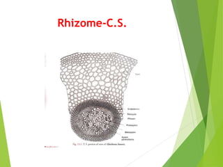
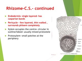
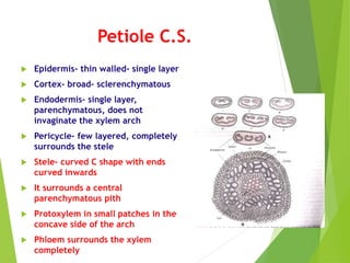
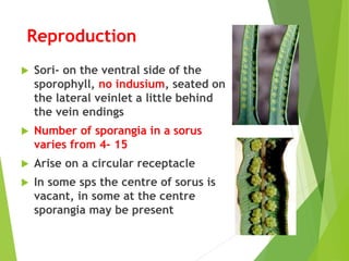
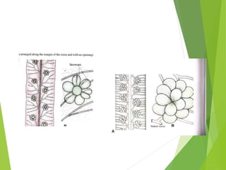
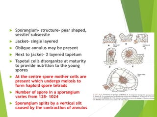
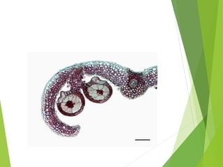
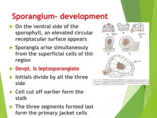
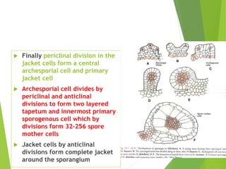
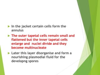
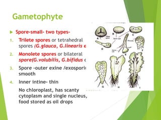
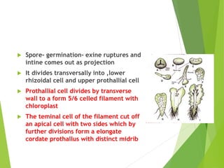
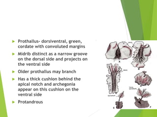
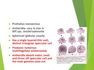
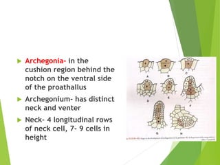
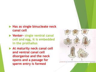
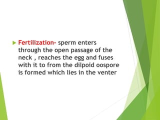
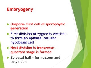
Gleichenia
Class - Leptosporangiatae
Order- Filicales
Family – Gleicheniaceae
Gleichenia
Gleichenia
 About- 130 sps
 In India- G.linearis, G.glauca
 G.glauca only in Eastern Himalayas
 G.linearis- wide distribution
 Usually in exposed regions, all are
xerophytes and terrestrial
Gleichenia
Sporophyte
 Has long, dichotomously branched,
prostrate , subterranean rhizome
bearing leaves on the upper surface and
adventitious roots on the under side
 Leaves occur in 3 rows though appear to
be in single row
 Leaves- a few feet to 50 mtrs in length
 Leaves show repeated forking-
pseudodichotomy as the rachis growth is
arrested and the pinnae below continue
to grow
 The leaflets lobed, the
incisions reach almost
the midrib so appear as
pinnules
 Has open venation
 Roots- branched-
arranged irregulary/ in
3 rows
Anatomy
 Rhizome-C.S.
 Epidermis- single layer of thin walled
colourless cells
 In some sclerenchymatous and brown
coloured
 No stomata
 Cortex – many layered, broad, may be fully
sclerenchymatous as in G.linearis or outer
sclerenchymatous and inner thin
parenchymatous region as in G.dichotoma
 Stele- protostelic except in G.pectinata
where it is amphiphloic siphonostele in the
internode and solenostele in node.
 In G.linearis and G.dichotoma – it is
mixed protostele
Rhizome-C.S.
Rhizome-C.S.- continued
 Endodermis- single layered- has
casparian bands
 Pericycle – few layered, thin walled ,
surrounds phloem completely
 Xylem occupies the centre- circular in
outline/lobed- usually mixed protostele
 Protoxylem- small patches at the
periphery
Petiole C.S.
 Epidermis- thin walled- single layer
 Cortex- broad- sclerenchymatous
 Endodermis- single layer,
parenchymatous, does not
invaginate the xylem arch
 Pericycle- few layered, completely
surrounds the stele
 Stele- curved C shape with ends
curved inwards
 It surrounds a central
parenchymatous pith
 Protoxylem in small patches in the
concave side of the arch
 Phloem surrounds the xylem
completely
 Pinnule – C.S.
 Has lower and upper epidermis, stomata
only in lower epidermis
 Mesophyll uniform homogenous- has
large air spaces and chlorenchymatous
 Stele- circular in outline
 Vascular bindles collateral
 Phloem faces lower epidermis
 Xylem exarch
Reproduction
 Sori- on the ventral side of the
sporophyll, no indusium, seated on
the lateral veinlet a little behind
the vein endings
 Number of sporangia in a sorus
varies from 4- 15
 Arise on a circular receptacle
 In some sps the centre of sorus is
vacant, in some at the centre
sporangia may be present
 Sporangium- structure- pear shaped,
sessile/ subsessile
 Jacket- single layered
 Oblique annulus may be present
 Next to jacket- 2 layered tapetum
 Tapetal cells disorganize at maturity
to provide nutrition to the young
spores
 At the centre spore mother cells are
present which undergo meiosis to
form haploid spore tetrads
 Number of spore in a sporangium
varies from 128- 1024
 Sporangium splits by a vertical slit
caused by the contraction of annulus
Sporangium- development
 On the ventral side of the
sporophyll, an elevated circular
receptacular surface appears
 Sporangia arise simultaneouly
from the superficial cells of this
region
 Devpt. Is leptosporangiate
 Initials divide by all the three
side
 Cell cut off earlier form the
stalk
 The three segments formed last
form the primary jacket cells
 Finally periclinal division in the
jacket cells form a central
archesporial cell and primary
jacket cell
 Archesporial cell divides by
periclinal and anticlinal
divisions to form two layered
tapetum and innermost primary
sporogenous cell which by
divisions form 32-256 spore
mother cells
 Jacket cells by anticlinal
divisions form complete jacket
around the sporangium
 In the jacket certain cells form the
annulus
 The outer tapetal cells remain small and
flattened but the inner tapetal cells
enlarge and nuclei divide and they
become multinucleate
 Later this layer disorganise and form a
nourishing plasmodial fluid for the
developng spores
Gametophyte
 Spore-small- two types-
1. Trilete spores or tetrahedral
spores (G.glauca, G.linearis etc
2. Monolete spores or bilateral
spore(G.volubilis, G.bifidus etc.)
3. Spore –outer exine /exosporium-
smooth
4. Inner intine- thin
No chloroplast, has scanty
cytoplasm and single nucleus,
food stored as oil drops
 Spore- germination- exine ruptures and
intine comes out as projection
 It divides transversally into ,lower
rhizoidal cell and upper prothallial cell
 Prothallial cell divides by transverse
wall to a form 5/6 celled filament with
chloroplast
 The teminal cell of the filament cut off
an apical cell with two sides which by
further divisions form a elongate
cordate prothallus with distinct midrib
 Prothallus- dorsiventral, green,
cordate with convoluted margins
 Midrib distinct as a narrow groove
on the dorsal side and projects on
the ventral side
 Older prothallus may branch
 Has a thick cushion behind the
apical notch and archegonia
appear on this cushion on the
ventral side
 Protandrous
 Prothallus monoecious
 Antheridia- vary in size in
diff.sps, sessile/subsessile
 Spherical/ globular usually
 Has a single layered thin wall,
distinct triangular opercular cell
 Produces numerous
multiflagellate antherozoids
 Antheridia absorb water, swell
and throw off opercular cell and
the male gametes ooze out
 Archegonia- in the
cushion region behind the
notch on the ventral side
of the proathallus
 Archegonium- has distinct
neck and venter
 Neck- 4 longitudinal rows
of neck cell, 7- 9 cells in
height
 Has as single binucleate neck
canal cell
 Venter- single ventral canal
cell and egg, it is embedded
in the prothallus
 At maturity neck canal cell
and ventral canal cell
disorganise and the neck
opens and a passage for
sperm entry is formed
 Fertilization- sperm enters
through the open passage of the
neck , reaches the egg and fuses
with it to from the dilpoid oospore
is formed which lies in the venter
Embryogeny
 Oospore- first cell of sporophytic
generation
 First division of zygote is vertical-
to form an epibasal cell and
hypobasal cell
 Next division is transverse-
quadrant stage is formed
 Epibasal half – forms stem and
cotyledon
 Hypobasal half- forms the foot and
root
 Foot- conspicuous and haustorial
 Primary root grows into soil and
primary cotyledon pushes upwards
through the notch
 Stem apex grows by a single apical
cell and finally an independent
sporophyte is formed

Gleichenia............................pptx

  • 1.
    Gleichenia Class - Leptosporangiatae Order-Filicales Family – Gleicheniaceae
  • 2.
  • 3.
    Gleichenia  About- 130sps  In India- G.linearis, G.glauca  G.glauca only in Eastern Himalayas  G.linearis- wide distribution  Usually in exposed regions, all are xerophytes and terrestrial
  • 4.
  • 5.
    Sporophyte  Has long,dichotomously branched, prostrate , subterranean rhizome bearing leaves on the upper surface and adventitious roots on the under side  Leaves occur in 3 rows though appear to be in single row  Leaves- a few feet to 50 mtrs in length  Leaves show repeated forking- pseudodichotomy as the rachis growth is arrested and the pinnae below continue to grow
  • 6.
     The leafletslobed, the incisions reach almost the midrib so appear as pinnules  Has open venation  Roots- branched- arranged irregulary/ in 3 rows
  • 7.
    Anatomy  Rhizome-C.S.  Epidermis-single layer of thin walled colourless cells  In some sclerenchymatous and brown coloured  No stomata  Cortex – many layered, broad, may be fully sclerenchymatous as in G.linearis or outer sclerenchymatous and inner thin parenchymatous region as in G.dichotoma  Stele- protostelic except in G.pectinata where it is amphiphloic siphonostele in the internode and solenostele in node.  In G.linearis and G.dichotoma – it is mixed protostele
  • 8.
  • 9.
    Rhizome-C.S.- continued  Endodermis-single layered- has casparian bands  Pericycle – few layered, thin walled , surrounds phloem completely  Xylem occupies the centre- circular in outline/lobed- usually mixed protostele  Protoxylem- small patches at the periphery
  • 10.
    Petiole C.S.  Epidermis-thin walled- single layer  Cortex- broad- sclerenchymatous  Endodermis- single layer, parenchymatous, does not invaginate the xylem arch  Pericycle- few layered, completely surrounds the stele  Stele- curved C shape with ends curved inwards  It surrounds a central parenchymatous pith  Protoxylem in small patches in the concave side of the arch  Phloem surrounds the xylem completely
  • 11.
     Pinnule –C.S.  Has lower and upper epidermis, stomata only in lower epidermis  Mesophyll uniform homogenous- has large air spaces and chlorenchymatous  Stele- circular in outline  Vascular bindles collateral  Phloem faces lower epidermis  Xylem exarch
  • 12.
    Reproduction  Sori- onthe ventral side of the sporophyll, no indusium, seated on the lateral veinlet a little behind the vein endings  Number of sporangia in a sorus varies from 4- 15  Arise on a circular receptacle  In some sps the centre of sorus is vacant, in some at the centre sporangia may be present
  • 14.
     Sporangium- structure-pear shaped, sessile/ subsessile  Jacket- single layered  Oblique annulus may be present  Next to jacket- 2 layered tapetum  Tapetal cells disorganize at maturity to provide nutrition to the young spores  At the centre spore mother cells are present which undergo meiosis to form haploid spore tetrads  Number of spore in a sporangium varies from 128- 1024  Sporangium splits by a vertical slit caused by the contraction of annulus
  • 16.
    Sporangium- development  Onthe ventral side of the sporophyll, an elevated circular receptacular surface appears  Sporangia arise simultaneouly from the superficial cells of this region  Devpt. Is leptosporangiate  Initials divide by all the three side  Cell cut off earlier form the stalk  The three segments formed last form the primary jacket cells
  • 17.
     Finally periclinaldivision in the jacket cells form a central archesporial cell and primary jacket cell  Archesporial cell divides by periclinal and anticlinal divisions to form two layered tapetum and innermost primary sporogenous cell which by divisions form 32-256 spore mother cells  Jacket cells by anticlinal divisions form complete jacket around the sporangium
  • 18.
     In thejacket certain cells form the annulus  The outer tapetal cells remain small and flattened but the inner tapetal cells enlarge and nuclei divide and they become multinucleate  Later this layer disorganise and form a nourishing plasmodial fluid for the developng spores
  • 19.
    Gametophyte  Spore-small- twotypes- 1. Trilete spores or tetrahedral spores (G.glauca, G.linearis etc 2. Monolete spores or bilateral spore(G.volubilis, G.bifidus etc.) 3. Spore –outer exine /exosporium- smooth 4. Inner intine- thin No chloroplast, has scanty cytoplasm and single nucleus, food stored as oil drops
  • 20.
     Spore- germination-exine ruptures and intine comes out as projection  It divides transversally into ,lower rhizoidal cell and upper prothallial cell  Prothallial cell divides by transverse wall to a form 5/6 celled filament with chloroplast  The teminal cell of the filament cut off an apical cell with two sides which by further divisions form a elongate cordate prothallus with distinct midrib
  • 21.
     Prothallus- dorsiventral,green, cordate with convoluted margins  Midrib distinct as a narrow groove on the dorsal side and projects on the ventral side  Older prothallus may branch  Has a thick cushion behind the apical notch and archegonia appear on this cushion on the ventral side  Protandrous
  • 22.
     Prothallus monoecious Antheridia- vary in size in diff.sps, sessile/subsessile  Spherical/ globular usually  Has a single layered thin wall, distinct triangular opercular cell  Produces numerous multiflagellate antherozoids  Antheridia absorb water, swell and throw off opercular cell and the male gametes ooze out
  • 23.
     Archegonia- inthe cushion region behind the notch on the ventral side of the proathallus  Archegonium- has distinct neck and venter  Neck- 4 longitudinal rows of neck cell, 7- 9 cells in height
  • 24.
     Has assingle binucleate neck canal cell  Venter- single ventral canal cell and egg, it is embedded in the prothallus  At maturity neck canal cell and ventral canal cell disorganise and the neck opens and a passage for sperm entry is formed
  • 25.
     Fertilization- spermenters through the open passage of the neck , reaches the egg and fuses with it to from the dilpoid oospore is formed which lies in the venter
  • 26.
    Embryogeny  Oospore- firstcell of sporophytic generation  First division of zygote is vertical- to form an epibasal cell and hypobasal cell  Next division is transverse- quadrant stage is formed  Epibasal half – forms stem and cotyledon
  • 27.
     Hypobasal half-forms the foot and root  Foot- conspicuous and haustorial  Primary root grows into soil and primary cotyledon pushes upwards through the notch  Stem apex grows by a single apical cell and finally an independent sporophyte is formed