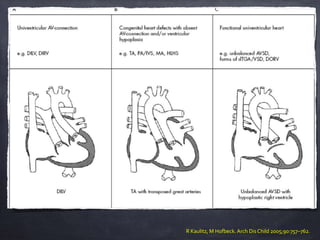
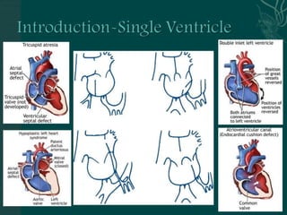
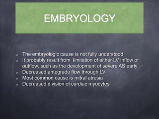
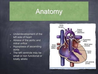
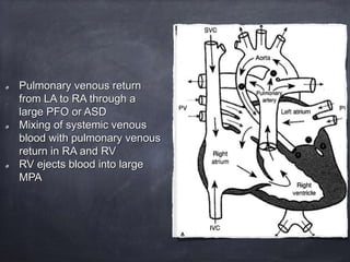
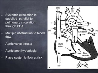
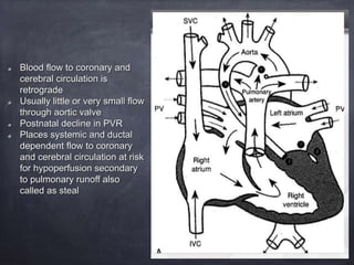
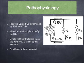
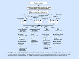
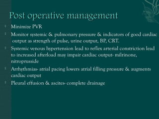
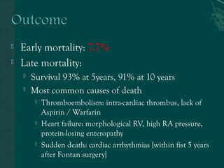
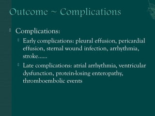
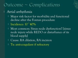
SINGLE VENTRICLE
BY: DR RICHA MALIK
MODERATOR: DR AJAY KUMAR
What is the ‘single ventricle’?
Prof. Richard van Praagh
‘One ventricular chamber receives blood from both
tricuspid and mitral valve or a common atrioventricular
valve’
Therefore, this excludes tricuspid atresia and mitral
atresia.
So !!! you are in doubt what the univentricular
physiology is.
Prof. Robert H Anderson emphasised that
‘the entire atrioventricular junction is connected to only one
chamber in the ventricular mass’
DILV
Absent of one atrioventricular connection
Common AV valve with one completely well-developed
ventricule
Only one fully developed ventricle
Heterotaxy syndrome
Rare miscellaneous forms
Univentricular heart
Congenital heart surgery nomenclature and database
Common inlet atrioventricular connection
DILV
DIRV
Absence of one atrioventricular connection
Tricuspid atresia
Mitral atresia
Common atrioventricular valve with one well-developed ventricle (unbalanced
AVSD)
Heterotaxia syndrome with only one fully developed ventricle
Other rare forms of univentricular heart
R Kaulitz, M Hofbeck. Arch Dis Child 2005;90:757–762.
Evolution of surgery for single
functioning ventricle
1940s Blalock-Taussig shunt
1952 PA banding by Muller WH and Dammann JF
1954 Shumacker and 1st cavopulmonary connection
1956 separation of single ventricle at Mayo Clinics
1971 Fontan’s and Kreutzer’s
EMBRYOLOGY
The embryologic cause is not fully understood
It probably result from limitation of either LV inflow or
outflow, such as the development of severe AS early
Decreased antegrade flow through LV
Most common cause is mitral atresia
Decreased division of cardiac myocytes
Genetics
Familial inheritance
A. Autosomal recessive and multifactorial inheritance have both been postulated
B. Sibling recurrence risk :0.5%
C. Sibling recurrence for all other cardiac malformation : 2.2%
Definable genetic disorder: 28%
- Turner syndrome
- Noonan syndrome
- Trisomy 13, 18, 21, or other micro deletion syndrome
Epidemiology
According to new England registry, there is reported incidence of
univerntricular heart to be 54 cases per million live births.
HLHS (hypoplastic left heart syndrome) the most common form of
single ventricle pathology- the 4th most common congenital heart
disease in neonate incidence is 2.3 cases per 10,000 live births
Tricuspid atresia the second most common subtype of univentricular
heart is thought to occur <1 for every 10,000 live births
23% of all neonatal mortality from CHD
Male predominance: 57-70%
Anatomy
Underdevelopment of the
left side of heart
Atresia of the aortic and
mitral orifice
Hypoplasia of ascending
aorta
The left ventricle may be
small or non functional or
totally atretic
Pulmonary venous return
from LA to RA through a
large PFO or ASD
Mixing of systemic venous
blood with pulmonary venous
return in RA and RV
RV ejects blood into large
MPA
Systemic circulation is
supplied parallel to
pulmonary circulation
through PDA
Multiple obstruction to blood
flow
- Aortic valve atresia
- Aortic arch hypoplasia
- Place systemic flow at risk
Blood flow to coronary and
cerebral circulation is
retrograde
Usually little or very small flow
through aortic valve
Postnatal decline in PVR
Places systemic and ductal
dependent flow to coronary
and cerebral circulation at risk
for hypoperfusion secondary
to pulmonary runoff also
called as steal
Pathophysiology
Relative Qp and Qs determined
by SVR and TVR
Ventricle must supply both Qp
and Qs
- Single right ventricle has twice
the work load of an in series
ventricle
- Significant volume overload
The aim of the initial management is to optimise Qp and
Qs in a manner that provides adequate end organ oxygen
delivery without overloading the single ventricle
The balancing act is only temporary not definitive
So optimal physiology of univentricular heat requires-
good ventricular function without AV valve regurgitation
and unrestrictive ASD and well balanced systemic and
pulmonary blood flow
Clinical features
Clinical manifestations hinge on the presence or absence of pulmonary
outflow obstruction. Without pulmonary stenosis, infants with low
pulmonary resistance present with signs and symptoms typical of large
left-to-right shunting, ie, congestive heart failure and failure to thrive
Because of increased pulmonary blood flow, cyanosis may not be
apparent. Aortic outflow obstruction may compound already excessive
pulmonary blood flow and worsen congestive heart failure. Some patients
(eg, type IV DILV) may have a preferential favourable stream of systemic
venous return to the pulmonary artery and pulmonary venous return to
aorta
In contrast, an unfavourable stream with a transposition-like
blood flow pattern may occur in patients with a right-sided
subaortic right ventricle and straddling right AV valve.
Cyanosis and systemic hypoxemia may result
In the settings of univentriclular heart, a certain degree of
pulmonary stenosis is physiologically desirable to prevent
pulmonary overcirculation. Severe pulmonary stenosis or
atresia may result in profound hypoxemia and cyanosis,
however.
Treatment options
Supportive therapy
- Only option till 25 years ago
- Is still main treatment option in many countries
Staged reconstruction
- StageI Norwood procedure
- Stage II bidirectional glenn or hemi-fontan
- Stage III Fontan
Transplant
- Mechanical assist devices
Mechanical assist device
The recent development
Devices have been used both as sub pulmonary pump to drive
blood forward through the pulmonary circuit and also in the
standard systemic ventriclular position to pull blood through Fontan
circuit by lowering left atrial pressure
Placement of these devices depends on underlying
hemodynamics, systemic ventricular failure VS elevation of PVR
Bridged to transplant, eventual destination therapy
The main pulmonary artery is divided, the proximal portion is
anastomosed to the ascending aorta, the aortic arch is repaired and
augmented, and pulmonary blood flow is maintained via a modified
Blalock-Taussig shunt or Gore-Tex shunt from the right ventricle.
The Norwood stage II procedure, performed before 6 months of
age, consists of a bidirectional
Glenn shunt or hemi-Fontan and closure of the Blalock- Taussig
shunt.
Between 18 months to 3 years go age ,stage III procedure
completes the total cavopulmonary Fon- tan by connection of the
inferior vena cava to the pulmonary artery.
Norwood Procedure
Post Norwood
Sano shunt
RV to PA conduit- introduced by Sano in 1990
The proposed solution to-
- Surgical manipulation of coronaries
- Pulmonary diastolic run off through MBT shunt,
causing steal phenomenon
Sano shunt
Development of fontan procedures
Developed in 1971 for tricuspid atresia, the Fontan procedure has undergone multiple
modifications to encompass several forms of palliative surgery that divert systemic
venous return to the pulmonary artery, usually without interposition of a subpulmonary
ventricle.
The classic Fontan involved a valved conduit between the right atrium and pulmonary
artery.
Currently, most adults will have had a modified Fontan, which consists of direct
anastomosis of the right atrium to pulmonary artery .
In 1987, de Leval et al proposed a major variation that consisted of an end-to-side
anastomosis of the superior vena cava to the undivided right pulmonary artery, a
composite intraatrial tunnel with the right atrial posterior wall, and a prosthetic patch to
channel the inferior vena cava to the transected superior vena cava.
Total cavopulmonary connections may also be performed as extracardiac
tunnels, with inferior vena caval flow directed to the pulmonary artery via an
external conduit. As in the intracardiac lateral tunnel, the superior vena cava
is anastomosed to the pulmonary artery.
In addition, Fontan pathways may be “fenestrated” by creation of an ASD in
the baffle or patch to provide an escape valve that allows right-to-left
shunting, which may be beneficial in early post op period as it leads to
augmentation of cardiac output and can decrease the high naval or right
atrial pressure.
Thank you for your patience

single ventricle physiology

  • 1.
    SINGLE VENTRICLE BY: DRRICHA MALIK MODERATOR: DR AJAY KUMAR
  • 2.
    What is the‘single ventricle’? Prof. Richard van Praagh ‘One ventricular chamber receives blood from both tricuspid and mitral valve or a common atrioventricular valve’ Therefore, this excludes tricuspid atresia and mitral atresia.
  • 3.
    So !!! youare in doubt what the univentricular physiology is. Prof. Robert H Anderson emphasised that ‘the entire atrioventricular junction is connected to only one chamber in the ventricular mass’ DILV Absent of one atrioventricular connection Common AV valve with one completely well-developed ventricule Only one fully developed ventricle Heterotaxy syndrome Rare miscellaneous forms
  • 4.
    Univentricular heart Congenital heartsurgery nomenclature and database Common inlet atrioventricular connection DILV DIRV Absence of one atrioventricular connection Tricuspid atresia Mitral atresia Common atrioventricular valve with one well-developed ventricle (unbalanced AVSD) Heterotaxia syndrome with only one fully developed ventricle Other rare forms of univentricular heart
  • 5.
    R Kaulitz, MHofbeck. Arch Dis Child 2005;90:757–762.
  • 7.
    Evolution of surgeryfor single functioning ventricle 1940s Blalock-Taussig shunt 1952 PA banding by Muller WH and Dammann JF 1954 Shumacker and 1st cavopulmonary connection 1956 separation of single ventricle at Mayo Clinics 1971 Fontan’s and Kreutzer’s
  • 8.
    EMBRYOLOGY The embryologic causeis not fully understood It probably result from limitation of either LV inflow or outflow, such as the development of severe AS early Decreased antegrade flow through LV Most common cause is mitral atresia Decreased division of cardiac myocytes
  • 9.
    Genetics Familial inheritance A. Autosomalrecessive and multifactorial inheritance have both been postulated B. Sibling recurrence risk :0.5% C. Sibling recurrence for all other cardiac malformation : 2.2% Definable genetic disorder: 28% - Turner syndrome - Noonan syndrome - Trisomy 13, 18, 21, or other micro deletion syndrome
  • 10.
    Epidemiology According to newEngland registry, there is reported incidence of univerntricular heart to be 54 cases per million live births. HLHS (hypoplastic left heart syndrome) the most common form of single ventricle pathology- the 4th most common congenital heart disease in neonate incidence is 2.3 cases per 10,000 live births Tricuspid atresia the second most common subtype of univentricular heart is thought to occur <1 for every 10,000 live births 23% of all neonatal mortality from CHD Male predominance: 57-70%
  • 11.
    Anatomy Underdevelopment of the leftside of heart Atresia of the aortic and mitral orifice Hypoplasia of ascending aorta The left ventricle may be small or non functional or totally atretic
  • 12.
    Pulmonary venous return fromLA to RA through a large PFO or ASD Mixing of systemic venous blood with pulmonary venous return in RA and RV RV ejects blood into large MPA
  • 13.
    Systemic circulation is suppliedparallel to pulmonary circulation through PDA Multiple obstruction to blood flow - Aortic valve atresia - Aortic arch hypoplasia - Place systemic flow at risk
  • 14.
    Blood flow tocoronary and cerebral circulation is retrograde Usually little or very small flow through aortic valve Postnatal decline in PVR Places systemic and ductal dependent flow to coronary and cerebral circulation at risk for hypoperfusion secondary to pulmonary runoff also called as steal
  • 15.
    Pathophysiology Relative Qp andQs determined by SVR and TVR Ventricle must supply both Qp and Qs - Single right ventricle has twice the work load of an in series ventricle - Significant volume overload
  • 16.
    The aim ofthe initial management is to optimise Qp and Qs in a manner that provides adequate end organ oxygen delivery without overloading the single ventricle The balancing act is only temporary not definitive So optimal physiology of univentricular heat requires- good ventricular function without AV valve regurgitation and unrestrictive ASD and well balanced systemic and pulmonary blood flow
  • 18.
    Clinical features Clinical manifestationshinge on the presence or absence of pulmonary outflow obstruction. Without pulmonary stenosis, infants with low pulmonary resistance present with signs and symptoms typical of large left-to-right shunting, ie, congestive heart failure and failure to thrive Because of increased pulmonary blood flow, cyanosis may not be apparent. Aortic outflow obstruction may compound already excessive pulmonary blood flow and worsen congestive heart failure. Some patients (eg, type IV DILV) may have a preferential favourable stream of systemic venous return to the pulmonary artery and pulmonary venous return to aorta
  • 19.
    In contrast, anunfavourable stream with a transposition-like blood flow pattern may occur in patients with a right-sided subaortic right ventricle and straddling right AV valve. Cyanosis and systemic hypoxemia may result In the settings of univentriclular heart, a certain degree of pulmonary stenosis is physiologically desirable to prevent pulmonary overcirculation. Severe pulmonary stenosis or atresia may result in profound hypoxemia and cyanosis, however.
  • 20.
    Treatment options Supportive therapy -Only option till 25 years ago - Is still main treatment option in many countries Staged reconstruction - StageI Norwood procedure - Stage II bidirectional glenn or hemi-fontan - Stage III Fontan Transplant - Mechanical assist devices
  • 21.
    Mechanical assist device Therecent development Devices have been used both as sub pulmonary pump to drive blood forward through the pulmonary circuit and also in the standard systemic ventriclular position to pull blood through Fontan circuit by lowering left atrial pressure Placement of these devices depends on underlying hemodynamics, systemic ventricular failure VS elevation of PVR Bridged to transplant, eventual destination therapy
  • 27.
    The main pulmonaryartery is divided, the proximal portion is anastomosed to the ascending aorta, the aortic arch is repaired and augmented, and pulmonary blood flow is maintained via a modified Blalock-Taussig shunt or Gore-Tex shunt from the right ventricle. The Norwood stage II procedure, performed before 6 months of age, consists of a bidirectional Glenn shunt or hemi-Fontan and closure of the Blalock- Taussig shunt. Between 18 months to 3 years go age ,stage III procedure completes the total cavopulmonary Fon- tan by connection of the inferior vena cava to the pulmonary artery. Norwood Procedure
  • 29.
  • 30.
    Sano shunt RV toPA conduit- introduced by Sano in 1990 The proposed solution to- - Surgical manipulation of coronaries - Pulmonary diastolic run off through MBT shunt, causing steal phenomenon
  • 31.
  • 39.
    Development of fontanprocedures Developed in 1971 for tricuspid atresia, the Fontan procedure has undergone multiple modifications to encompass several forms of palliative surgery that divert systemic venous return to the pulmonary artery, usually without interposition of a subpulmonary ventricle. The classic Fontan involved a valved conduit between the right atrium and pulmonary artery. Currently, most adults will have had a modified Fontan, which consists of direct anastomosis of the right atrium to pulmonary artery . In 1987, de Leval et al proposed a major variation that consisted of an end-to-side anastomosis of the superior vena cava to the undivided right pulmonary artery, a composite intraatrial tunnel with the right atrial posterior wall, and a prosthetic patch to channel the inferior vena cava to the transected superior vena cava.
  • 40.
    Total cavopulmonary connectionsmay also be performed as extracardiac tunnels, with inferior vena caval flow directed to the pulmonary artery via an external conduit. As in the intracardiac lateral tunnel, the superior vena cava is anastomosed to the pulmonary artery. In addition, Fontan pathways may be “fenestrated” by creation of an ASD in the baffle or patch to provide an escape valve that allows right-to-left shunting, which may be beneficial in early post op period as it leads to augmentation of cardiac output and can decrease the high naval or right atrial pressure.
  • 54.
    Thank you foryour patience