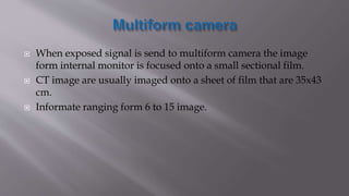
BY:BISHAL KHANAL
 The internal structure of an object can be reconstructed from multiple
projection.
 A large number of data along a thin line of 2D section is acquired to
reconstruct the structure within that slice.
 This involves five main stages:-
1) Scanning phase and data acquisition system (DAS).
2) Pre-processing of raw data.
3) Image reconstruction.
4) Linear attenuation coefficient of each pixel is converted into Hounsfield
unit (HU).
5) Display and Recording.
Scanning phase
 After the patient lies in the gantry the following steps
are done such as
a. Field of view is selected.
b. FOV cuts into thinner and smaller slices.
c. Placing a matrix grid on thin slices.
d. Scanning of selected slice from multiple projection and
acquire data.
e. Select next slice and repeat scanning.
 It revolves tube along with detector.
 It consist of detector which converts intensity
information into digital signal.

 The computer hardware and software convert the raw data of CT into CT image
through the process called image reconstruction.
 It is done by filter back projection.
 In this pre-processed data are sending into array process by the help of filter back
projection.
 ARRAY PROCESS:-it is small computer in the host processer which does the
mathematical calculation.
 Function:-
 The main function is the mathematical manuplation of data very quickely.
 It help in reconstruction of linear attuation coefficient.
 Design of array process:-
 High speed memory.
 High speed bus.

TISSUES CT NUMBER RANGE IN HU
 Air
 Lungs
 Fat
 Water
 Muscle
 Soft tissue
 Cortical bone
 Trabecular bone
 -1000
 -900 to -300
 -120 to –80
 0
 10 to 30
 10 to 30
 50 to 100
 500 to 1000
 Digital imaging is based on following
Matrix size
𝑀 =
𝐹𝑂𝑉
𝑝𝑖𝑥𝑒𝑙 𝑠𝑖𝑧𝑒
Pixel bit depth
It is the ability of pixel to show different shades of grey.
We can differentiate the shades of grey by window width(W) and
window level(L).
 Window width (W)
 It decides the shades.
 It depends on contrast.
 Narrow window width having
high contrast wider window width
having low contrast.
 Decreasing the window width,
increasing the contrast of the
image so good for looking at
differences in soft tissues.
 Increasing the window width
allows structures with a large pixel
range to be viewed.
 Window level(L)
 It is the mid-point of the
window width.
 It identifies the type of tissues.
 It controls the CT number
which determine the position
of window.
 Recording of the image produce by CT scanner is usually done
with the multi-format camera.
 A more recently introduced recording system uses a laser beam
to directly record the image on the system.
 When exposed signal is send to multiform camera the image
form internal monitor is focused onto a small sectional film.
 CT image are usually imaged onto a sheet of film that are 35x43
cm.
 Informate ranging form 6 to 15 image.
 Instead of exposing the film to an image displayed on an internal
television monitor, a laser camera uses a digital.
 Image data directly and scan the film link by line , bulding of the
image gradually.
 A laser beam is scanned over the film and intensity of lesser
beam is determined by the pixel value at that point in the image
data.
 The narrow beam gemeratry of leaser beam create less scattered
light in the system and therefore laser camera produces film
image with more contrast than do multiformate camera.

Basic principle of C.T.

  • 1.
  • 2.
     The internalstructure of an object can be reconstructed from multiple projection.  A large number of data along a thin line of 2D section is acquired to reconstruct the structure within that slice.  This involves five main stages:- 1) Scanning phase and data acquisition system (DAS). 2) Pre-processing of raw data. 3) Image reconstruction. 4) Linear attenuation coefficient of each pixel is converted into Hounsfield unit (HU). 5) Display and Recording.
  • 3.
    Scanning phase  Afterthe patient lies in the gantry the following steps are done such as a. Field of view is selected. b. FOV cuts into thinner and smaller slices. c. Placing a matrix grid on thin slices. d. Scanning of selected slice from multiple projection and acquire data. e. Select next slice and repeat scanning.
  • 4.
     It revolvestube along with detector.  It consist of detector which converts intensity information into digital signal.
  • 5.
  • 6.
     The computerhardware and software convert the raw data of CT into CT image through the process called image reconstruction.  It is done by filter back projection.  In this pre-processed data are sending into array process by the help of filter back projection.  ARRAY PROCESS:-it is small computer in the host processer which does the mathematical calculation.  Function:-  The main function is the mathematical manuplation of data very quickely.  It help in reconstruction of linear attuation coefficient.  Design of array process:-  High speed memory.  High speed bus.
  • 7.
  • 8.
    TISSUES CT NUMBERRANGE IN HU  Air  Lungs  Fat  Water  Muscle  Soft tissue  Cortical bone  Trabecular bone  -1000  -900 to -300  -120 to –80  0  10 to 30  10 to 30  50 to 100  500 to 1000
  • 9.
     Digital imagingis based on following Matrix size 𝑀 = 𝐹𝑂𝑉 𝑝𝑖𝑥𝑒𝑙 𝑠𝑖𝑧𝑒 Pixel bit depth It is the ability of pixel to show different shades of grey. We can differentiate the shades of grey by window width(W) and window level(L).
  • 10.
     Window width(W)  It decides the shades.  It depends on contrast.  Narrow window width having high contrast wider window width having low contrast.  Decreasing the window width, increasing the contrast of the image so good for looking at differences in soft tissues.  Increasing the window width allows structures with a large pixel range to be viewed.  Window level(L)  It is the mid-point of the window width.  It identifies the type of tissues.  It controls the CT number which determine the position of window.
  • 11.
     Recording ofthe image produce by CT scanner is usually done with the multi-format camera.  A more recently introduced recording system uses a laser beam to directly record the image on the system.
  • 12.
     When exposedsignal is send to multiform camera the image form internal monitor is focused onto a small sectional film.  CT image are usually imaged onto a sheet of film that are 35x43 cm.  Informate ranging form 6 to 15 image.
  • 13.
     Instead ofexposing the film to an image displayed on an internal television monitor, a laser camera uses a digital.  Image data directly and scan the film link by line , bulding of the image gradually.  A laser beam is scanned over the film and intensity of lesser beam is determined by the pixel value at that point in the image data.  The narrow beam gemeratry of leaser beam create less scattered light in the system and therefore laser camera produces film image with more contrast than do multiformate camera.