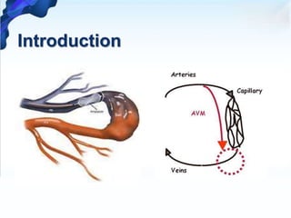
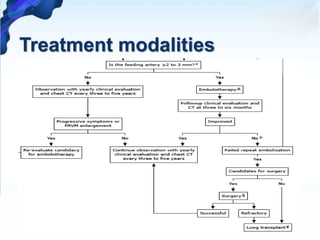
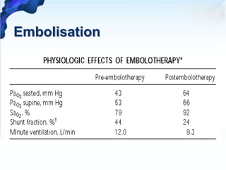
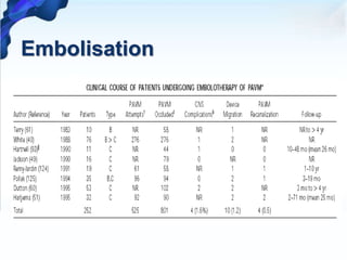
1. Pulmonary arteriovenous malformations (PAVMs) are rare vascular anomalies where abnormal dilated vessels provide a right-to-left shunt between the pulmonary artery and vein.
2. PAVMs are usually diagnosed through imaging like chest X-ray, CT, or MRI which show dilated vessels. Right-to-left shunting can be detected using echocardiography, oxygen studies, or pulmonary angiography.
3. Treatment involves embolization to occlude the abnormal vessels which successfully treats over 99% of PAVMs. Surgery is an alternative for cases that cannot be embolized or if embolization fails.
Imaging plays an important role in diagnosis and formulating differential diagnosis in case of Solitary pulmonary nodule. It helps in differentiating and predicting benign and malignant nodules.
Role of hrct in interstitial lung diseases pk uploadDr pradeep Kumar
Role of hrct in interstitial lung diseases pk , This is best powerpoint slides presentation including Latest American thoracic society and fleishners society guidelines . this includes radiographic images a well HRCT chest findings of various ILD. This will help alot for md pg radiology resident and radiologist. Thanks
Solitary pulmonary nodules radiology ppt is very good power point presentation from various source radiology assistant and latest guidelines. this power-point also includes many sign with multiple xray, ct and mri images. this will help alot. Thanks.
How to Make a Field invisible in Odoo 17Celine George
It is possible to hide or invisible some fields in odoo. Commonly using “invisible” attribute in the field definition to invisible the fields. This slide will show how to make a field invisible in odoo 17.
Introduction to AI for Nonprofits with Tapp NetworkTechSoup
Dive into the world of AI! Experts Jon Hill and Tareq Monaur will guide you through AI's role in enhancing nonprofit websites and basic marketing strategies, making it easy to understand and apply.
Francesca Gottschalk - How can education support child empowerment.pptxEduSkills OECD
Francesca Gottschalk from the OECD’s Centre for Educational Research and Innovation presents at the Ask an Expert Webinar: How can education support child empowerment?
Welcome to TechSoup New Member Orientation and Q&A (May 2024).pdfTechSoup
In this webinar you will learn how your organization can access TechSoup's wide variety of product discount and donation programs. From hardware to software, we'll give you a tour of the tools available to help your nonprofit with productivity, collaboration, financial management, donor tracking, security, and more.
Biological screening of herbal drugs: Introduction and Need for
Phyto-Pharmacological Screening, New Strategies for evaluating
Natural Products, In vitro evaluation techniques for Antioxidants, Antimicrobial and Anticancer drugs. In vivo evaluation techniques
for Anti-inflammatory, Antiulcer, Anticancer, Wound healing, Antidiabetic, Hepatoprotective, Cardio protective, Diuretics and
Antifertility, Toxicity studies as per OECD guidelines
Honest Reviews of Tim Han LMA Course Program.pptxtimhan337
Personal development courses are widely available today, with each one promising life-changing outcomes. Tim Han’s Life Mastery Achievers (LMA) Course has drawn a lot of interest. In addition to offering my frank assessment of Success Insider’s LMA Course, this piece examines the course’s effects via a variety of Tim Han LMA course reviews and Success Insider comments.
The French Revolution, which began in 1789, was a period of radical social and political upheaval in France. It marked the decline of absolute monarchies, the rise of secular and democratic republics, and the eventual rise of Napoleon Bonaparte. This revolutionary period is crucial in understanding the transition from feudalism to modernity in Europe.
For more information, visit-www.vavaclasses.com
The Roman Empire A Historical Colossus.pdfkaushalkr1407
The Roman Empire, a vast and enduring power, stands as one of history's most remarkable civilizations, leaving an indelible imprint on the world. It emerged from the Roman Republic, transitioning into an imperial powerhouse under the leadership of Augustus Caesar in 27 BCE. This transformation marked the beginning of an era defined by unprecedented territorial expansion, architectural marvels, and profound cultural influence.
The empire's roots lie in the city of Rome, founded, according to legend, by Romulus in 753 BCE. Over centuries, Rome evolved from a small settlement to a formidable republic, characterized by a complex political system with elected officials and checks on power. However, internal strife, class conflicts, and military ambitions paved the way for the end of the Republic. Julius Caesar’s dictatorship and subsequent assassination in 44 BCE created a power vacuum, leading to a civil war. Octavian, later Augustus, emerged victorious, heralding the Roman Empire’s birth.
Under Augustus, the empire experienced the Pax Romana, a 200-year period of relative peace and stability. Augustus reformed the military, established efficient administrative systems, and initiated grand construction projects. The empire's borders expanded, encompassing territories from Britain to Egypt and from Spain to the Euphrates. Roman legions, renowned for their discipline and engineering prowess, secured and maintained these vast territories, building roads, fortifications, and cities that facilitated control and integration.
The Roman Empire’s society was hierarchical, with a rigid class system. At the top were the patricians, wealthy elites who held significant political power. Below them were the plebeians, free citizens with limited political influence, and the vast numbers of slaves who formed the backbone of the economy. The family unit was central, governed by the paterfamilias, the male head who held absolute authority.
Culturally, the Romans were eclectic, absorbing and adapting elements from the civilizations they encountered, particularly the Greeks. Roman art, literature, and philosophy reflected this synthesis, creating a rich cultural tapestry. Latin, the Roman language, became the lingua franca of the Western world, influencing numerous modern languages.
Roman architecture and engineering achievements were monumental. They perfected the arch, vault, and dome, constructing enduring structures like the Colosseum, Pantheon, and aqueducts. These engineering marvels not only showcased Roman ingenuity but also served practical purposes, from public entertainment to water supply.
A Strategic Approach: GenAI in EducationPeter Windle
Artificial Intelligence (AI) technologies such as Generative AI, Image Generators and Large Language Models have had a dramatic impact on teaching, learning and assessment over the past 18 months. The most immediate threat AI posed was to Academic Integrity with Higher Education Institutes (HEIs) focusing their efforts on combating the use of GenAI in assessment. Guidelines were developed for staff and students, policies put in place too. Innovative educators have forged paths in the use of Generative AI for teaching, learning and assessments leading to pockets of transformation springing up across HEIs, often with little or no top-down guidance, support or direction.
This Gasta posits a strategic approach to integrating AI into HEIs to prepare staff, students and the curriculum for an evolving world and workplace. We will highlight the advantages of working with these technologies beyond the realm of teaching, learning and assessment by considering prompt engineering skills, industry impact, curriculum changes, and the need for staff upskilling. In contrast, not engaging strategically with Generative AI poses risks, including falling behind peers, missed opportunities and failing to ensure our graduates remain employable. The rapid evolution of AI technologies necessitates a proactive and strategic approach if we are to remain relevant.
2. Introduction
• Rare vascular anomalies of the lung, in which
abnormal dilated vessels provide a right-to-left shunt
between the pulmonary artery and vein.
• Also known as pulmonary arteriovenous fistulae,
pulmonary arteriovenous aneurysms, cavernous
angiomas of the lung, and pulmonary telangiectases
• First described by T. Churton in a 12 year old boy
with epistaxis and hemoptysis.
3. Introduction
• They are generally considered direct high flow, low-
resistance fistulous connections between the
pulmonary arteries and veins.
• Important consideration in the differential diagnosis
of common pulmonary problems, including
hypoxemia, pulmonary nodules, and hemoptysis.
• Mostly congenital in nature however they may be
acquired due number of secondary causes.
5. Epidemiology
• Female predilection with around 1.5 to 1.8:1
though more common in the male infants in
neonatal age.
• The estimated incidence is thought to be around
2-3 per 100,000.
• Around 10% of cases of PAVM are identified in
infancy or childhood, followed by a gradual increase
in the incidence through the fifth and sixth decades.
• Approximately 70% of the cases of PAVM are
associated with HHT.
6. Etiology
• Mostly congenital in nature and due to HHT in
majority of cases
• Most common acquired cause is hepatic cirrhosis
• They are also associated with surgeries for CHD
including classic glenn shunt
• HHT has an autosomal dominant inheritance
• Endoglin, the most abundant TGFb binding protein
that is found on endothelial cells, has been identified
as the HHT gene mapping to locus 9q3, and is
presently referred to as the gene for HHT 1.
7. Etiology
• Hereditary hemorrhagic telangiectasia
• Hepatic cirrhosis
• Penetrating chest trauma or congenital heart surgery
• Mitral stenosis
• Schistosomiasis
• Actinomycosis
• Fanconi's syndrome
• Metastatic thyroid carcinoma
• Status post surgery for congenital heart disease
• Idiopathic
8. Natural History
• True mortality and morbidity data are unknown
• Mortality is usually attributed to the complications of
PAVM like stroke and brain abscess, as well as
hypoxemic respiratory failure and life-threatening
hemoptysis or hemothorax.
• Most PAVMs remain stable in size. However,
approximately 25 percent will enlarge slowly, usually
at a rate of 0.3 to 2 mm/year.
• PAVMs do not spontaneously resolve.
9. Pathology
• Analogus to arterio-venous malformations else
where in the body having an afferent feeder artery
and a efferent draining artery.
• PAVM may appear macroscopically as a large, single
or multi-lobulated sac, a plexiform mass of dilated
vascular channels, or a dilated and tortuous direct
anastomosis between an artery and vein.
• usually thin-walled, consisting of a single layer of
endothelium and variable amounts of connective
tissue stroma
10. Pathology
• PAVMs are supplied by pulmonary arteries in about
95 percent of cases and are usually drained by
pulmonary veins; however, they may occasionally be
fed by systemic arteries (ie, the bronchial artery)
and/or drain into the left atrium or inferior vena
cava.
• When PAVMs are fed by systemic arteries, hereditary
hemorrhagic telangiectasia (HHT) is usually not the
cause of the PAVM
11. Pathology
• Most solitary PAVMs are seen in bilateral lower
lobes, the left lower lobe being the most common
location, followed by right lower lobe, left upper
lobe, right middle lobe, and right upper lobe
• PAVMs are usually found in close proximity to the
visceral pleura or embedded in the outer third of
lung parenchyma.
12. Classification
• Pathological classification
• simple type: commonest; has a single segmental
artery feeding the malformation; the feeding
segmental artery may have multiple subsegmental
branches that feed the malformation, but must have
only one single segmental level
• complex type: have multiple segmental feeding
arteries (~20%)
• diffuse type: rare (~5% of lesions); the diffuse form
of the disease is characterised by hundreds of
malformations; some patients can have combination
of simple and complex AVMs within a diffuse lesion
13. Classification
• Anatwabi et al in 1965
• group I: multiple small arteriovenous fistulas without
aneurysm
• group II: large arteriovenous aneurysm
• group III
– large arteriovenous aneurysm (central)
– large arteriovenous aneurysm with anomalous venous drainage
– multiple small arteriovenous fistulae with anomalous venous
drainage
• group IV
– large venous aneurysm with systemic arterial communication
– large venous aneurysm without fistula
• group V: anomalous venous drainage with fistulae
14. Pathophysiology
• In contrast to systemic arteriovenous malformation,
PAVMs do not affect cardiac haemodynamics.
Cardiac output, cardiac index, pulmonary capillary
wedge pressure, heart rate, blood pressure, and the
electrocardiogram are usually within normal limits.
• Degree of shunt is what determines the clinical
effects on the patient.
• The peripheral oxygen saturation is low and as
expected does not normalise with 100% oxygen.
15. Clinical Features
• pulmonary symptoms in only 20 to 65 percent
(approximately 40 percent) and the remainder are
asymptomatic, typically found incidentally on chest
imaging.
• The most common pulmonary symptoms are dyspnea in
13 to 56 percent and hemoptysis in 7 to 30 percent
• Patients with underlying HHT often shows symptoms
attributable to this disorder including epistaxis and
mucocutaneous telangiectases
16. Clinical Features
• PAVM, and is seen in almost all patients who have
associated clubbing
• it has been noted clinically that symptoms such as
dyspnea are sometimes strikingly minimal when
compared with associated signs such as cyanosis and
clubbing
• patients also have platypnea
• This phenomenon is believed to be secondary to a
decrease in blood flow through PAVM in the dependent
portions of the lungs upon assuming the supine position.
17. Clinical Features
• Epistaxis, which is caused by bleeding from mucosal
telangiectases and reflects the high incidence of HHT in
patients with PAVM.
• Epistaxis is characteristically spontaneous or precipitated
by minor trauma. Epistaxis is an early symptom specially
in patients with HHT.
• Bleeding from telangiectases on the skin and in the GIT is
seen in patients with PAVM and HHT.
• The incidence of gastrointestinal hemorrhage in patients
with HHT is 15 to 30%.
18. Clinical Features
• In a sizeable number of patients (43%–67%), a history of
neurological symptoms—that is, headache, vertigo,
paresis, numbness, paresthaesia, syncope, or confusion
can be found.
• The most common physical findings are cyanosis,
clubbing, and pulmonary vascular bruit.
• Pulmonary bruit is increased by inspiration and the
Muller manoeuvre, this is caused by an increase in the
pulmonary blood flow and decreased by expiration and
the Valsalva manoeuvre, by decreasing venous return to
the lung.
19. Diagnosis
• PAVMs should always be suspected in patients who
present with unexplained dyspnea or hypoxemia as
well as in patients with nodules and a history of a
stroke or brain abscess
• The classical triad of dyspnea, cyanosis and clubbing
may be found few patients.
• Diagnosis rests on radiological demonstration of
malformation, documentation of the right to left
shunt in the setting of clinical scenario.
21. Chest X Ray
• Classic roentgenographic appearance of a PAVM is
that of a round or oval mass of uniform density,
frequently lobulated but sharply defined, more
commonly in the lower lobes, and ranging from 1 to
5 cm in diameter.
• Patients with microvascular telangiectases may have
normal chest radiographs, or only a vague increase in
pulmonary vascular markings.
23. ABG and Orthodexia
• Patients with diffuse PAVMs are uniformly hypoxemic
with a mean arterial oxygen tension (PaO2) of about
47 mmHg.
• Orthodeoxia is the laboratory correlate of platypnea.
It is defined as a decrease in the oxyhemoglobin
saturation by 2 percent or more when rising from the
supine to the upright position
24. Echocardiography
• Transthoracic contrast echocardiography — TTCE (also
known as “bubble study”) is the preferred initial test.
• TTCE identifies PAVM-associated shunt with a sensitivity,
specificity, positive predictive value, and negative
predictive value of 100, 49, 32, and 100 percent,
respectively.
• TTCE involves the injection of echocardiographic
contrast, usually 10 to 20 mL of agitated saline into a
peripheral vein while simultaneously imaging the right
and left atria.
• The contrast is normally visualized in the right atrium
soon after injection and should not be visualized in the
left cardiac chambers at all
25. Echocardiography
When microbubbles are seen in the left atrium
• within one cardiac cycle of their appearance in the
right atrium, this is typically associated with an intra-
cardiac shunt
• within three to eight cycles is consistent with an
intra-pulmonary shunt
• within one to three cardiac cycles the location of the
shunt is indeterminate
26. Echocardiography
• Grade 0 – Grade 0 refers to no bubbles reaching the
left ventricle (ie, no right-to-left shunt).
• Grade 1 – Grade 1 refers to fewer than 30 bubbles
seen in the left ventricle. should undergo yearly
observation.
• Grade 2 or 3 – Grade 2 shunt (30 to 100 bubbles in
the left ventricle) or grade 3 shunt (>100 bubbles in
the left ventricle)
27. 100% Oxygenation
• It can be determined by the 100 percent oxygen
method, which involves measuring the arterial
oxygen tension (PaO2) and saturation (SaO2) and the
arterial carbon dioxide tension (PaCO2) after
breathing 100 percent oxygen through a mouthpiece
or airtight mask for 15 to 20 minutes and then using
those values to calculate the shunt fraction.
• A shunt fraction of >5 percent is considered
abnormal.
28. Exercise Tolerance
• Symptom-limited incremental exercise resulted in a
decrease in SaO2 from 86% at rest to 73% with peak
exercise. Despite this impressive degree of
desaturation, exercise capacity was generally well
preserved with 60% patients achieving 70% of
predicted maximal workload.
• 6 minute walk test may also be done.
29. Chest CT
• CT is often the diagnostic imaging modality of choice.
• The characteristic presentation of a PAVM on non-
contrast CT is a homogeneous, well-circumscribed,
non-calcified nodule up to several centimeters in
diameter or the presence of a serpiginous mass
connected with blood vessels
• Contrast injection demonstrates enhancement of the
feeding artery, the aneurysmal part, and the draining
vein on early-phase sequences.
30. Chest CT
• If the CT scan shows one or more PAVM with a
feeding artery diameter (FAD) ≥2 to 3 mm diameter,
the patient should be referred for pulmonary
angiography and potential embolotherapy.
• If the CT scan shows PAVMs with a FAD <2mm, in
most patients pulmonary angiography may be
deferred unless patients have clinical features
suggestive of symptomatic PAVM.
• If the CT scan is negative for PAVM and shunt is
present on TTCE, microscopic PAVMs may be
responsible for the shunt
32. Pulmonary Angiogram
• Pulmonary angiography is the gold standard test for
defining the vascular anatomy of PAVMs that are
identified on prior CT as suitable for embolotherapy.
• During angiographic testing, contrast should be
directly injected into the feeding artery or a distal
pulmonary artery (ie, hyper-selective angiography) to
accurately define the angioarchitecture of individual
lesions.
35. MRI
• Three-dimensional contrast–enhanced MR
angiography has is considered the MR technique of
choice for imaging vascular structures in the thorax
10. Most lesions within the lung have a relatively
long relaxation time and produce medium to high
intensity signals. Lesions with rapid blood flow within
result in a signal void and produce low intensity
signals
36. Treatment modalities
• Surgical resection was first reported in 1942 and was
the mainstay of treatment till 1980s
• Successful percutaneous embolization by Taylor in
1978 opened up new therapeutic modality.
• Percutaneous embolization is now preferred
modality of choice.
• May use
– Balloon embolization
– Coil embolization
38. Surgery
• Surgical resection of PAVMs is indicated in patients
who fail embolotherapy, develop serious bleeding
complication despite embolotherapy, have
intrapleural rupture of the PAVM, or have
untreatable contrast allergy and lesions not
amenable to embolotherapy.
• Different surgical techniques have been employed
which include local excision, segmental resection,
lobectomy, ligation, and even pneumonectomy.
39. Surgery
• Lung conserving resection, local resection, or
segmentectomy is the procedure of choice whenever
possible. Staged bilateral thoracotomies were
performed in a case of an extensive bilateral PAVM.
• PAVM surgery has the same risk as any other thoracic
surgery procedure, but when properly performed in
well selected patients, it results in minimal morbidity
and mortality
41. Embolisation
• embolotherapy is the mainstay of treatment as most
PAVMs (>99 percent) can be successfully treated with
this therapy.
• PAVMs with FAD ≥3 mm are targeted for
embolization and smaller PAVMs are embolized, if
technically feasible.
• distal lesions with hyperacute branching of the
pulmonary artery or of the feeding artery itself may
make it technically difficult to access the PAVM for
coil placement
42. Embolisation
• most patients who undergo embolotherapy,
symptoms improve and complications are avoided.
• However, in a small percentage (<20 percent) the
pulmonary arteriovenous malformation (PAVM) will
not be successfully occluded, the PAVM recanalizes,
or a new PAVM develops.