Bronchiectasis is a condition defined as abnormal, irreversible dilation of the bronchi. It can be caused by primary infections, impaired mucus clearance, immunodeficiencies, hyperimmune responses, bronchial obstructions, autoimmune diseases, and developmental defects. The main pathogenesis involves bacterial infections destroying the bronchial wall structures through proteases and toxins. This leads to dilation of the bronchi and accumulation of thick, purulent material. Bronchiectasis can occur in several syndromes as well, including Kartagener's syndrome and Williams-Campbell syndrome. Tuberculosis is noted as a cause of predominantly upper lobe bronchiectasis, where the initial infection causes bronchial swelling and narrowing
Bronchiectasis refers to the congenital/acquired irreversible airway dilation that involves the bronchi/bronchioles in either a focal or a diffuse manner.
It is a pulmonary disease related to chronic infections in the background of inability of respiratory mucosa to clear the infections and impaired ciliary function.
It is chronic disease with high morbidity and mortality
Lecture slides about bronchiectasis with contents including definition, causes, pathogenesis and pathology, and how to make diagnosis. Treatment for bronchiectasis is presented separately.
These lecture notes were prepared by Dr. Hamdi Turkey- Pulmonologist- Department of internal medicine - Taiz university
Do Not Forget To Visit Our Pages On Facebook on the following Links:
https://www.facebook.com/groups/569435236444761/
AND
https://www.facebook.com/groups/690331650977113/
PATHOGENESIS OF BRONCHIECTASIS BY DR BASHIR AHMED DAR ASSOCIATE PROFESSOR MED...Prof Dr Bashir Ahmed Dar
Dr Bashir Ahmed Dar associate professor medicine chinkipora sopore kashmir presently working in malaysia speaks about bronchiectasis.Bronchiectasis which is defined as the irreversible dilatation of the cartilage-containing airways bronchi or bronchioles.
An approach to Interstitial Lung Disease / Diffuse Parenchymal Lung DiseaseThomas Kurian
YouTube link: https://youtu.be/gPr31qrivUc
An approach to Diffuse Parenchymal Lung disease / Interstitial Lung disease with emphasis on the idiopathic causes.
DYSPNOEA IS DEFINED AS THE UNDUE AWARENESS OF UNPLEASANT BREATHING.WHEN THERE IS AMIS MATCH BETWEEN THE AFFERENT VENTILATORY SIGNALS AND THE EFFERENT RESPIRATORY SIGNALS IN THE BRAIN WE MAY GET AN UNIGNORABLE FEELING FOR NEED OF MORE AND MORE OXYGEN.
Dr. Md. Khairul Hassan Jessy
Associate Professor, Respiratory Medicine
National Institute of Diseases of the Chest and Hospital (NIDCH), Mohakhali, Dhaka.
Acknowledment:
Davidson’s Principles and Practice of Medicine
Abnormal fluid accumulation in potential space in between parietal and visceral pleurae – there is imbalance between formation and absorption in response to injury, inflammation or both locally and systematically
This is a case study on Viral Pneumonia where a patient came with fever, generalised bodyache and fatigue but was undiagnosed , but when she suddenly, developed respiratory distress, desaturated,then the whole story got changed.so, may this study be of some help to you all!
PNEUMONIA,
DEFINITION
Pneumonia is an infection of the pulmonary parenchyma.
To the pathologist, pneumonia is an infection of the alveoli ,distal airways, and interstitium of the lung that is manifested by increased weight of the lungs, replacement of normal lung’s sponginess by consolidation ,and alveoli filled with white blood cells ,red blood cells and fibrin .To the clinician, pneumonia is a constellation of symptoms and signs in combination with at least one opacity on CXR.
Epidemiology
Between 5 and 10 million cases of infectious pneumonia occur annually in the United States and result in more than 1 million hospitalizations.
Pneumonia is a leading cause of death worldwide, the sixth leading cause of death in the United States, and the most common lethal infectious disease.
Bronchiectasis and Role of Surgical Management.pptxRohanReddy66
The pathophysiology and management aspects of Brtonchiectasis are outlined; emphasis on indications of surgery, types of surgery and their implications.
Bronchiectasis is a long-term condition where the airways of the lungs become abnormally widened, leading to a build-up of excess mucus that can make the lungs more vulnerable to infection.
In bronchiectasis , one or more of bronchi are abnormally widened . Damage caused to the lungs by bronchiectasis is permanent.
Bronchiectasis – first described- rené Laennec (inventor – stethoscope).
Introduction
Definition
Pathway of odontogenic infection
Classification
Maxillary space infection
Mandibular space infection
Ludwigs angina
Cavernous sinus thrombophlebitis
Occurrence of infectious disease is determined by interaction of host , the microorganism and the environment
In healthy state there is balance among these factors and when the balance is lost disease occurs
Most odontogenic infections arise as a sequel of pulp necrosis caused by caries, trauma, periodontitis
Definition : the fascial spaces are the potential spaces between the various layers of fascia normally filled with loose connective tissue and bounded by anatomical barriers , usually of bone , muscle or fascial layers.
(Moore – 1975)
Invasion of dental pulp by bacteria after decay of a tooth
inflammation edema and lack of blood supply
Venous congestion ,pulpal tissue death
Reservoir for bacterial growth
Periodic egress of bacteria into surrounding alveolar Acute stage
in acute stage ,infection spreading in the soft tissues can take the following forms of in the clinical situation
Abscess
Cellulitis
Fulminating infections
TEST BANK for Operations Management, 14th Edition by William J. Stevenson, Ve...kevinkariuki227
TEST BANK for Operations Management, 14th Edition by William J. Stevenson, Verified Chapters 1 - 19, Complete Newest Version.pdf
TEST BANK for Operations Management, 14th Edition by William J. Stevenson, Verified Chapters 1 - 19, Complete Newest Version.pdf
The prostate is an exocrine gland of the male mammalian reproductive system
It is a walnut-sized gland that forms part of the male reproductive system and is located in front of the rectum and just below the urinary bladder
Function is to store and secrete a clear, slightly alkaline fluid that constitutes 10-30% of the volume of the seminal fluid that along with the spermatozoa, constitutes semen
A healthy human prostate measures (4cm-vertical, by 3cm-horizontal, 2cm ant-post ).
It surrounds the urethra just below the urinary bladder. It has anterior, median, posterior and two lateral lobes
It’s work is regulated by androgens which are responsible for male sex characteristics
Generalised disease of the prostate due to hormonal derangement which leads to non malignant enlargement of the gland (increase in the number of epithelial cells and stromal tissue)to cause compression of the urethra leading to symptoms (LUTS
micro teaching on communication m.sc nursing.pdfAnurag Sharma
Microteaching is a unique model of practice teaching. It is a viable instrument for the. desired change in the teaching behavior or the behavior potential which, in specified types of real. classroom situations, tends to facilitate the achievement of specified types of objectives.
Lung Cancer: Artificial Intelligence, Synergetics, Complex System Analysis, S...Oleg Kshivets
RESULTS: Overall life span (LS) was 2252.1±1742.5 days and cumulative 5-year survival (5YS) reached 73.2%, 10 years – 64.8%, 20 years – 42.5%. 513 LCP lived more than 5 years (LS=3124.6±1525.6 days), 148 LCP – more than 10 years (LS=5054.4±1504.1 days).199 LCP died because of LC (LS=562.7±374.5 days). 5YS of LCP after bi/lobectomies was significantly superior in comparison with LCP after pneumonectomies (78.1% vs.63.7%, P=0.00001 by log-rank test). AT significantly improved 5YS (66.3% vs. 34.8%) (P=0.00000 by log-rank test) only for LCP with N1-2. Cox modeling displayed that 5YS of LCP significantly depended on: phase transition (PT) early-invasive LC in terms of synergetics, PT N0—N12, cell ratio factors (ratio between cancer cells- CC and blood cells subpopulations), G1-3, histology, glucose, AT, blood cell circuit, prothrombin index, heparin tolerance, recalcification time (P=0.000-0.038). Neural networks, genetic algorithm selection and bootstrap simulation revealed relationships between 5YS and PT early-invasive LC (rank=1), PT N0—N12 (rank=2), thrombocytes/CC (3), erythrocytes/CC (4), eosinophils/CC (5), healthy cells/CC (6), lymphocytes/CC (7), segmented neutrophils/CC (8), stick neutrophils/CC (9), monocytes/CC (10); leucocytes/CC (11). Correct prediction of 5YS was 100% by neural networks computing (area under ROC curve=1.0; error=0.0).
CONCLUSIONS: 5YS of LCP after radical procedures significantly depended on: 1) PT early-invasive cancer; 2) PT N0--N12; 3) cell ratio factors; 4) blood cell circuit; 5) biochemical factors; 6) hemostasis system; 7) AT; 8) LC characteristics; 9) LC cell dynamics; 10) surgery type: lobectomy/pneumonectomy; 11) anthropometric data. Optimal diagnosis and treatment strategies for LC are: 1) screening and early detection of LC; 2) availability of experienced thoracic surgeons because of complexity of radical procedures; 3) aggressive en block surgery and adequate lymph node dissection for completeness; 4) precise prediction; 5) adjuvant chemoimmunoradiotherapy for LCP with unfavorable prognosis.
- Video recording of this lecture in English language: https://youtu.be/lK81BzxMqdo
- Video recording of this lecture in Arabic language: https://youtu.be/Ve4P0COk9OI
- Link to download the book free: https://nephrotube.blogspot.com/p/nephrotube-nephrology-books.html
- Link to NephroTube website: www.NephroTube.com
- Link to NephroTube social media accounts: https://nephrotube.blogspot.com/p/join-nephrotube-on-social-media.html
MANAGEMENT OF ATRIOVENTRICULAR CONDUCTION BLOCK.pdfJim Jacob Roy
Cardiac conduction defects can occur due to various causes.
Atrioventricular conduction blocks ( AV blocks ) are classified into 3 types.
This document describes the acute management of AV block.
New Directions in Targeted Therapeutic Approaches for Older Adults With Mantl...i3 Health
i3 Health is pleased to make the speaker slides from this activity available for use as a non-accredited self-study or teaching resource.
This slide deck presented by Dr. Kami Maddocks, Professor-Clinical in the Division of Hematology and
Associate Division Director for Ambulatory Operations
The Ohio State University Comprehensive Cancer Center, will provide insight into new directions in targeted therapeutic approaches for older adults with mantle cell lymphoma.
STATEMENT OF NEED
Mantle cell lymphoma (MCL) is a rare, aggressive B-cell non-Hodgkin lymphoma (NHL) accounting for 5% to 7% of all lymphomas. Its prognosis ranges from indolent disease that does not require treatment for years to very aggressive disease, which is associated with poor survival (Silkenstedt et al, 2021). Typically, MCL is diagnosed at advanced stage and in older patients who cannot tolerate intensive therapy (NCCN, 2022). Although recent advances have slightly increased remission rates, recurrence and relapse remain very common, leading to a median overall survival between 3 and 6 years (LLS, 2021). Though there are several effective options, progress is still needed towards establishing an accepted frontline approach for MCL (Castellino et al, 2022). Treatment selection and management of MCL are complicated by the heterogeneity of prognosis, advanced age and comorbidities of patients, and lack of an established standard approach for treatment, making it vital that clinicians be familiar with the latest research and advances in this area. In this activity chaired by Michael Wang, MD, Professor in the Department of Lymphoma & Myeloma at MD Anderson Cancer Center, expert faculty will discuss prognostic factors informing treatment, the promising results of recent trials in new therapeutic approaches, and the implications of treatment resistance in therapeutic selection for MCL.
Target Audience
Hematology/oncology fellows, attending faculty, and other health care professionals involved in the treatment of patients with mantle cell lymphoma (MCL).
Learning Objectives
1.) Identify clinical and biological prognostic factors that can guide treatment decision making for older adults with MCL
2.) Evaluate emerging data on targeted therapeutic approaches for treatment-naive and relapsed/refractory MCL and their applicability to older adults
3.) Assess mechanisms of resistance to targeted therapies for MCL and their implications for treatment selection
Phone Us ❤85270-49040❤ #ℂall #gIRLS In Surat By Surat @ℂall @Girls Hotel With...
lung bronchiectasis pulmonology ( respiratory medicine )
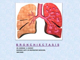
1. B R O N C H I E C T A S I S
DR. SHRIRAM .S. SHENOY
RESIDENT, DEPT. OF RESPIRATORY MEDICINE,
SBKS MIRC.
2. AIM OF THIS PRESENTATION:
DEFINITION OF BRONCHIECTASIS
TYPES OF BRONCHIECTASIS
EITIOPATHOGENESIS OF BRONCHIECTASIS
3. DEFINITION:
• Bronchiectasis is a morphological term used
to describe abnormal irreversibly dilated and
often thick-walled bronchi.
• represents the end stage of a variety of
pathologic processes that cause destruction of
the bronchial wall and its surrounding
supporting tissues.
15. AETIOPATHOGENESIS:
• Bronchiectasis may be caused by
I) Primary infective insult
II) Primary impairment of mucous clearance
III) Immunodeficiency syndrome
a) Congenital b) acquired
IV) Hyperimmune response
16. AETIOPATHOGENESIS:
• V) Infection secondary to bronchial
obstruction
A) Intraluminal B) Extraluminal
VI) Autoimmune disease
VII) Inhalational/aspiration injury
VIII) Developmental defects
A) Structural B) Biochemical
19. • Primary ciliary dyskinesia
DNAI1, DNAH5, and DNAH11
• respiratory distress in neonates,
• recurrent respiratory tract infections
• Bronchiectasis
• Situs inversus, heterotaxia, infertility, and
hydrocephalus, singly or in various combinations.
• Noone et al. showed that bronchiectasis was
seen in 98 percent of cases of PCD.
20. IMMUNODEFICIENCY SYNDROME
• A) CONGENITAL:
• Common varied congenital immunodeficiency
• Selective immunoglobulin deficiency
• Functional immune deficiency
• B) ACQUIRED:
• Secondary hypogammaglobulinaemia
• Human immunodeficiency virus infection
22. SECONDARY TO BRONCHIAL
OBSTRUCTION
A) Intraluminal
• Slow-growing tumour
• Aspirated foreign body
• B) Extraluminal:
• Lymphadenopathy
• Growth on the thoracic wall
27. • Micro-organisms such as staphylococcus
aureus , klebsiella, psuedomonas aeruginosa,
haemophilus influenzae etc.
protease and other toxins
28. Up-regulation of neutrophils
(Elastase and Matrix metallo-protinases)
Destruction of wall structures- cartilage,muscle,elastic tissue
and replaced by fibrous connective tissue
Dilatation of the bronchi leading to accumulation of the
thick pool of purulent material and occlusion due to
fibrous thickening
Increased vascularity due to enlargement of bronchial
arteries and increased anastomosis between bronchial
artery and pulmonary artery
38. Tuberculosis as a Cause of Upper Lobe
Bronchiectasis
DAVID SALKIN,
M.D., San
Fernando
LEVEL OF
EVIDENCE:
A
This article
examines the
association
between
tuberculosis as a
cause of
predominantly
upper lobe
bronciectasis.
By serial bronchography
and bronchoscopy the
author has observed
clinically the formation
of bronchiectasis from
tuberculous bronchitis
in the lower lobes. It
was noted that there
was mural infiltration of
the diseased bronchus,
swelling of the
mucosa, and
pronounced decrease in
the size of the lumen, in
some cases to the point
of occlusion. Then, as
healing occurred, the
weakened bronchus
became
irregular and dilated