
Downloaded 64 times

![ The release of inflammatory mediators, elastases, and
collagenases leads to inflammation and destruction of
elastic and muscular components of bronchial walls.
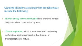
Bronchiectasis associated with bronchial obstruction may
have a focal distribution distal to the site of obstruction
[ Foreign body] .
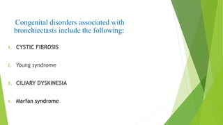
Bronchiectasis associated with underlying disease is likely
to be diffuse [ Cystic Fibrosis].](https://image.slidesharecdn.com/dryusufimranbronchiectasis-140620153650-phpapp02/85/Dr-yusuf-imran-bronchiectasis-4-320.jpg)
![Common infectious causes includes:
1. Severe pneumonia, especially viral, [inadequately treated]
2. Measles, tuberculosis, pertussis, Adenovirus, Mycobacterium
avium, and Aspergillus fumigatus infections.
3. HIV infection](https://image.slidesharecdn.com/dryusufimranbronchiectasis-140620153650-phpapp02/85/Dr-yusuf-imran-bronchiectasis-6-320.jpg)


This document discusses bronchiectasis in children. Bronchiectasis is the irreversible dilatation of the bronchi due to destruction of elastic and muscular components of the bronchial walls. It generally results from obstruction and inflammation of the airways leading to chronic infection and recruitment of inflammatory cells. The inflammatory mediators released destroy bronchial walls causing bronchiectasis. Common causes include severe pneumonia, measles, tuberculosis, cystic fibrosis, and primary ciliary dyskinesia. Patients present with recurrent cough and purulent sputum. Diagnosis involves chest X-ray, CT scan, and sputum/swab cultures. Treatment consists of antibiotics, airway clearance techniques, and addressing underlying disorders. Surgery


