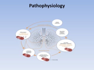
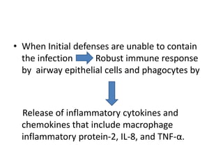
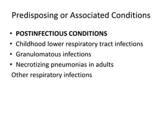
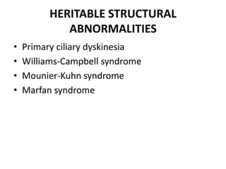
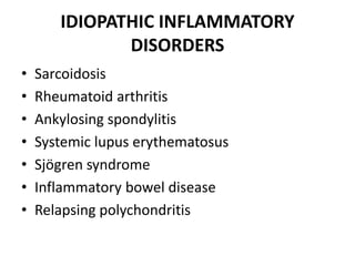
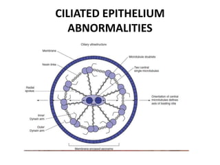
Bronchiectasis has a variety of etiologies including prior lung infections, genetic disorders affecting cilia or cartilage, and inflammatory conditions. The pathophysiology involves a vicious cycle of infection and inflammation that destroys the bronchial walls. Chronic neutrophilic inflammation from bacterial toxins causes tissue damage through proteolytic enzymes. This further impairs mucociliary clearance and predisposes the individual to recurrent infections that perpetuate the cycle of inflammation and lung damage leading to bronchiectasis.
Lecture slides about bronchiectasis with contents including definition, causes, pathogenesis and pathology, and how to make diagnosis. Treatment for bronchiectasis is presented separately.
Bronchiectasis ( Bronchos- airways ; ectasia- dilatation) is a morphological term used to describe abnormal irreversibly dilated and often thick walled bronchi.
Bronchiectasis represents the end stage of variety of pathological precesses that cause destruction of bronchial wall and its surrounding tissues.
PATHOGENESIS OF BRONCHIECTASIS BY DR BASHIR AHMED DAR ASSOCIATE PROFESSOR MED...Prof Dr Bashir Ahmed Dar
Dr Bashir Ahmed Dar associate professor medicine chinkipora sopore kashmir presently working in malaysia speaks about bronchiectasis.Bronchiectasis which is defined as the irreversible dilatation of the cartilage-containing airways bronchi or bronchioles.
Pleural effusion may be defined figuratively as the juice, oozing from the leaky lingerie of the lung. However the text book definition is the abnormal accumulation of fluid in the pleural space due to disturbances in the forces that keep the pleural fluid economy in equilibrium...
Pneumonia is an inflammatory condition of the lung affecting primarily the small air sacs known as alveoli. Typically symptoms include some combination of productive or dry cough, chest pain, fever, and trouble breathing. Severity is variable.
Pneumonia is usually caused by infection with viruses or bacteria and less commonly by other microorganisms, certain medications and conditions such as autoimmune diseases. Risk factors include cystic fibrosis, chronic obstructive pulmonary disease (COPD), asthma, diabetes, heart failure, a history of smoking, a poor ability to cough such as following a stroke, and a weak immune system. Diagnosis is often based on the symptoms and physical examination. Chest X-ray, blood tests, and culture of the sputum may help confirm the diagnosis. The disease may be classified by where it was acquired with community, hospital, or health care associated pneumonia.
Vaccines to prevent certain types of pneumonia are available. Other methods of prevention include handwashing and not smoking. Treatment depends on the underlying cause. Pneumonia believed to be due to bacteria is treated with antibiotics. If the pneumonia is severe, the affected person is generally hospitalized. Oxygen therapy may be used if oxygen levels are low.
Pneumonia affects approximately 450 million people globally (7% of the population) and results in about four million deaths per year. Pneumonia was regarded by William Osler in the 19th century as "the captain of the men of death". With the introduction of antibiotics and vaccines in the 20th century, survival improved. Nevertheless, in developing countries, and among the very old, the very young, and the chronically ill, pneumonia remains a leading cause of death. Pneumonia often shortens suffering among those already close to death and has thus been called "the old man's friend"
Bronchiectasis refers to the congenital/acquired irreversible airway dilation that involves the bronchi/bronchioles in either a focal or a diffuse manner.
It is a pulmonary disease related to chronic infections in the background of inability of respiratory mucosa to clear the infections and impaired ciliary function.
It is chronic disease with high morbidity and mortality
These lecture notes were prepared by Dr. Hamdi Turkey- Pulmonologist- Department of internal medicine - Taiz university
Do Not Forget To Visit Our Pages On Facebook on the following Links:
https://www.facebook.com/groups/569435236444761/
AND
https://www.facebook.com/groups/690331650977113/
PNEUMONIA,
DEFINITION
Pneumonia is an infection of the pulmonary parenchyma.
To the pathologist, pneumonia is an infection of the alveoli ,distal airways, and interstitium of the lung that is manifested by increased weight of the lungs, replacement of normal lung’s sponginess by consolidation ,and alveoli filled with white blood cells ,red blood cells and fibrin .To the clinician, pneumonia is a constellation of symptoms and signs in combination with at least one opacity on CXR.
Epidemiology
Between 5 and 10 million cases of infectious pneumonia occur annually in the United States and result in more than 1 million hospitalizations.
Pneumonia is a leading cause of death worldwide, the sixth leading cause of death in the United States, and the most common lethal infectious disease.
Bronchiectasis is a disease in which there is permanent enlargement of parts of the airways of the lung.[5] Symptoms typically include a chronic cough with mucus production.[3] Other symptoms include shortness of breath, coughing up blood, and chest pain.[2] Wheezing and nail clubbing may also occur.[2] Those with the disease often get frequent lung infections.[8]
Bronchiectasis may result from a number of infectious and acquired causes, including pneumonia, tuberculosis, immune system problems, as well as the genetic disorder cystic fibrosis.[11][3][12] Cystic fibrosis eventually results in severe bronchiectasis in nearly all cases.[13] The cause in 10–50% of those without cystic fibrosis is unknown.[3] The mechanism of disease is breakdown of the airways due to an excessive inflammatory response.[3] Involved airways (bronchi) become enlarged and thus less able to clear secretions.[3] These secretions increase the amount of bacteria in the lungs, resulting in airway blockage and further breakdown of the airways.[3] It is classified as an obstructive lung disease, along with chronic obstructive pulmonary disease and asthma.[14] The diagnosis is suspected based on symptoms and confirmed using computed tomography.[7] Cultures of the mucus produced may be useful to determine treatment in those who have acute worsening and at least once a year
These simplified slides by Dr. Sidra Arshad present an overview of the non-respiratory functions of the respiratory tract.
Learning objectives:
1. Enlist the non-respiratory functions of the respiratory tract
2. Briefly explain how these functions are carried out
3. Discuss the significance of dead space
4. Differentiate between minute ventilation and alveolar ventilation
5. Describe the cough and sneeze reflexes
Study Resources:
1. Chapter 39, Guyton and Hall Textbook of Medical Physiology, 14th edition
2. Chapter 34, Ganong’s Review of Medical Physiology, 26th edition
3. Chapter 17, Human Physiology by Lauralee Sherwood, 9th edition
4. Non-respiratory functions of the lungs https://academic.oup.com/bjaed/article/13/3/98/278874
Ozempic: Preoperative Management of Patients on GLP-1 Receptor Agonists Saeid Safari
Preoperative Management of Patients on GLP-1 Receptor Agonists like Ozempic and Semiglutide
ASA GUIDELINE
NYSORA Guideline
2 Case Reports of Gastric Ultrasound
New Directions in Targeted Therapeutic Approaches for Older Adults With Mantl...i3 Health
i3 Health is pleased to make the speaker slides from this activity available for use as a non-accredited self-study or teaching resource.
This slide deck presented by Dr. Kami Maddocks, Professor-Clinical in the Division of Hematology and
Associate Division Director for Ambulatory Operations
The Ohio State University Comprehensive Cancer Center, will provide insight into new directions in targeted therapeutic approaches for older adults with mantle cell lymphoma.
STATEMENT OF NEED
Mantle cell lymphoma (MCL) is a rare, aggressive B-cell non-Hodgkin lymphoma (NHL) accounting for 5% to 7% of all lymphomas. Its prognosis ranges from indolent disease that does not require treatment for years to very aggressive disease, which is associated with poor survival (Silkenstedt et al, 2021). Typically, MCL is diagnosed at advanced stage and in older patients who cannot tolerate intensive therapy (NCCN, 2022). Although recent advances have slightly increased remission rates, recurrence and relapse remain very common, leading to a median overall survival between 3 and 6 years (LLS, 2021). Though there are several effective options, progress is still needed towards establishing an accepted frontline approach for MCL (Castellino et al, 2022). Treatment selection and management of MCL are complicated by the heterogeneity of prognosis, advanced age and comorbidities of patients, and lack of an established standard approach for treatment, making it vital that clinicians be familiar with the latest research and advances in this area. In this activity chaired by Michael Wang, MD, Professor in the Department of Lymphoma & Myeloma at MD Anderson Cancer Center, expert faculty will discuss prognostic factors informing treatment, the promising results of recent trials in new therapeutic approaches, and the implications of treatment resistance in therapeutic selection for MCL.
Target Audience
Hematology/oncology fellows, attending faculty, and other health care professionals involved in the treatment of patients with mantle cell lymphoma (MCL).
Learning Objectives
1.) Identify clinical and biological prognostic factors that can guide treatment decision making for older adults with MCL
2.) Evaluate emerging data on targeted therapeutic approaches for treatment-naive and relapsed/refractory MCL and their applicability to older adults
3.) Assess mechanisms of resistance to targeted therapies for MCL and their implications for treatment selection
Pulmonary Thromboembolism - etilogy, types, medical- Surgical and nursing man...VarunMahajani
Disruption of blood supply to lung alveoli due to blockage of one or more pulmonary blood vessels is called as Pulmonary thromboembolism. In this presentation we will discuss its causes, types and its management in depth.
Anti ulcer drugs and their Advance pharmacology ||
Anti-ulcer drugs are medications used to prevent and treat ulcers in the stomach and upper part of the small intestine (duodenal ulcers). These ulcers are often caused by an imbalance between stomach acid and the mucosal lining, which protects the stomach lining.
||Scope: Overview of various classes of anti-ulcer drugs, their mechanisms of action, indications, side effects, and clinical considerations.
Title: Sense of Smell
Presenter: Dr. Faiza, Assistant Professor of Physiology
Qualifications:
MBBS (Best Graduate, AIMC Lahore)
FCPS Physiology
ICMT, CHPE, DHPE (STMU)
MPH (GC University, Faisalabad)
MBA (Virtual University of Pakistan)
Learning Objectives:
Describe the primary categories of smells and the concept of odor blindness.
Explain the structure and location of the olfactory membrane and mucosa, including the types and roles of cells involved in olfaction.
Describe the pathway and mechanisms of olfactory signal transmission from the olfactory receptors to the brain.
Illustrate the biochemical cascade triggered by odorant binding to olfactory receptors, including the role of G-proteins and second messengers in generating an action potential.
Identify different types of olfactory disorders such as anosmia, hyposmia, hyperosmia, and dysosmia, including their potential causes.
Key Topics:
Olfactory Genes:
3% of the human genome accounts for olfactory genes.
400 genes for odorant receptors.
Olfactory Membrane:
Located in the superior part of the nasal cavity.
Medially: Folds downward along the superior septum.
Laterally: Folds over the superior turbinate and upper surface of the middle turbinate.
Total surface area: 5-10 square centimeters.
Olfactory Mucosa:
Olfactory Cells: Bipolar nerve cells derived from the CNS (100 million), with 4-25 olfactory cilia per cell.
Sustentacular Cells: Produce mucus and maintain ionic and molecular environment.
Basal Cells: Replace worn-out olfactory cells with an average lifespan of 1-2 months.
Bowman’s Gland: Secretes mucus.
Stimulation of Olfactory Cells:
Odorant dissolves in mucus and attaches to receptors on olfactory cilia.
Involves a cascade effect through G-proteins and second messengers, leading to depolarization and action potential generation in the olfactory nerve.
Quality of a Good Odorant:
Small (3-20 Carbon atoms), volatile, water-soluble, and lipid-soluble.
Facilitated by odorant-binding proteins in mucus.
Membrane Potential and Action Potential:
Resting membrane potential: -55mV.
Action potential frequency in the olfactory nerve increases with odorant strength.
Adaptation Towards the Sense of Smell:
Rapid adaptation within the first second, with further slow adaptation.
Psychological adaptation greater than receptor adaptation, involving feedback inhibition from the central nervous system.
Primary Sensations of Smell:
Camphoraceous, Musky, Floral, Pepperminty, Ethereal, Pungent, Putrid.
Odor Detection Threshold:
Examples: Hydrogen sulfide (0.0005 ppm), Methyl-mercaptan (0.002 ppm).
Some toxic substances are odorless at lethal concentrations.
Characteristics of Smell:
Odor blindness for single substances due to lack of appropriate receptor protein.
Behavioral and emotional influences of smell.
Transmission of Olfactory Signals:
From olfactory cells to glomeruli in the olfactory bulb, involving lateral inhibition.
Primitive, less old, and new olfactory systems with different path
Factory Supply Best Quality Pmk Oil CAS 28578–16–7 PMK Powder in Stockrebeccabio
Factory Supply Best Quality Pmk Oil CAS 28578–16–7 PMK Powder in Stock
Telegram: bmksupplier
signal: +85264872720
threema: TUD4A6YC
You can contact me on Telegram or Threema
Communicate promptly and reply
Free of customs clearance, Double Clearance 100% pass delivery to USA, Canada, Spain, Germany, Netherland, Poland, Italy, Sweden, UK, Czech Republic, Australia, Mexico, Russia, Ukraine, Kazakhstan.Door to door service
Hot Selling Organic intermediates
Report Back from SGO 2024: What’s the Latest in Cervical Cancer?bkling
Are you curious about what’s new in cervical cancer research or unsure what the findings mean? Join Dr. Emily Ko, a gynecologic oncologist at Penn Medicine, to learn about the latest updates from the Society of Gynecologic Oncology (SGO) 2024 Annual Meeting on Women’s Cancer. Dr. Ko will discuss what the research presented at the conference means for you and answer your questions about the new developments.
Couples presenting to the infertility clinic- Do they really have infertility...Sujoy Dasgupta
Dr Sujoy Dasgupta presented the study on "Couples presenting to the infertility clinic- Do they really have infertility? – The unexplored stories of non-consummation" in the 13th Congress of the Asia Pacific Initiative on Reproduction (ASPIRE 2024) at Manila on 24 May, 2024.
MANAGEMENT OF ATRIOVENTRICULAR CONDUCTION BLOCK.pdfJim Jacob Roy
Cardiac conduction defects can occur due to various causes.
Atrioventricular conduction blocks ( AV blocks ) are classified into 3 types.
This document describes the acute management of AV block.
2. What is bronchiectasis ?
• Bronchiectasis is derived from the Greek
words:
Bronckos – airway
Ectasis – widening
• It is a chronic lung condition defined as the
abnormal irreversible dilatation of the
bronchi,where the elastic and muscular tissue
is destroyed by acute or chronic inflammation
and infection.
3. • Bronchiectasis represents the end stage of a
variety of pathologic processes that cause
destruction of the bronchial wall and its
surrounding supporting tissues.
• Etiologies include prior lung infection, systemic
inflammatory disorders, and genetic disorders of
host defense’
• However, bronchiectasis is considered to be
idiopathic in up to half of the affected individuals.
4. Pathophysiology
• As bronchiectasis is an acquired disorder, its
pathophysiology is commonly described as
distinct phases of infection and chronic
inflammation.
• The interaction between these phases
establishes a vicious circle in which the end
result is the destruction of the bronchi and the
accompanying clinical symptoms.
5. • Various mechanisms operate to produce
permanent, pathologic dilation and damage of
the airways.
• They may be in of in terms of traction,
pulsion, and weakened tensile strength of
the airways.
• In most cases the pathogenesis becomes
invariably linked with and propelled by the
destructive effects of chronic infection.
6. • As the lung undergoes fibrotic changes
consequent to disorders such as sarcoidosis,
interstitial lung disorders or infections such as
tuberculosis, local retractile forces result in
fixed dilation of the airways, or “traction”
bronchiectasis.
• The prototypic “pulsion” bronchiectasis
permanent airway dilatation as a result of
intense inflammation originating in the lumen
7. • The first stage in the development of
bronchiectasis -is an initial infective insult to the
airways, which triggers a mucociliary response.
• Micro-organisms trigger the release of toxins and
an inflammatory response within the airways.
• This inflammatory response includes the release
of neutrophils, lymphocytes and macrophages
within the bronchial lumen.
8. • Neutrophils also alter the function of the cilial
epithelium, leading to changes in cilial beat
frequency and mucous gland hypersecretion.
Both processes compromise mucociliary
clearance.
• Weakness of the airways-eg postinfectious
bronchiectasis
10. MICROBILOGY
“vicious circle” phase of bronchiectasis is dominated
by the influx of neutrophils. Neutrophils enter the airway lumina through gaps
between epithelial cells ,Neutrophils are attracted by the release of
chemokines such as in IL-8 and leukotriene B4 (LTB4) from macrophages, and
IL-17 from Th17 cells ,
11. • When Initial defenses are unable to contain
the infection Robust immune response
by airway epithelial cells and phagocytes by
Release of inflammatory cytokines and
chemokines that include macrophage
inflammatory protein-2, IL-8, and TNF-α.
12. • Airway infiltration by predominantly neutrophils,
macrophages and lymphocytes causes damage to
the airway epithelium
Through the release of various proteolytic
enzymes such as neutrophil elastase and
metalloproteinases
• Erosion of mucosal barriers, creating
microabscesses that can harbor bacteria
19. CYSTIC FIBROSIS(mucoviscidosis)
• CF is caused by a mutation in chromosome 7
q -CFTR gene.
• It is the most common autosomal recessive
disorder among whites
• The CFTR protein produced by this gene
regulates the movement of chloride and
sodium ions across epithelial cell membranes
20. • Cl ion help to lyse mucus
• Pt wil hv recurrent pneumonia
• Mcc-staphylococcus aureus
• Psueudomonas aeroginosa – with the help of
biofilm formation
• Burkholderia cepacia –pneumonia mc associated
with increased mortality
• CF and CF variants are one of the most common
causes of bronchiectasis
21.
22. Youngs syndrome
It is seen only in males ,the genetic basis for
Young syndrome is not know- Possible
mechanism is ciliary dysfunction
clinical features of Youngs syndrome
• Bronchiectasis and recurrent LRTI
• sinusitis
• Infertility-obstructive azoospermia
23. DISORDERS OF IMMUNITY
• Primary diseases that result in
immunodeficiency may devolve from
mutations that impair B or T lymphocytes and
cause abnormal humoral immunity,cellular
immunity, or both
• Common variable immunodeficiency, or
acquired hypogammaglobulinemia is the most
frequent syndrome recognized in this group of
diseases.
24. Acquired Hypogammaglobulinemia
• Although there are normal numbers of
circulating B lymphocytes, they fail to
differentiate into antibodyproducing cells.
• This results in particular vulnerability to
infections with encapsulated bacteria such
• S. pneumoniae,
• H. influenzae,
• S. aureus, and P. aeruginosa.
25. • Other disorders of immunity include
• Hyper-IgM or hyper-IgE(Job syndrome)
• Impaired production of cytokines secreted by
type 1 helper T cells such as interferon-γ (IFN-
γ) and TNF-α, cytokines
• They are known to be important in controlling
such infections.
• Increase the susceptibility of individuals to
pyogenic bacteria and NTM organisms
26. Thymic hypoplasia resulting in abnormal
cellular immunity (DiGeorge syndrome)
severe combined immunodeficiency
syndrome, “bare lymphocyte” syndrome,
Wiskott-Aldrich syndrome (an X-linked
recessive illness associated with small
platelets and eczema)
Ataxia-telangiectasia syndrome
28. • Each ciliated epithelial cell possesses
approximately 200 cilia. The direction of ciliary
beating is determined by the orientation of
the central pair of microtubules.
• Dysfunction of the ciliary apparatus may
involve a variety of structural abnormalities in
the cilia or disorganization of the ciliary axes.
29. • A variety of abnormalities have been
described
• Including the complete or partial absence of
outer or inner dynein arms
• A lack of radial spokes
• Disordered microtubule arrangements,ciliary
disorientation etc
30. • PCD (primary ciliary dyskinasia )with situs
inversus universalis is known as Kartagener
syndrome
• The patients have a marked tendency toward
colonization and infection with H. influenzae.
31. 1
• Ineffective beating of the ciliated cells
2
• Stagnation and accumulation of mucus
3
• early-onset refractory or recurrent infectionsof the upper and lower respiratory tract
Bronchiectasis is a common sequela of PCD and it typically involve the
dependent zones, including the lower lobes, right middle
lobe, and/or the lingular segment of the left upper lobe
32. Bronchiectasis: Kartagener syndrome.-
CXR-shows dextrocardia with basal predominant linear opacities -bronchial
wall thickening and bronchiectasis.
Axial chest CT shows dextrocardia and severe, cystic bronchiectasis
33. BRONCHIAL CARTILAGE OR ELASTIC
FIBER DEFECTS
• Mounier-Kuhn syndrome, or
congenitaltracheobronchomegaly
• It is a rare disorder associated with gross
enlargementor dilation of the trachea and segmental
bronchi
• The underlying defect is atrophy and even absence of
elastic fibers and smooth muscle tissues of the large
airways.
• In addition, primary or secondary atrophy of the
connective tissue between the rings may result in
outpouchings or diverticula, potentially serving as
reservoirs for recurrent infections
34. Congenital tracheobronchomegaly (Mounier-Kuhn syndrome) with
bronchiectasis. This 73-year-old woman has had recurring
respiratory infections throughout her adult life
• B-The dilated trachea with prominent cartilaginous rings is
confirmed on a CT scan (between arrows).
• C, Not only is the trachea Enlarged, but the main-stem bronchi
are dilated (between arrows).
35. CT images show multifocal, thin-walled cystic bronchiectasis associated
with dilated trachea and central bronchi
36. • Mounier-Kuhn patients may present in their
early years or as late as the fourth decade
with recurring lower respiratory infections
• In advanced stages, airway collapsibility may
result in severe airflow obstruction.
37. Williams-Campbell syndrome
• Williams-Campbell syndrome or congenital
bronchial cartilage deficiency syndrome-It is
another rare disorder that tends to present
early in life with recurring infection and
bronchiectasis.
• The absence of cartilage from the segmental
to the first few generations of the
subsegmental airways is the typical finding in
Williams-Campbell syndrome
38. Williams-Campbell syndrome
This 50-year-old man had a lifelong history of recurring
respiratory infections and productive cough.
The airways are massively dilated with collections of
respiratory secretion spooling in some of the cystic spaces.
39. CONNECTIVE TISSUE ABNORMALITIES
• Marfan syndrome- due to mutation/defect in the
genes that produce fibrillin 1 (FBN1) gene.
• Morphologic anomalies seen with Marfan
syndrome have been traced to increased localized
production of TGF-β, a cytokine that increases
susceptibility to mycobacteria.
• Marfan syndrome has been linked to middle lobe
hypoplasia because the right middle lobe is the
most commonly affected lobe in NTM-associated
bronchiectasis.
40. IDIOPATHIC INFLAMMATORY
DISORDERS
• Mcc- Sarcoidosis
• Mechanisms -diffuse parenchymal scarring resulting in
traction and airway distortion, endobronchial
granulomatous inflammation including stricture with
poststenotic infection, or compression secondary to
hypertrophic peribronchial lymphadenopathy.
• Bronchiectasis has been seen in 20% to 35% of RA
patients undergoing HRCT scanning
• Potential causal mechanisms include increased
propensity for infections,either intrinsic to RA or
secondary to steroid or cytotoxictherapy
41. Axial chest CT through the upper lobes in a patient with sarcoidosis
shows biapical bronchiectasis with architectural distortion.
43. Inflammatory bowel disease &
Bronchiectasis
common epithelial targets
of autoimmunity -inhaled or
ingested.
cryptogenic
infection that
incites both
airway and
intestinal
inflammation
ulcerative colitis-
develop after
therapeutic
colectomy
44. Relapsing polychondritis – It is identified essentially
as progressive inflammation, weakness, and
deformity of cartilaginous structures, including the
ears, nose, larynx, and tracheobronchial
tree.Respiratory involvement is a common.
And is a major cause of mortality.
Bronchiectasis in such patients may bedue to
primary bronchial damage and/or recurrent
infection.
Bronchial
cartilage
inflammation
Airway
collapse
Airflow
limitation
45. Relapsing polychondritis. A, Axial inspiratory chest CT shows thickening of
the anterior two thirds of the trachea (arrowheads).
B, Axial chest CT performed following a forced vital capacity maneuver
shows excessive trachea collapse
46. ASPIRATION
1. Direct spillage of secretions from the
oropharynx, containing a plethora of
microorganisms, including microaerophilic and
anaerobic bacteria
2.Materials refluxed from the esophagus and/or
stomach-contain food particles, hydrochloric
acid Biliary or pancreatic secretions, and
microbes indigenous to the gut, including
Helicobacter pylori.
47. When Laryngeal protective functions are imperfect,
“microaspiration”
MCC is GERD , others including
(1) Depressed sensorium
(2) Altered brain-stem function
(3) Altered laryngeal structure/ Function
(4) Esophageal disorders -dysmotility, obstruction by tumors
or strictures, muscular dystrophy, achalasia,
tracheoesophageal fistulas, or lower esophageal sphincter
incompetence .
(5) Gastric dysfunction (dysmotility or outlet obstruction).
48. • Also chronic coughing, which stresses and dilates the
diaphragmatic ring, might disrupt the lower
esophageal sphincter and subject the esophagus to
distending forces
• An additional factor that could contribute to
gastroesophageal reflux disease is the medications
employed for pulmonary disorders, including
anticholinergics, β2-agonists, theophylline, and
corticosteroids, all of which impair lower esophageal
sphincter function and broadspectrum antibiotics,
which alter gastroesophageal flora.
49. Toxic inhalation / Thermal injury
• Acute and chronic inflammation of the
tracheobronchial tree, bronchiolitis, bronchiolitis
obliterans, and diffuse alveolar damage may be a
consequence of exposure to toxic metal fumes-
Aluminum,cadmium,chromium, nickel
• Toxic gases like ammonia, chlorine,
phosgene, sulfur dioxide
• Bronchiectasis may ensue because of either infectious
complications of the exposure, denuding of the ciliated
epithelium, or progressive fibrosis.
• chronic airway damage and bronchiectasis may evolve
following thermal or smoke injury.
50. POSTOBSTRUCTIVE DISORDERS
Foreign bodies/Tumors
Airway obstruction
Poor drainage
Recurrent/chronic infection
Bronchiectasis.
The more common tumor types include bronchogenic carcinomas particularly
the squamous cell variety,carcinoid tumors
51. • Extrinsic airway compression due most often
to hypertrophic lymphadenitis from
granulomatous diseases such as sarcoidosis or
infections, including tuberculosis or
histoplasmosis, may severely narrow or even
occlude large airways
• Focal bronchiectasis is seen particularly
those with disease limited to only one region/
one segment/ one lobe or even one lung
52. Postobstructive bronchiectasis. A–C, Axial chest CT shows severe narrowing of the
left lower lobe bronchus (arrow), due to carcinoid tumor.
Multifocal bronchiectatic consolidation (arrowheads) is present throughout the
left lower lobe
53. ALLERGIC BRONCHOPULMONARY
ASPERGILLOSIS
• Allergic bronchopulmonary
aspergillosis
• inhaled Aspergillus -lodged in
the airways.
• proliferating fungi form large
mucoid
Mucoid plugs
• medium-sized
bronchi
inflammation and
distention • thin-walled
bronchiectasis of the
central airways
characteristic
of ABPA
54. • In ABPA there are intense, immunologically
• mediated reactions to inhaled Aspergillus that
has
• lodged in the airways. The onglomerates that
fill the central airways; a
• sequela of this airway inflammatory process
and mucoid
• impaction is bronchiectasis
55. • Also in long-standing, inadequately controlled
ABPA/M. In such cases, extensive fibrosis and
airway distortion may evolve because of
uncontrolled inflammation.
In these cases the patients may acquire
secondary airway pathogens including
P. aeruginosa or other gram-negative bacilli as
well as NTM.
In these “burned-out” cases, the patients may
notdemonstrate asthma, eosinophilia,or elevated
levels of I
58. IDIOPATHIC BRONCHIECTASIS
• Idiopathic is estimated to account for 25% to 50%
of cases
• It often has a characteristic phenotype of bilateral
lower lobe bronchiectasis and chronic
rhinosinusitis
• Genotyping studies of class I and class IImajor
histocompatibility complex molecules indicated
that allelic polymorphism for HLA-B (HLA-B5 and
HLA-B52), HLA-C (HLA-Cw*03 and and HLA-
DR/DQ (HLA-DR1/DQ5) are associated with
idiopathic bronchiectasis.
59. MISCELLANEOUS
• AIDS bronchiectasis has been identifiedin a
significant proportion of those undergoing CT
scans, including children
• Probable pathogenesis involves severe,
chronic, and recurrent infections with a
variety of opportunistic pathogens
• Also oxidative damage associated with
infection
60. • Yellow nail syndrome is an uncommon disorder
marked by the triad of: yellow, thick, dystrophic
nails; chronic lymphedema of the face, hands,
and lower extremities and pleural effusions.
• Recurrent sinusitis and lower respiratory tract
infections are common
• Abnormal lymphatic structure, increased vascular
permeability, deficient immunoglobulin
production, and/or ciliary dysfunction.
61. • Radiation therapy- typically delivered for
carcinoma of the breast or mediastinal tumors
including lymphomas may result in profound
damage to the central airways.
• Focal damage to the cartilage and mucosa of
the airways leading to patulous distention and
irregularities of the major bronchi in the field
of irradiation
62. .
• Result in profound damage to the central
airways.
.
• Focal damage to the cartilage and mucosa of
the airways
.
• patulous distention and irregularities of the
major bronchi in the field of irradiation
Radiation therapy
63. CT scan shows dense fibrosis and bronchiectasis
in the radiation field
non–small cell carcinoma of the lung. Pt had
received radiation therapy to the right hilar region
approximately 18 months previously
64. • Cylindrical bronchiectasis is described as
failure of the involved airways to taper
progressively in their distal course.
• Usually, in this condition the bronchial walls
are smooth or regular
66. CLASSIFICATION
• Varicoid bronchiectasis is an allusion to
varicose veins and is marked by irregular
dilation, narrowing and outpouching of the
airways
• Saccular bronchiectasis- also known as cystic
bronchiectasis, includes focal or cystic
distortion of the distal airways
67. • It may be isolated or may be more
confluent,producing the appearance of
bronchiectatic consolidation and volume loss
• Cylindrical bronchiectasis is described as
failure of the
• involved airways to taper progressively in their
distal course