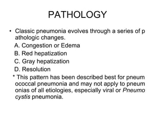
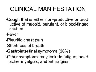
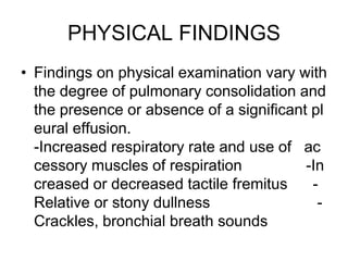
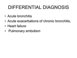
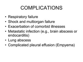
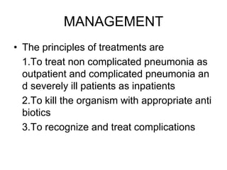
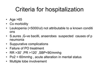
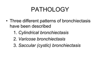
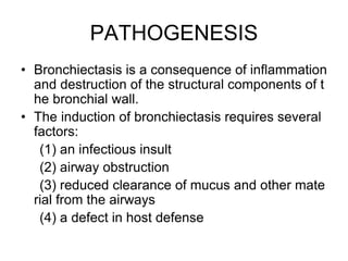
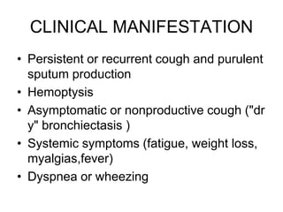
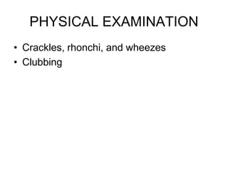
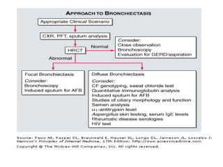
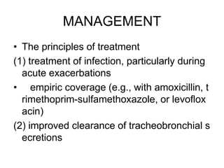
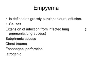
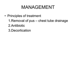
PNEUMONIA,
DEFINITION
Pneumonia is an infection of the pulmonary parenchyma.
To the pathologist, pneumonia is an infection of the alveoli ,distal airways, and interstitium of the lung that is manifested by increased weight of the lungs, replacement of normal lung’s sponginess by consolidation ,and alveoli filled with white blood cells ,red blood cells and fibrin .To the clinician, pneumonia is a constellation of symptoms and signs in combination with at least one opacity on CXR.
Epidemiology
Between 5 and 10 million cases of infectious pneumonia occur annually in the United States and result in more than 1 million hospitalizations.
Pneumonia is a leading cause of death worldwide, the sixth leading cause of death in the United States, and the most common lethal infectious disease.
2. PNEUMONIA
DEFINITION
• Pneumonia is an infection of the pulmonary par
enchyma.
• To the pathologist, pneumonia is an infection of t
he alveoli ,distal airways, and interstitium of the l
ung that is manifested by increased weight of th
e lungs, replacement of normal lung’s spongines
s by consolidation ,and alveoli filled with white bl
ood cells ,red blood cells and fibrin .
3. • To the clinician, pneumonia is a constellation of
symptoms and signs in combination with at least
one opacity on CXR.
Epidemiology
• Between 5 and 10 million cases of infectious pne
umonia occur annually in the United States and r
esult in more than 1 million hospitalizations.
• Pneumonia is a leading cause of death worldwid
e, the sixth leading cause of death in the United
States, and the most common lethal infectious di
sease.
4. CLASSIFICATION
• ETIOLOGIC
1.Infections 2.Inhala
tion of gastric contents 3.Immunological reactio
ns 4.Inhalation of other toxic substanc
es
• ANATOMIC
1.Lobar or segmental : process is confined to th
ese division of the lung
2.Bronchopneumonia : small areas of the lung al
veoli and lobule around small terminal bronchi ar
e affected
5. • Revised classification system
1.Community-acquired pneumonia (CAP)
–includes
- Cases of infectious pneumonia in patients l
iving independently in the community
- Patients who have been hospitalized for ot
her reasons for less than 48 hours before t
he development of respiratory symptoms
6. 2.Health care–associated pneumonia (HCAP)
-Patients who have previously been hospitalized
for at least 2 days within the 90 days before infe
ction
-Patients from nursing homes who received intrav
enous antibiotic therapy, chemotherapy, or woun
d care within the past 30 days
-Patients from hemodialysis centers
-Patients contracting pneumonia greater than 48
hours after the institution of endotracheal intubat
ion and mechanical ventilation
a. Hospital-acquired pneumonia (HAP)
b. Ventilator-associated pneumonia (VAP).
7. PATHOPHISIOLOGY
• Pneumonia results from the proliferation of microbial pat
hogens at the alveolar level and the host's response to t
hose pathogens.
1.Microorganism
• How do micro organisms gain access to the lower respir
atory tract ?
a .Aspiration from the oropharynx or nasopharynx
b .Direct inhalation from ambient air
c .Haematogenous dissemination
d .Penetrating chest trauma
e .Local spread from contagious site
8. 2 . Host defence
• What are the host defence mechanisms?
A. mechanical factors
- Hairs and turbinates of the nares & airway cilia
-The gag reflex and
-The cough mechanism
B. Cellular immunity
- Alveolar macrophage and neutrophils
C. Humoral immunity
- IgG and IgA
9. PATHOLOGY
• Classic pneumonia evolves through a series of p
athologic changes.
A. Congestion or Edema
B. Red hepatization
C. Gray hepatization
D. Resolution
* This pattern has been described best for pneum
ococcal pneumonia and may not apply to pneum
onias of all etiologies, especially viral or Pneumo
cystis pneumonia.
10. CLINICAL MANIFESTATION
-Cough that is either non-productive or prod
uctive of mucoid, purulent, or blood-tinged
sputum
-Fever
-Pleuritic chest pain
-Shortness of breath
-Gastrointestinal symptoms (20%)
-Other symptoms may include fatigue, head
ache, myalgias, and arthralgias.
11. PHYSICAL FINDINGS
• Findings on physical examination vary with
the degree of pulmonary consolidation and
the presence or absence of a significant pl
eural effusion.
-Increased respiratory rate and use of ac
cessory muscles of respiration -In
creased or decreased tactile fremitus -
Relative or stony dullness -
Crackles, bronchial breath sounds
15. MANAGEMENT
• The principles of treatments are
1.To treat non complicated pneumonia as
outpatient and complicated pneumonia an
d severely ill patients as inpatients
2.To kill the organism with appropriate anti
biotics
3.To recognize and treat complications
16. Criteria for hospitalization
• Age >65
• Co morbidity
• Leukopenia (<5000/ul) not attributable to a known conditi
ons
• S.aures ,G-ve bacilli, anaerobes suspected causes of p
neumonia
• Suppurative complications
• Failure of PO treatment
• RR >30’ ,PR >120’ ,SBP<90/mmhg
• Po2 < 60mmhg , acute alteration in mental status
• Multiple lobe involvement
17. FOLLOW UP
• Fever and leukocytosis usually resolve wit
hin 2 and 4 days, respectively .
• Physical findings may persist longer.
• Chest radiographic abnormalities are slow
est to resolve and may require 4–12 week
s to clear.
18. Failure to improve
• Noninfectious conditions
• Correct diagnosis but that a different patho
gen
• The pathogen may be resistant to the drug
selected
• Wrong drug or the correct drug at the wron
g dose or frequency of administration
• Nosocomial superinfections
20. LUNG ABCESS
• Is defined as pulmonary parenchymal necrosis and cavit
ation resulting from infection.
Conditions that predispose to lung abscess
• any cause of aspiration or decreased ciliary action
reduced levels of consciousness
alcoholism
seizure disorders
general anesthesia
cerebrovascular accidents
drug addiction
22. MICROBIOLOGY
• Anaerobic bacteria ( most common )-strept
occoci, H.influenza
• Aerobic or facultative bacteria - S. aureus,
Klebsiella pneumoniae, Nocardia sp., and
gram-negative organisms
• Fungi
• parasites
23. CLINICAL MANIFESTATION
• Clinical course
-acute presentations - aerobic bacteria
-evolve over an extended period of time -
anaerobic infection
• Presentation
-Asymptomatic
-Symptomatic - cough, copious foul smelli
ng purulent sputum production, pleuritic ch
est pain, fever, and hemoptysis
24. PHYSICAL EXAMINATION
• Unrevealing
• Fetid breath and poor dentition
• Dullness to percussion
• Reduced breath sounds or criptation
• Clubbing or hypertrophic pulmonary osteo
arthropathy
25. INVESTIGATION
• Chest radiograph - cavity with an air-fluid level, with or wi
thout surrounding infiltrate
-Lung abscess occur commonly in dependent segment
s of the lung
.The posterior segments of the upper lobes and
.The superior segments of the lower lobes
• CT of the chest - to define the size and location of the ab
scess
• Gram stain and culture of sputum
• Blood culture
• Pleural fluid cultures
• Bronchoscopy
27. MANAGEMENT
The principles of treatments are
1.To kill the organism with appropriate antibiotics
- Clindamycin (150 mg–300 mg every 6 h) 4-8wks
- Metronidazole 400mg tid plus penicillin
2. To drain the pus -Postural
-Bronchoscopical
3.Surgical resection of affected segment
Indications
-refractory hemoptysis
-inadequate response to medical therapy
-Empyema or bronchopleural fistula
-the need for a tissue diagnosis when there is concern fo
r a noninfectious etiology (lung ca).
28. BRONCHECTASIS
• is an abnormal and permanent dilatation of
bronchi.
• It may be either focal, involving airways su
pplying a limited region of pulmonary pare
nchyma, or diffuse, involving airways in a
more widespread distribution .
29. PATHOLOGY
• Three different patterns of bronchiectasis
have been described
1. Cylindrical bronchiectasis
2. Varicose bronchiectasis
3. Saccular (cystic) bronchiectasis
30. PATHOGENESIS
• Bronchiectasis is a consequence of inflammation
and destruction of the structural components of t
he bronchial wall.
• The induction of bronchiectasis requires several
factors:
(1) an infectious insult
(2) airway obstruction
(3) reduced clearance of mucus and other mate
rial from the airways
(4) a defect in host defense
31. CLINICAL MANIFESTATION
• Persistent or recurrent cough and purulent
sputum production
• Hemoptysis
• Asymptomatic or nonproductive cough ("dr
y" bronchiectasis )
• Systemic symptoms (fatigue, weight loss,
myalgias,fever)
• Dyspnea or wheezing
34. MANAGEMENT
• The principles of treatment
(1) treatment of infection, particularly during
acute exacerbations
• empiric coverage (e.g., with amoxicillin, t
rimethoprim-sulfamethoxazole, or levoflox
acin)
(2) improved clearance of tracheobronchial s
ecretions
35. (3) reduction of inflammation; and
(4) treatment of an identifiable underlying pr
oblem
36. Empyema
• Is defined as grossly purulent pleural effusion.
• Causes
Extension of infection from infected lung (
pnemonia,lung abcess)
Subphrenic abcess
Chest trauma
Esophageal perforation
Iatrogenic
37. MANAGEMENT
• Principles of treatment
1.Removal of pus – chest tube drainage
2.Antibiotic
3.Decortication