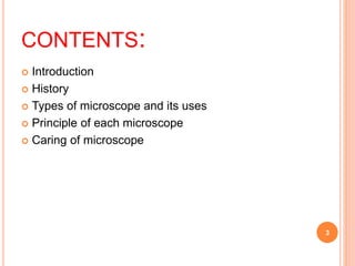
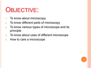
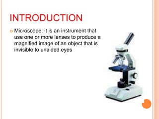
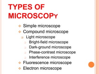
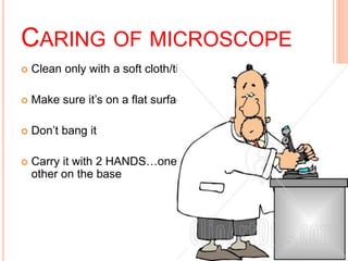
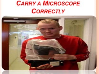
MICROSCOPY
Presenter:- Neha
Nayansha
CONTENTS:
 Introduction
 History
 Types of microscope and its uses
 Principle of each microscope
 Caring of microscope
3
OBJECTIVE:
1. To know about microscopy
2. To know different parts of microscopy
3. To know various types of microscope and its
principle
4. To know about uses of different microscope
5. How to care a microscope
4
THE HISTORY
INTRODUCTION
 Microscope: it is an instrument that
use one or more lenses to produce a
magnified image of an object that is
invisible to unaided eyes
 Reflected light: light source above the sample and
what is seen though the view point are light waves
that have reflected off the sample
o Transmitted light : light source below the
microscope stage and sends light upwards towards
the sample and up to the viewing points
 Magnification: a measure of the ability of a lens or
other optical instrument to magnify expressed as
the ratio of the size of the image to that of an object
That means an object of any size is magnified to form
an enlarged image
 Focus:
 Principle axis:
Eyepiece
Body Tube
Revolving Nosepiece
Arm
Objective Lens
Stage
Stage
Clips Coarse Focus
Fine Focus
Base
Diaphragm
Light
APPLICATION
o Industrial microscope
o Professional biological microscope
o Digital microscope
o Educational microscope
o Metullargic microscope
o Student microscope
o Children microscope
o Forensic microscope
TYPES OF
MICROSCOPY
 Simple microscope
 Compound microscope
 Light microscope
 Bright-field microscope
 Dark-ground microscope
 Phase-contrast microscope
 Interference microscope
 Fluorescence microscope
 Electron microscope
SIMPLE MICROSCOPY
 It is a simple magnifying hand lens.
 Magnification power ranges from 2 to 20
LIGHT MICROSCOPE
 Use natural or transmitted light as a source of light
 Resolving power is an important component of light
microscope
 dependent on the wavelength of light used to
illuminate the object and on the numerical aperture
of microscope
14
Bright –field microscope
o Ordinary light microscope
o Most common form of light microscope
o Consists of compound lens system
o Used for demonstration of structural detail
o For demonstrating of motility of flagellated bacteria
o Eg : E .coli
o Also used for measuring approximate size of bacteria ,
fungi in stained preparation
PRINCIPLE:
 light is emitted from source
of light to condenser lens
which contain aperture
diaphragm to control and
focus light on specimen
then the light is passed
through specimen and
reach the objective lens
where the light is magnified
and transmit it to eyepiece
some of the light is
absorbed by stain,
pigmentation and this
contrast allow us to see the
object
Dark ground microscope
o Special type of compound microscope
o Use a dark field condenser with a central circular stop
o Use reflected light
o Used for demonstration of very thin bacteria
o Eg: treponema pallidum
o Also used for demonstration of motility of flagellated
bacteria and protozoa
PRINCIPLE:
Light rays falling on the
Object are reflected or
scattered onto the object
lens within the result that
microorganisms appear
bright against dark
background
PHASE CONTRAST
MICROSCOPE
 Special condenser and objective are used
 Used for examining the internal structure of a living
cell
 Examination of living microorganism
19
PRINCIPLE:
using a specific optical
system it converts
differences in phase in
an organism into
differences in intensity
of light thereby
producing
light and dark contrast
in image
INTERFERENCE
MICROSCOPE
 Another specialized application of light microscope
 Used for demonstrating cell organelles
 Useful for quantitative measurement of the chemical
constituents of the cell as protein , lipids
FLOURSCENCE
MICROSCOPE
o Use ultraviolet light
o Used for demonstration of antigen of a pathogen by
direct fluorescece antibody test eg : cornybacterium
diptheriae,n.gonorhoea
o Also for estimation of antibodies in the serum by
indirect antibody fluorescece test eg : syphills
,brucellosis
PRINCIPLE:
 The specimen stained
with Fluorescent dye
when exposed to
ultraviolet light result
emmision of longer
wavelength Bacteria
stained with fluorescent
dye appear as a brightly
glowing against dark
background
.
ELECTRON MICROSCOPY
o Use beam of electron instead of beam of light
o Resolving power of microscope is extremely high
because it use electron of wavelength 0.005nm
o Used for rapid detection of viruses specially for
noncultivable virus
o Ultrastructural study of various microorganisms
Principle:
A beam of electron is
focused by an
electromagnet when a
beam of electron
passes through an
object ,the electrons
get scattered producing
an image
Shadow casting : by deposition of thin layer of
platinum on the microorganism to be examined
which on bombardment with electron beam scatters
the electron and produces an image that is focused
CARING OF MICROSCOPE
 Clean only with a soft cloth/tissue
 Make sure it’s on a flat surface
 Don’t bang it
 Carry it with 2 HANDS…one on the arm and the
other on the base
28
CARRY A MICROSCOPE
CORRECTLY
29
Dr.T.V.Rao
MD
REFERENCES:
 Textbook of microbiology and immunology
Subhash Chandra Parija
 Textbook of microbiology P Chakraborty
Microscope history types principles.pptx

Microscope history types principles.pptx

  • 2.
  • 3.
    CONTENTS:  Introduction  History Types of microscope and its uses  Principle of each microscope  Caring of microscope 3
  • 4.
    OBJECTIVE: 1. To knowabout microscopy 2. To know different parts of microscopy 3. To know various types of microscope and its principle 4. To know about uses of different microscope 5. How to care a microscope 4
  • 5.
  • 6.
    INTRODUCTION  Microscope: itis an instrument that use one or more lenses to produce a magnified image of an object that is invisible to unaided eyes
  • 7.
     Reflected light:light source above the sample and what is seen though the view point are light waves that have reflected off the sample o Transmitted light : light source below the microscope stage and sends light upwards towards the sample and up to the viewing points
  • 8.
     Magnification: ameasure of the ability of a lens or other optical instrument to magnify expressed as the ratio of the size of the image to that of an object That means an object of any size is magnified to form an enlarged image  Focus:  Principle axis:
  • 9.
    Eyepiece Body Tube Revolving Nosepiece Arm ObjectiveLens Stage Stage Clips Coarse Focus Fine Focus Base Diaphragm Light
  • 10.
    APPLICATION o Industrial microscope oProfessional biological microscope o Digital microscope o Educational microscope o Metullargic microscope o Student microscope o Children microscope o Forensic microscope
  • 11.
    TYPES OF MICROSCOPY  Simplemicroscope  Compound microscope  Light microscope  Bright-field microscope  Dark-ground microscope  Phase-contrast microscope  Interference microscope  Fluorescence microscope  Electron microscope
  • 12.
    SIMPLE MICROSCOPY  Itis a simple magnifying hand lens.  Magnification power ranges from 2 to 20
  • 14.
    LIGHT MICROSCOPE  Usenatural or transmitted light as a source of light  Resolving power is an important component of light microscope  dependent on the wavelength of light used to illuminate the object and on the numerical aperture of microscope 14
  • 15.
    Bright –field microscope oOrdinary light microscope o Most common form of light microscope o Consists of compound lens system o Used for demonstration of structural detail o For demonstrating of motility of flagellated bacteria o Eg : E .coli o Also used for measuring approximate size of bacteria , fungi in stained preparation
  • 16.
    PRINCIPLE:  light isemitted from source of light to condenser lens which contain aperture diaphragm to control and focus light on specimen then the light is passed through specimen and reach the objective lens where the light is magnified and transmit it to eyepiece some of the light is absorbed by stain, pigmentation and this contrast allow us to see the object
  • 17.
    Dark ground microscope oSpecial type of compound microscope o Use a dark field condenser with a central circular stop o Use reflected light o Used for demonstration of very thin bacteria o Eg: treponema pallidum o Also used for demonstration of motility of flagellated bacteria and protozoa
  • 18.
    PRINCIPLE: Light rays fallingon the Object are reflected or scattered onto the object lens within the result that microorganisms appear bright against dark background
  • 19.
    PHASE CONTRAST MICROSCOPE  Specialcondenser and objective are used  Used for examining the internal structure of a living cell  Examination of living microorganism 19
  • 20.
    PRINCIPLE: using a specificoptical system it converts differences in phase in an organism into differences in intensity of light thereby producing light and dark contrast in image
  • 21.
    INTERFERENCE MICROSCOPE  Another specializedapplication of light microscope  Used for demonstrating cell organelles  Useful for quantitative measurement of the chemical constituents of the cell as protein , lipids
  • 22.
    FLOURSCENCE MICROSCOPE o Use ultravioletlight o Used for demonstration of antigen of a pathogen by direct fluorescece antibody test eg : cornybacterium diptheriae,n.gonorhoea o Also for estimation of antibodies in the serum by indirect antibody fluorescece test eg : syphills ,brucellosis
  • 23.
    PRINCIPLE:  The specimenstained with Fluorescent dye when exposed to ultraviolet light result emmision of longer wavelength Bacteria stained with fluorescent dye appear as a brightly glowing against dark background
  • 24.
    . ELECTRON MICROSCOPY o Usebeam of electron instead of beam of light o Resolving power of microscope is extremely high because it use electron of wavelength 0.005nm o Used for rapid detection of viruses specially for noncultivable virus o Ultrastructural study of various microorganisms
  • 25.
    Principle: A beam ofelectron is focused by an electromagnet when a beam of electron passes through an object ,the electrons get scattered producing an image
  • 26.
    Shadow casting :by deposition of thin layer of platinum on the microorganism to be examined which on bombardment with electron beam scatters the electron and produces an image that is focused
  • 28.
    CARING OF MICROSCOPE Clean only with a soft cloth/tissue  Make sure it’s on a flat surface  Don’t bang it  Carry it with 2 HANDS…one on the arm and the other on the base 28
  • 29.
  • 30.
    REFERENCES:  Textbook ofmicrobiology and immunology Subhash Chandra Parija  Textbook of microbiology P Chakraborty