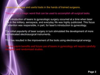
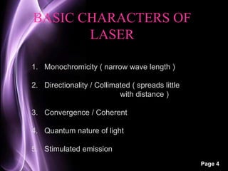
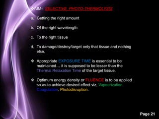
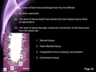
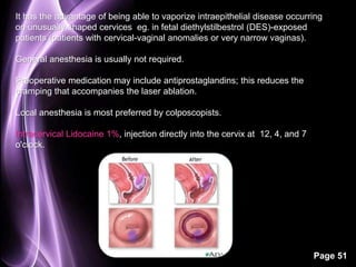
Page 1 
LASERS IN GYNAECOLOGY 
BY- DR. RIDHI KATHURIA
Page 2 
L LIGHT 
A AMPLIFICATION by 
S STIMULATED 
E EMISSION of 
R RADIATION
Page 3 
Lasers are unique and useful tools in the hands of trained surgeons. 
They are not a magic wand that can be used to accomplish all surgical tasks. 
The introduction of lasers to gynecologic surgery occurred at a time when laser 
use in the military, aerospace, and everyday life was highly publicized. This focus 
of attention was responsible, in part, for laser's introduction to gynecology. 
The initial popularity of laser surgery in turn stimulated the development of more 
sophisticated electrosurgical instruments. 
This has resulted in the improvement of the tools using electrosurgical energy. 
The long-term benefits and future use of lasers in gynecology will require carefully 
designed and randomized studies.
Page 4 
BASIC CHARACTERS OF 
LASER 
1. Monochromicity ( narrow wave length ) 
2. Directionality / Collimated ( spreads little 
with distance ) 
3. Convergence / Coherent 
4. Quantum nature of light 
5. Stimulated emission
ᴥ 1917, ALBERT EINSTEIN predicted & described process 
of stimulated emission of radiation. 
ᴥ 1953, CHARLES TOWNER & COLLG. produced device c/d 
‘’MASER’’, which could amplify microwaves by stimulated 
emission of radiation. 
ᴥ 1960, MAIMAN constructed first working laser, with a ruby 
crystal with wavelength of 690 nm. It was stimulated to emit 
laser energy by pumping with light from xenon flash lamp. 
ᴥ 1964, PATEL developed CO2 laser generating energy at 
wavelength 10,600 nm. 
Page 5
ᴥ KAPLAN & COLLG, first used laser in gyn surgery, by 
doing CO2 laser vaporization of infected cervical tissue. 
ᴥ During late 1970’s laser was coupled with 
laproscope, performing the 1st successful surgery of its 
kind. 
Page 6
Page 7 
LASER COMPONENTS 
OPTICAL 
RESONATOR 
• TOTALLY 
REFLECTIVE 
• PARTIALLY 
REFLECTIVE 
GAIN MEDIUIM 
• SOLID 
• LIQUID 
• SEMICONDUCTOR 
PUMPING 
PROCESS 
• LIGHT FLASH 
• ELECTRICAL 
ENERGY
Laser light is extremely bright, of similar color and direction, and is 
most often described in terms of its wave characteristics or 
WAVELENGTH. 
Wavelength is the distance between two successive crests or 
troughs, and wavelength determines the color of the light. 
Wavelengths are usually measured in Microns or Nanometers 
(nm) 
Page 8 
One micron = 1/1,000 mm. 
1 nm =1/1,000,000 mm. 
Electromagnetic waves are often referred to as light, although 
visible light occupies only a small portion of the electromagnetic 
spectrum.
Page 9 
An atom is composed of a positively charged nucleus 
surrounded by electrons that orbit this nucleus. 
Each electron orbit can be described in terms of energy levels. 
As orbital distance from the nucleus increases, energy level 
increases. 
As per the laws of quantum mechanics, 
When an electron moves from a higher-energy orbit to a lower-energy 
orbit, the atom loses a specific amount of energy in the 
form of a PHOTON, which also has a specific wavelength.
Lasers contain what is referred to as an Active Lasing 
Medium: a collection of atoms or molecules that are housed in 
an optically resonant cavity. 
This cavity, often cylindrical, is designed so that light of a 
specific wavelength will resonate between the closed ends of 
the cavity. 
The active medium is stimulated, by an external energy 
source, viz electricity or light, which stimulates the active lasing 
medium to higher energy levels. 
As decay back to resting energy levels occurs, photons of 
energy are released into the optical cavity. 
Electrons that are still in excited higher orbital energy levels 
can also be stimulated by bombardment with these newly 
released photons so that they undergo identical decay and emit 
an identical photon. 
Page 10 
This is called STIMULATED EMISSION, because one 
photon has stimulated the production of another photon.
Page 11
Page 12 
Optical cavity of the laser tube is closed at each end by a mirror. 
One of these mirrors is Totally Reflective, but the other is Semitransparent. 
Although photon direction in the tube is random, a certain number of photons will 
be emitted in the axis of the optical cavity. 
The others are focused by the mirrors so that most are resonating or bouncing back 
and forth along the axis of the cavity. 
Some of these photons emerge through the semitransparent mirror and are 
emitted from the laser as the monochromatic parallel coherent laser beam. 
Coherent means that the waves are all in phase or perfectly aligned. 
The laser thus creates a light that travels in a tight beam over long distances.
Page 13 
•LASER POWER is measured in terms of WATTS, and 
LASER ENERGY is expressed in terms of JOULES. 
•One Joule Equals 1 W Of Power Applied For 1 Second. 
•Lasers are named for the active medium contained in their optical 
cavities. Many different lasers have been constructed for diverse uses. 
•In gynecologic surgery, the CO2 and YAG lasers are the most commonly 
used. 
•Despite many efforts, fiber-optic delivery systems have never been 
satisfactorily adapted to CO2 laser energy. 
However, other lasers produce wavelengths that are conducted along 
quartz fibers. 
•The use of the fiber is of great advantage in endoscopic surgery 
because it can be passed down the channel of an operative telescope, 
suction irrigator probe, or other hollow channel within the instrument.
For any given power setting of the laser, the concentration 
or density of the power is greater as the spot size becomes 
smaller, and conversely, as the spot size (tissue impact 
area) is enlarged, the power density decreases. 
By focusing or defocusing the laser energy, it is used as a 
cutting or coagulating tool. 
THE CONCENTRATION OF LASER ENERGY AT ITS 
FOCAL POINT IS REFERRED TO AS ITS POWER 
DENSITY (PD) AND IS EXPRESSED AS WATTS PER 
SQUARE CENTIMETER. 
Page 14
Page 15 
►Although the photons of laser light are parallel, laser 
energy is not completely uniform throughout the cross-sectional 
diameter of the beam . 
►The term TRANSVERSE ELECTROMAGNETIC MODE 
(TEM) refers to the energy distribution of the cross-sectional 
diameter. 
For example, in most CO2 surgical lasers, the energy 
distribution is greatest at the center of the beam and 
decreases toward the periphery. 
►If one graphs the energy distribution of the cross-sectional 
beam diameter of the CO2 laser, the resultant 
curve is bell-shaped. 
►This Gaussian distribution does not hold true in fiber-optic 
lasers, YAG, Potassium-titanyl-phosphate (KTP), and 
Argon, in which energy distribution tends to be more 
uniform.
 Effect of lasers on tissue is dependent on the duration that the tissue is 
exposed to laser energy. 
Page 16 
In CONTINUOUS WAVE (CW) mode delivery, the laser oscillates 
constantly and delivers non varying power to the tissue. 
A PULSED LASER can deliver energy in the form of a single pulse or a 
train of pulses. The duration of a pulse is typically less than 0.25 second. 
Pulsing the laser to the tissue for a short duration is useful to control the 
delivery and power of the beam. Significantly higher power density can cut 
tissue with minimal coagulation necrosis at the margins.
Q-SWITCHING LASER is a laser that reduces its pulse time and 
significantly increases its peak power. 
In this mode, laser radiation is released in a short burst of laser 
light with high peak power. 
When the shutter of the optical cavity is rapidly opened, the laser 
energy is discharged in an extremely short period of time (10-6 to10-9 
seconds). 
Page 17 
Laser energy of 107 watts can be obtained using this mode. 
Q-switched laser pulse is used when it is critical to minimize 
adjacent tissue effects. 
Pulsing lasers at extremely high peak power (e.g., 500 to 2500 
pulses/second) permit cutting tissue with lower water content.
Page 18
Page 19
Page 20
Page 21 
AIM- SELECTIVE PHOTO-THERMOLYSIS 
a. Getting the right amount 
b. Of the right wavelength 
c. To the right tissue 
d. To damage/destroy/target only that tissue and nothing 
else. 
 Appropriate EXPOSURE TIME is essential to be 
maintained… it is supposed to be lesser than the 
Thermal Relaxation Time of the target tissue. 
 Optimum energy density or FLUENCE is to be applied 
so as to achieve desired effect viz, Vapourization, 
Coagulation, Photodisruption.
Page 22 
Three zones of laser tissue damage that may be defined: 
(i) the area vaporized, 
(ii) the area of tissue death that results from the heated tissue short 
of vaporization, 
(iii) the area of tissue damage caused by conduction of the heat away 
from the lased site 
1. Normal tissue 
2. Heat affected tissue 
3. Coagulated tissue showing vacuolisation 
4. Carbonised tissue
Page 23
 Laser energy interacts with tissue causing biologic, photochemical, or thermal 
reactions. 
 Laser energy is absorbed, scattered, or affected by the thermal conductivity 
and local circulation of the tissue. 
 Primary tissue effect of the surgical lasers used in gynecology is thermal. 
The beam profile for any given power setting can be altered by changing the 
spot size diameter. 
Soft tissue is about 80% water by volume. CO2 laser is highly absorbed by 
water, limiting its penetration to the surface of the tissue, where it can be 
monitored visually. 
Page 24
Page 25 
 Deep penetration of this laser energy into tissue is minimal as long as 
intracellular and extracellular water remains to be vaporized. 
 When heat is delivered by the laser beam, the tissue's temperature is 
elevated. 
 When tissue temperature is elevated to 57°C, irreversible damage to the 
cell's proteins occurs, and the cell dies. 
 Between 57°C and 100°C, there will be tissue death without vaporization. 
 Above 100°C, vaporization occurs, with conversion of the fluid content of 
tissue to vapor and the other cellular components being converted to smoke. 
This product is referred to as the LASER PLUME. 
When the plume is evacuated, it debrides the area and effectively removes 
the tissue. Additional tissue damage results from lateral conduction of heat away 
from the laser impact site. The amount of damage caused by heat conduction is 
directly proportional to the amount of time spent in lasing.
Page 26
 Several advantages of CO2 laser over a knife and electrosurgical instruments 
are obvious. 
Page 27 
Laser surgery is performed with no contact to the tissue. No bacteria is 
transferred from the surgical instrument to the tissue. 
 The laser beam sterilizes the operative field as it vaporizes it. 
 Because it removes tissue with vaporization and evacuation, the suctioned 
plume allows the tissue base to heal without a devitalized tissue covering. 
 Postoperative pain is reduced because nerve endings are sealed by the 
beam. 
 Coagulating lasers can be used in patients with bleeding disorders. The laser 
can be delivered for easy access to confined areas with a clear field of view.
The Helium Neon laser (HeNe) is a gas laser with a wavelength of 633 nm in the 
red portion of the visible spectrum. 
Page 28 
It is a low-powered laser with output ranging from 1 to 100 mW. 
The most popular HeNe lasers produces red light. Other colored helium neon 
lasers include green with a wavelength of 543 nm, yellow at 594 nm, and orange 
at 612 nm. 
Helium neon laser is used as an aiming beam for lasers that have non visible 
wavelengths, such as carbon dioxide. 
When aligned with the non visible laser, it defines the laser's impact site for the 
surgeon. This alignment of these two laser beams is checked before treatment 
begins.
• The typical helium-neon laser consists 
of three components: the laser tube, a 
high-voltage power supply, and structural 
packaging. 
• The laser tube consists of a sealed glass 
tube which contains the laser gas, 
electrodes, and mirrors. 
Page 29 
• Depending on the power output of the 
laser, the tube may vary in size. 
• The laser gas is a mixture of helium and 
neon in proportions of between 5:1 and 
14:1, respectively. 
• Electrodes, situated near each end of the 
tube, discharge electricity through the gas. 
• Mirrors, located at each end of the tube, 
increase efficiency.
Page 30 
• IN HELIUM-NEON LASERS, THE NEON ATOMS ARE THE SOURCE 
OF LASER LIGHT. 
• Because stimulated emission only takes place when there are excited 
neon atoms available, the process will quickly come to an end unless the 
neon atoms are replenished with energy. 
• The helium atoms in the laser gas carry out the process of re-energizing 
the neon. Helium is perfect for this task because it has a meta-stable state 
(does not decay as quickly) corresponding to the energy required to re-energize 
the neon 
• Therefore, not only do the helium atoms have the proper energy to re-energize 
the neon, they can hold onto that energy long enough to transfer 
it.
Page 31 
o Nd:YAG (Neodymium-doped Yttrium Aluminum Garnet; Nd:Y3Al5O12) is 
a crystal that is used as a lasing medium for solid state lasers. 
o YAG laser energy is able to penetrate tissue to a much greater depth. 
oUnlike CO2 laser, Nd:YAG laser is poorly absorbed by water. The YAG is often 
used in a liquid medium. 
o Because it penetrates water so well, the YAG is not a good laser for 
vaporization. It is an effective coagulator, however, as it passes deeper into 
tissue before absorption occurs. 
oYAG energy scatters in tissue, so thermal damage is greater than with CO2 
laser. 
o It is absorbed with a power density that is maximum below the tissue surface. 
This limits the ability to visually monitor the YAG laser's depth of penetration and 
stresses the importance of the surgeon to understand the physics of each laser.
o YAG laser can be transmitted through quartz fibers and fluids. 
Page 32 
o The depth of penetration provides excellent hemostasis. 
oGoldrath and colleagues in 1981 recognized this and used 
Nd:YAG laser for the first hysteroscopic endometrial ablations. 
oThis laser is also used for removal of hair and vascular lesions 
in a Q-switched delivery.
 Produces a blue-green light that has two distinct bands at 488 nm and 
515 nm. 
 Referred to as colored lasers because they are in the visible spectrum. 
 Can be transmitted through water or quartz fibers of 200 to 600 micron 
diameter. 
Page 33 
 Hemoglobin and other deeply 
colored pigments absorb their 
wavelengths and are responsible 
for their use in vascular and 
pigmented lesions.
 Penetration into skin, typically 1 to 2 mm, is affected by the 
concentration of skin pigments. 
 It produce a moderate scatter 100 times that of CO2 laser. 
This results in an excellent hemostasis. 
Because of the scatter, they have minimal cutting ability. 
The depth of tissue injury is roughly correlated with the laser 
power settings. 
Page 34
ᴥ KTP-532 laser derives its name from the potassium (K), titanyl (T), and 
phosphate (P) crystal used to double the frequency and halve the 
wavelength of the YAG laser. 
Page 35 
ᴥ 532-nm wavelength is emerald green. 
ᴥ Tissue effects are very similar to those produced by the argon laser. 
ᴥThe KTP-532 is used to vaporize or to coagulate, and energy emerging 
from the end of the fiber can be used to cut tissue. 
ᴥTheir greatest application has been in endoscopic surgery, because the 
delivery systems for their energy can be passed down the operative 
channel of an endoscope.
Page 36 
 This laser's wavelength is 2,100 nm. 
 It is pulsed at 350 to 700 microseconds at repeating intervals of 5 to 20 
Hz . 
 The Ho-YAG laser's depth of penetration in tissue is 0.4 mm. 
 Like CO2 laser, this laser is absorbed at the superficial layers of tissue. 
 Vaporization speed, coagulation depth, and hemostasis can be precisely 
controlled. 
 These properties can be particularly useful in gynecologic endoscopy.
 Laser treatment can be delivered through small channels to 
areas of difficult access. 
 Of equal interest is its ability to limit the lateral spread of the 
thermal energy. 
 The risk of injury to bowel, ureters, blood vessels, and nerves 
because of any lateral thermal spread is reduced. 
Page 37
Page 38 
ARGON-PUMPED RHODAMINE-B-DYE 
LASERS are tuned to produce a specific 
wavelength of light. 
They are delivered to cells that have been 
sensitized with HEMATOPORPHYRIN 
derivatives (HPDs). 
Certain HPD drugs concentrate in 
malignant tissue and are activated by 
specific laser wavelengths. 
This results in a cytotoxic effect on the 
tumor. 
The release of singlet oxygen results in the 
death of the cancer cells.
Page 39 
o The laser beam must be delivered 
from its source to the tissue. 
o Carbon dioxide laser, the first laser 
used in gynecology, is delivered to 
tissue through air in an articulated 
arm composed of hollow tubes and a 
series of internal mirrors. 
o The mirrors are precisely aligned to 
keep the laser beam directed along 
the path of the arm. 
o At the distal end a lens made of a 
special compound focuses the laser 
beam.
 Delivery unit used to aim and control the laser beam is called 
a micromanipulator. 
 This system allows the surgeon to precisely deliver the laser 
to the tissue. 
Page 40 
 It is attached to the microscope for easy access by the 
surgeon. The beam is reflected to the tissue by a mirror 
controlled by a joystick. 
 A Colposcope is used for the operating microscope and used 
to treat the cervix and vulva. 
Micromanipulators can be fitted with focusing lenses similar to 
a zoom lens of a camera. This allows the spot to be varied in 
size without changing the optical focus for the surgeon.
 This system is composed of a focusing lens contained within a 
metal tube that is held in the hand. 
 The lens typically has a short focal length, capable of producing 
a very small 0.2-mm spot size. 
Page 41 
 With this narrow beam, the laser is used as a cutting 
instrument. 
By pulling the hand piece away from the tissue, the surgeon 
creates a larger spot size used for vaporizing tissue. 
 Cutting and coagulating external genital surgery became its 
principal application
Page 42 
 Next advancement was the use of lasers in laparoscopy. 
 CO2 laser was delivered through large channels in a single 
puncture operative laparoscope. 
 The large channels were necessary to transmit the beam and 
avoid reflections from the channel walls. 
 Rigid wave guides were introduced that could also be used 
through ancillary 3 mm or larger channels.
 Fibre delivery is less cumbersome and produces less plume and more 
hemostasis. 
Page 43 
 It requires smaller channels. 
 The aiming beam is easily seen. 
 This mode of delivery is especially useful in hysteroscopy because 
fiber lasers could be used through fluid distending media. 
 Neodymium and holmium YAG are delivered through optical quartz 
fibers 100 to 600 microns in diameter. 
 At the tip of the fiber, the beam diverges, so power density is greatest 
just off the tip of the fiber. 
The beam also can also be defocused by moving the tip of the fiber 
farther away from the target tissue.
Page 44
‡ First use of lasers in gynecology was to treat CERVICAL 
INTRAEPITHELIAL NEOPLASIA (CIN) and VISIBLE CONDYLOMATA of 
the lower reproductive tract. 
Page 45 
‡ The precise cutting and ablative properties of CO2 laser prompted an 
interest in this surgery. 
‡ It was successfully used in the 1980s, and CO2 laser was enthusiastically 
received by many gynecologists. 
‡ Colposcopy provided an accurate means of identifying the location and 
size of the lesions, and CO2 laser provided unparalleled precision to treat 
these conditions. 
‡ Micromanipulator delivery is most popular, although another alternative is 
using the laser hand piece while visualizing the surgical field through a 
colposcope.
Page 46 
 CIN / Squamous Intraepithelial Lesions (SILs), has been treated 
successfully by either in situ destruction or excision. 
 In 1968, Palucek and Townsend reported a series in which cryosurgery was 
used to destroy CIN. 
 From the beginning, however, the use of destructive procedures that did not 
produce a pathologic specimen evoked strong criticism, fearing that lack of a 
complete pathologic examination of the target tissue could result in a missed 
diagnosis of invasion. 
 In the late 1960s, Cartier (Paris) developed a cutting loop electrode with a 
wire diameter of 200 micron that, when properly used, produced only a very 
small amount of thermal damage in the excised specimen. 
 A larger wire loop was designed for excisional cervical surgery in the United 
States. Its use, known as a Loop Electrode Excision Procedure (LEEP) or a 
Large Loop Excision Of Transformation Zone (LLETZ) procedure, provided a 
tissue specimen as an alternative to ablation procedures.
 Although CIN has been described as a noninvasive surface phenomenon, 
it is well known that the process involves the endocervical crypts. 
 The work of Te Linde and others pointed out that the endocervical crypts 
were involved in CIN, but it 
was Anderson and Hartley in 1980 who really 
emphasized the therapeutic implications; 
namely, adequate depth of destruction or 
excision is required to achieve satisfactory 
results. 
 Anderson and Hartley measured the depth of 
crypt involvement and found that 99% of CIN 
extends no further than 4 mm from the surface 
into the cervical crypt. 
Page 47
 If an area of intraepithelial neoplasia on the cervix was either excised or 
vaporized to a depth greater than 4 mm, virtually all of the neoplastic process 
would be treated. 
 This concept of depth-of-crypt involvement is extremely important as it 
dictates a measurable depth of destruction to be achieved by vaporization of 
the transformation zone if a surgical specimen is not to be removed by 
conization 
 It follows that if the lesion of intraepithelial neoplasia is visualized in its 
entirety (i.e., if the borders of the lesion are clearly seen on the cervix, and the 
lesion does not extend up into the canal), then it can be biopsied and correctly 
diagnosed as intraepithelial neoplasia. 
Page 48 
 This assumption provides the justification for all nonexcisional, ablative 
procedures used to destroy cervical intraepithelial disease. 
 Laser ablation by virtue of its ability to be delivered under colposcopic 
magnification and applied according to a patient's specific, unique anatomy is a 
valuable tool.
Page 49 
Cervical conization has been defined as an 
operation that removes a volume of tissue from 
the central longitudinal axis of the cervix 
including the external os & certain length of 
endocervical canal. 
Actual shape of the volume of tissue removed 
should be determined by the distribution of the 
lesion and not by some preconceived 
geometric shape. 
Conization, then, is a generic term, and does not necessarily imply that 
a perfect cone-shaped defect has been produced in the cervix. 
Cones can be asymmetric- short, long, thin, cylindrical, or any other 
shape that accomplishes the intended goal of complete removal of the 
lesion and the tissue at risk.
Page 50 
A number of techniques have been used to perform conization operations. 
In laser surgery, the end result is the same. If the CIN lesion cannot be entirely 
visualized or a neoplasm cannot be properly diagnosed for any reason, the CIN 
should not be treated by an ablative procedure. 
PRE REQUISITES 
a. Lesion must be completely visualized. 
b. Transformation zone must be completely seen. 
c. No doubt as to the intraepithelial nature of the disease. 
d. Adenocarcinoma of the endocervical canal must be ruled 
out.
It has the advantage of being able to vaporize intraepithelial disease occurring 
on unusually shaped cervices eg. in fetal diethylstilbestrol (DES)-exposed 
patients (patients with cervical-vaginal anomalies or very narrow vaginas). 
Page 51 
General anesthesia is usually not required. 
Preoperative medication may include antiprostaglandins; this reduces the 
cramping that accompanies the laser ablation. 
Local anesthesia is most preferred by colposcopists. 
Intracervical Lidocaine 1%, injection directly into the cervix at 12, 4, and 7 
o'clock.
Page 52 
No special prep is necessary. 
The cervix is cleaned with a 4% acetic acid solution, and the lesion to be treated 
is identified. 
Margin of the transformation zone is outlined by a series of vaporization craters, 
using short bursts of laser energy . 
The entire transformation zone is included in the area to be vaporized. 
The dots are connected so that the area to be treated is completely outlined. 
Spot size of 2 mm is used for the vaporization operation. 
Vaporization is carried to 7 mm. 
Depth can be measured with a calibrated measuring device.
It is more accurate to divide the cervix into sections and then destroy the tissue 
section by section. 
In this way, part of the normal cervical anatomy is preserved throughout the 
operation, and even though the topography of the cervix may change, the exact 
depth of destruction can always be measured for each section. 
The margin peripheral to the transformation zone is treated for a distance of 2 to 
5 mm. 
Page 53
Page 54 
The endocervical canal is exposed with a 
cotton applicator for examination. 
A wet applicator is used within the canal to 
limit the laser beam from vaporizing the 
upper canal. 
POST OP INSTRUCTIONS 
Patients undergoing laser surgery to the cervix usually require very little 
postoperative care. 
Postoperative follow-up in 2 to 4 weeks. 
Coitus is avoided for at least 4 weeks. 
Temperature elevations above 38°C and bleeding heavier than her normal 
menses are reported. About 15% of these patients will have some small amount 
of vaginal bleeding. Excessive bleeding is encountered in only about 2% of the 
patients. 
The patient is allowed to return to work within 48 hours.
Page 55 
1. There is less bleeding than with a scalpel. 
2. There is less tissue damage than with the electric cautery. 
3. The procedure has advantages over cryosurgery in that it can be 
done with much more precision, and the results are better for 
lesions of all sizes. 
4. Morbidity is low. 
5. All types and extents of intraepithelial neoplasia can be treated. 
6. The most conservative procedures can be performed by using 
combinations of vaporization and excision.
Page 56 
• Unlike the cervix, the vulva is often the site of multifocal disease. 
• Vulvar laser surgery should be done with the aid of the colposcope. 
• It is also imperative that the surgeon performing laser surgery on the vulva or 
the vagina has a working knowledge of vulvovaginal histology and of the 
variations in thickness of normal and abnormal neoplastic vulvovaginal 
epithelium.
Page 57 
ANATOMY OF THE 
VUVLAR SKIN 
 Vulvar skin is composed of two layers, EPIDERMIS and DERMIS. 
 Margin between epidermis and dermis is an irregular one because of the RETE 
RIDGES. 
Between the rete ridges are projections of dermis known as DERMAL PAPILLAE. 
The dermis can be divided into two layers: the Superficial Papillary Layer and the 
Deep Reticular Layer. 
There are also skin appendages, such as 
Pilosebaceous follicles; 
Eccrine Sweat glands; 
Apocrine glands; 
which project deep into the dermis.
VIN, & Human Papillomavirus (HPV), may involve skin appendages 
and epidermis. 
Although the thickness of the epidermis may be a fraction of a 
millimeter, the dermis may measure 7 to 8 mm in thickness, and skin 
appendages may penetrate the full thickness. 
It is the goal of the laser surgeon to remove the involved epidermis 
and a portion of the skin appendage that may also be involved in the 
disease process 
Page 58
 Unlike cervical vaporization, the depth of vaporization on the vulva 
cannot be measured. 
Page 59 
 REID 1991 suggested 4 Surgical Planes as follows:- 
a. Epidermis down to the basement membrane. 
b. Extending into the papillary layer of the dermis so that the laser 
surgeon removes both the epidermis and the papillary dermis. 
c. Extends upto the reticular dermis and uncovers the coarse 
collagen bundles that can be seen through the colposcope as 
grayish white fibers. 
d. Complete removal of the skin right down to the underlying 
subdermal fat. 
If this level is reached, healing must take place from the periphery, or 
a graft must be supplied.
Genital warts are soft growths on the skin and mucus membranes of the 
genitals. 
Page 60 
Most commonly caused by HPV. 
They may be found on the penis, vulva, urethra, vagina, cervix and 
in & around the anus. 
Genital warts are a sexually transmitted infection (STI). 
Certain types of HPV can lead to pre cancerous changes- cervical cancer, or 
anal cancer.
• It involves the epidermis and may also involve the 
superficial portions of skin appendages. 
• Only the warty lesion itself plus the surrounding 
epidermal margin need to be destroyed 
• Each condylomata should be identified and the laser beam directed this 
target. 
•Laser vaporizes the condylomata rapidly with the beam. The char and debris 
are wiped away, and the proper surgical level is colposcopically identified. 
Page 61 
• Vaporization is done upto the papillary dermis (second surgical 
plane). 
•Cold normal saline is used to cool the vulvar skin, because cooling helps 
reduce the heat diffusion and, therefore, the resultant tissue injury lateral to 
the laser impact site.
It is impossible to colposcopically differentiate 
between some forms of HPV lesions that occur on the 
vulva and significant VIN. 
Therefore, it is extremely important to obtain a biopsy 
specimen of the vulva in as many areas as necessary 
to correctly identify the pathology before laser surgery. 
When laser surgery is used, the vulva is recolposcoped, and 4% acetic acid is applied 
to demonstrate areas and borders of neoplastic or viral involvement. 
Page 62 
The larger areas are outlined in the manner described for CIN. 
If a lesion is large, it may be divided into smaller areas for lasing, because this 
provides a more accurate approach.
As soon as the laser beam is passed over the surface of the tissue, all landmarks 
disappear. 
Proper identification and marking of the limits of the disease by the laser is a highly 
necessary step in treatment. 
Although destruction must be carried to the third surgical plane (i.e., the reticular 
dermis), the first step in vulvar laser surgery always involves identifying the papillary 
dermis. 
When the tiny micropapules of this layer are seen, the laser surgeon again ablates 
through this most superficial dermis, wiping away char and identifying the underlying 
reticular dermis. 
Page 63 
A. Pt after 4 wks of therapy. 
B. Pt after 6 wks of therapy
Often intermittent pulses of laser energy can improve surgical control of the laser 
beam. 
Instead of reducing the power of the beam, it is possible to use the mechanical timer 
on the laser console to reduce exposure by delivering one-tenth-second to one-twentieth- 
Page 64 
second bursts of laser energy. 
This technique allows the surgeon time to react to the microsurgical review of the 
tissue and more easily control the rate of energy delivery, thus too-deep penetration, 
char, and residual thermal damage are also reduced to a minimum.
Page 65
Page 66 
POST - OP CARE 
1. Foley catheter may be required to avoid the immediate discomfort caused by 
urinary salts on the denuded vulvar surface. 
Catheters can be placed either through the urethra or suprapubically. 
2. An antibacterial cream or ointment, such as Sulfadine Cream or Bacitracin 
Ointment, is applied to the lased tissue to protect the raw surface from 
agglutination and to help prevent superficial infection. 
3. The patient must be given instructions to keep the vulvar folds separated, and 
the application of this medication helps in this respect. 
4. An ice pack is placed on the vulva when the patient leaves the operating room. 
5. Sitz baths begin the first day after laser surgery and are continued two times a 
day until the patient is no longer uncomfortable and the vulvar area is well on the 
way to reepithelialization. 
6. The patient is seen in a week to ensure there is no agglutination of vulvar folds.
Vaginal Intraepithelial Neoplasia (VAIN) and associated HPV infections 
are among the most difficult lower reproductive tract intraepithelial 
neoplasias to treat for a number of reasons: 
i. The vagina has a large surface area, which is difficult to visualize 
Page 67 
colposcopically. 
ii. There are many rugae and folds in the vagina. 
iii. The angle of the vaginal axis makes it difficult to treat by 
perpendicular beam impact. 
iv. The vaginal fornices are difficult to stabilize because they are quite 
distensible, and the cervix may hide portions of the fornix. 
v. The colposcopic appearance of VAIN varies greatly and often goes 
unrecognized even by the expert colposcopist.
VAIN does not demonstrate mosaic patterns except in areas of 
Adenosis. 
Often one sees coarse punctation, but the lesion may vary in color 
from a pale grayish white to the intense whiteness produced by 
hyperkeratosis. 
Page 68 
The lesions are multifocal and the borders are usually distinct, 
although the lesions may be serpiginous. 
VAIN May Appear As An Iodine-free Zone. 
In the postmenopausal patient, it is helpful to treat the patient with 
topical estrogen for 2 weeks before examination for optimal accuracy 
of the iodine test.
When the vaginal epithelium is destroyed, there 
must be visual identification of the underlying 
lamina propria because, as it is impossible to 
accurately measure the depth of destruction in this 
very irregular area. 
Because the vaginal squamous epithelium is only a 
fraction of a millimeter thick and there are no skin 
appendages in the vagina, ablation is a superficial 
operation. 
Surgeon often uses the speculum to one side and shoots through the open sides to 
manipulate the target into a position that is perpendicular to the laser beam. 
The vaginal rugae must also be ironed out to ensure even laser energy application. 
All of this is & thus most patients with VAIN require general anesthesia on an 
outpatient basis. 
Page 69
Laser beam is rapidly passed over the area, and the epidermis is lifted 
away from the underlying lamina propria. 
A wet sponge or cotton swab is used to wipe away char and coagulated 
epithelium. 
Often the tops of rugae are removed, but the troughs or valleys in 
between still contain viable dysplastic epithelium. 
With proper use of the speculum and other instruments, this problem is 
easily overcome. 
Page 70 
Adequate margins of 5 mm or more are used.
Page 71 
POST - OP CARE 
 Much of the discomfort encountered in postoperative vaginal laser surgery 
stems from laser wounds in the vaginal introitus. 
 Sitz baths . 
 Protective vulvar creams. 
 Daily vaginal applications of either Estrogen or Sulfa Cream or of some 
other bacteriostatic preparation. 
With extensive laser treatment, vaginal walls can touch each other. The 
patient should also be examined at intervals to make sure that vaginal 
coaptation is not occurring. 
Vaginal healing usually takes place from the periphery of the wound 
because of the lack of skin appendages in vaginal mucosa. 
If the laser has been thorough and the denuded area is large, healing is 
often delayed, and granulation tissue may result. This granulation tissue can 
be removed and the wound then treated with Silver Nitrate.
Page 72
Page 73 
procedure percentage 
1. Fulguration / Excision of endometriosis 68 % 
2. Lysis of adhesions 62 % 
3. Ovarian Cystectomy 31 % 
4. Division of Uterosacral Ligaments 30 % 
5. Presacral Neurectomy 10 % 
6. salpingo-oophorectomy 22 % 
7. Removal of Ectopic Pregnancy 10 % 
8. Myomectomy 4 % 
9. Neo salpingostomy 4 % 
10. LAVH 18 % 
11. Retropubic Urethropexy 10 % 
12. Sacral Colpopexy 3 %
Advantages of laser surgery in the abdomen include precision, 
limited adjacent tissue damage, rapid healing, minimal scarring, 
and the ability to treat areas of difficult access from a remote site. 
An example of the minimal trauma of laser on tissue is the research 
work of Bern, who demonstrated the ability to remove intracellular 
chromosomal material with the carbon dioxide laser without 
destroying the cell. 
Page 74
THE DELIVERY OF LASER UNDER MICROSCOPIC CONTROL WAS 
USED FIRST IN OPEN ABDOMINAL INFERTILITY SURGERY 
Page 75 
1) Adhesiolysis 
2) Fenestration of paraovarian and polycystic cysts 
3) Vaporization of endometriosis 
4) Fimbrioplasty 
5) Neosalpingostomy 
6) Resection of ectopic pregnancy 
7) Tubal anastomosis 
8) Tubal implant 
9) Myomectomy 
10)Metroplasty
Page 76 
ADVANTAGES 
Greatest magnification 
Spot-size control 
Ease of deep pelvic surgery 
Instrument-free field 
Constant focus 
Remote control 
DISADVANTAGES 
Delivery angle restricted 
Difficult to change spot 
Larger spot size with a greater margin of injury
Page 77 
ADVANTAGES 
Can be used without magnification 
Develops smallest spot size possible 
Multiple angle of delivery possible 
Focus and defocus is easy 
Highest power density for minimal tissue injury 
DISADVANTAGES 
Cumbersome equipment 
Shallow depth of focus 
Difficult to maintain precise focus 
Handpiece must be in field
Page 78 
 Unipolar electrocoagulation 
 Bipolar electrocoagulation 
 Laser coagulation 
 Extracorporeal ligatures 
 Extracorporeal sutures 
 Intracorporeal sutures 
 Endocoagulation (Semm) 
 Hemoclip
It is important that surgeons, nurses, and all those associated with 
laser surgery have an understanding of the potential health and safety 
hazards associated with the use of medical laser systems and the 
precautions needed to use them safely. 
Laser injury and potential hazards can be considered related to the 
organ systems at risk. 
Page 79
EYE is considered to be the organ most vulnerable to damage by 
laser. 
The eye collects and concentrates light energy on the retina. 
It has a natural protective mechanism in the lid and tearing reflexes. 
However, many lasers are extremely intense and are delivered so 
rapidly that injury can occur. 
Lasers in the visible and near infrared bands will be transmitted 
through the eye to the retina. 
With sufficient intensity, they can cause visual loss. 
Page 80
Protective measures for eye protection include spectacles, goggles, or coverall 
goggles to filter out the specific laser wavelength while transmitting visible light. 
Colposcopes and operative lenses of laparoscopes shield the laser surgeon's 
eyes from CO2 laser injury when delivered through these systems. 
The fiber-optic lasers that penetrate water and clear glass also penetrate the 
eye. 
In endoscopic surgery, special glasses or filters specific for the laser are used for 
protection. 
The optical filter is coupled to the eyepiece of the telescope and can be activated 
when the surgeon steps on the foot switch. 
Page 81 
As a general rule, eye protectors are required for work with lasers.
Page 82 
SKIN is considerably less vulnerable to laser injury than the 
eye. 
Skin injury can also occur from ignition of flammable material 
by the laser beam. 
Laser hazards to operating room personnel and patient 
includes accidental exposure from misdirection of the laser 
beam. 
The risk of this happening can be reduced by using the 
Standby Mode when not actually operating.
Electrical hazards of laser equipment are similar to other 
types of electrical or electronic equipment. 
There is no unique electrical safety problem associated with 
laser use. 
Vaporization of tissue produces smoke with potential health 
hazards. 
Careful evacuation and filtration of the laser plume will reduce 
any hazard of laser smoke inhalation. 
The evacuation of the plume is most effective if the tip of the 
tube is within 2 cm of the tissue being treated. 
It is important to remove smoke from the closed abdominal 
cavity during laparoscopic surgery. This allows the surgeon to 
see clearly as well as removing it from inhalation by the 
operating room personnel. 
Special high-efficiency masks provide additional respiratory 
protection. 
Page 83
 Understand the physics and tissue effects of the laser one is 
using. 
Page 84 
Implement precautions needed for laser safety. 
 Set the time the laser is exposed to tissue for the optimum 
effect. 
 Biopsy tissue before coagulation or vaporization if 
intraepithelial stage is in doubt. 
 Select appropriate backstops if laser beam can continue 
through the tissue being treated.
Page 85

Lasers

  • 1.
    Page 1 LASERSIN GYNAECOLOGY BY- DR. RIDHI KATHURIA
  • 2.
    Page 2 LLIGHT A AMPLIFICATION by S STIMULATED E EMISSION of R RADIATION
  • 3.
    Page 3 Lasersare unique and useful tools in the hands of trained surgeons. They are not a magic wand that can be used to accomplish all surgical tasks. The introduction of lasers to gynecologic surgery occurred at a time when laser use in the military, aerospace, and everyday life was highly publicized. This focus of attention was responsible, in part, for laser's introduction to gynecology. The initial popularity of laser surgery in turn stimulated the development of more sophisticated electrosurgical instruments. This has resulted in the improvement of the tools using electrosurgical energy. The long-term benefits and future use of lasers in gynecology will require carefully designed and randomized studies.
  • 4.
    Page 4 BASICCHARACTERS OF LASER 1. Monochromicity ( narrow wave length ) 2. Directionality / Collimated ( spreads little with distance ) 3. Convergence / Coherent 4. Quantum nature of light 5. Stimulated emission
  • 5.
    ᴥ 1917, ALBERTEINSTEIN predicted & described process of stimulated emission of radiation. ᴥ 1953, CHARLES TOWNER & COLLG. produced device c/d ‘’MASER’’, which could amplify microwaves by stimulated emission of radiation. ᴥ 1960, MAIMAN constructed first working laser, with a ruby crystal with wavelength of 690 nm. It was stimulated to emit laser energy by pumping with light from xenon flash lamp. ᴥ 1964, PATEL developed CO2 laser generating energy at wavelength 10,600 nm. Page 5
  • 6.
    ᴥ KAPLAN &COLLG, first used laser in gyn surgery, by doing CO2 laser vaporization of infected cervical tissue. ᴥ During late 1970’s laser was coupled with laproscope, performing the 1st successful surgery of its kind. Page 6
  • 7.
    Page 7 LASERCOMPONENTS OPTICAL RESONATOR • TOTALLY REFLECTIVE • PARTIALLY REFLECTIVE GAIN MEDIUIM • SOLID • LIQUID • SEMICONDUCTOR PUMPING PROCESS • LIGHT FLASH • ELECTRICAL ENERGY
  • 8.
    Laser light isextremely bright, of similar color and direction, and is most often described in terms of its wave characteristics or WAVELENGTH. Wavelength is the distance between two successive crests or troughs, and wavelength determines the color of the light. Wavelengths are usually measured in Microns or Nanometers (nm) Page 8 One micron = 1/1,000 mm. 1 nm =1/1,000,000 mm. Electromagnetic waves are often referred to as light, although visible light occupies only a small portion of the electromagnetic spectrum.
  • 9.
    Page 9 Anatom is composed of a positively charged nucleus surrounded by electrons that orbit this nucleus. Each electron orbit can be described in terms of energy levels. As orbital distance from the nucleus increases, energy level increases. As per the laws of quantum mechanics, When an electron moves from a higher-energy orbit to a lower-energy orbit, the atom loses a specific amount of energy in the form of a PHOTON, which also has a specific wavelength.
  • 10.
    Lasers contain whatis referred to as an Active Lasing Medium: a collection of atoms or molecules that are housed in an optically resonant cavity. This cavity, often cylindrical, is designed so that light of a specific wavelength will resonate between the closed ends of the cavity. The active medium is stimulated, by an external energy source, viz electricity or light, which stimulates the active lasing medium to higher energy levels. As decay back to resting energy levels occurs, photons of energy are released into the optical cavity. Electrons that are still in excited higher orbital energy levels can also be stimulated by bombardment with these newly released photons so that they undergo identical decay and emit an identical photon. Page 10 This is called STIMULATED EMISSION, because one photon has stimulated the production of another photon.
  • 11.
  • 12.
    Page 12 Opticalcavity of the laser tube is closed at each end by a mirror. One of these mirrors is Totally Reflective, but the other is Semitransparent. Although photon direction in the tube is random, a certain number of photons will be emitted in the axis of the optical cavity. The others are focused by the mirrors so that most are resonating or bouncing back and forth along the axis of the cavity. Some of these photons emerge through the semitransparent mirror and are emitted from the laser as the monochromatic parallel coherent laser beam. Coherent means that the waves are all in phase or perfectly aligned. The laser thus creates a light that travels in a tight beam over long distances.
  • 13.
    Page 13 •LASERPOWER is measured in terms of WATTS, and LASER ENERGY is expressed in terms of JOULES. •One Joule Equals 1 W Of Power Applied For 1 Second. •Lasers are named for the active medium contained in their optical cavities. Many different lasers have been constructed for diverse uses. •In gynecologic surgery, the CO2 and YAG lasers are the most commonly used. •Despite many efforts, fiber-optic delivery systems have never been satisfactorily adapted to CO2 laser energy. However, other lasers produce wavelengths that are conducted along quartz fibers. •The use of the fiber is of great advantage in endoscopic surgery because it can be passed down the channel of an operative telescope, suction irrigator probe, or other hollow channel within the instrument.
  • 14.
    For any givenpower setting of the laser, the concentration or density of the power is greater as the spot size becomes smaller, and conversely, as the spot size (tissue impact area) is enlarged, the power density decreases. By focusing or defocusing the laser energy, it is used as a cutting or coagulating tool. THE CONCENTRATION OF LASER ENERGY AT ITS FOCAL POINT IS REFERRED TO AS ITS POWER DENSITY (PD) AND IS EXPRESSED AS WATTS PER SQUARE CENTIMETER. Page 14
  • 15.
    Page 15 ►Althoughthe photons of laser light are parallel, laser energy is not completely uniform throughout the cross-sectional diameter of the beam . ►The term TRANSVERSE ELECTROMAGNETIC MODE (TEM) refers to the energy distribution of the cross-sectional diameter. For example, in most CO2 surgical lasers, the energy distribution is greatest at the center of the beam and decreases toward the periphery. ►If one graphs the energy distribution of the cross-sectional beam diameter of the CO2 laser, the resultant curve is bell-shaped. ►This Gaussian distribution does not hold true in fiber-optic lasers, YAG, Potassium-titanyl-phosphate (KTP), and Argon, in which energy distribution tends to be more uniform.
  • 16.
     Effect oflasers on tissue is dependent on the duration that the tissue is exposed to laser energy. Page 16 In CONTINUOUS WAVE (CW) mode delivery, the laser oscillates constantly and delivers non varying power to the tissue. A PULSED LASER can deliver energy in the form of a single pulse or a train of pulses. The duration of a pulse is typically less than 0.25 second. Pulsing the laser to the tissue for a short duration is useful to control the delivery and power of the beam. Significantly higher power density can cut tissue with minimal coagulation necrosis at the margins.
  • 17.
    Q-SWITCHING LASER isa laser that reduces its pulse time and significantly increases its peak power. In this mode, laser radiation is released in a short burst of laser light with high peak power. When the shutter of the optical cavity is rapidly opened, the laser energy is discharged in an extremely short period of time (10-6 to10-9 seconds). Page 17 Laser energy of 107 watts can be obtained using this mode. Q-switched laser pulse is used when it is critical to minimize adjacent tissue effects. Pulsing lasers at extremely high peak power (e.g., 500 to 2500 pulses/second) permit cutting tissue with lower water content.
  • 18.
  • 19.
  • 20.
  • 21.
    Page 21 AIM-SELECTIVE PHOTO-THERMOLYSIS a. Getting the right amount b. Of the right wavelength c. To the right tissue d. To damage/destroy/target only that tissue and nothing else.  Appropriate EXPOSURE TIME is essential to be maintained… it is supposed to be lesser than the Thermal Relaxation Time of the target tissue.  Optimum energy density or FLUENCE is to be applied so as to achieve desired effect viz, Vapourization, Coagulation, Photodisruption.
  • 22.
    Page 22 Threezones of laser tissue damage that may be defined: (i) the area vaporized, (ii) the area of tissue death that results from the heated tissue short of vaporization, (iii) the area of tissue damage caused by conduction of the heat away from the lased site 1. Normal tissue 2. Heat affected tissue 3. Coagulated tissue showing vacuolisation 4. Carbonised tissue
  • 23.
  • 24.
     Laser energyinteracts with tissue causing biologic, photochemical, or thermal reactions.  Laser energy is absorbed, scattered, or affected by the thermal conductivity and local circulation of the tissue.  Primary tissue effect of the surgical lasers used in gynecology is thermal. The beam profile for any given power setting can be altered by changing the spot size diameter. Soft tissue is about 80% water by volume. CO2 laser is highly absorbed by water, limiting its penetration to the surface of the tissue, where it can be monitored visually. Page 24
  • 25.
    Page 25 Deep penetration of this laser energy into tissue is minimal as long as intracellular and extracellular water remains to be vaporized.  When heat is delivered by the laser beam, the tissue's temperature is elevated.  When tissue temperature is elevated to 57°C, irreversible damage to the cell's proteins occurs, and the cell dies.  Between 57°C and 100°C, there will be tissue death without vaporization.  Above 100°C, vaporization occurs, with conversion of the fluid content of tissue to vapor and the other cellular components being converted to smoke. This product is referred to as the LASER PLUME. When the plume is evacuated, it debrides the area and effectively removes the tissue. Additional tissue damage results from lateral conduction of heat away from the laser impact site. The amount of damage caused by heat conduction is directly proportional to the amount of time spent in lasing.
  • 26.
  • 27.
     Several advantagesof CO2 laser over a knife and electrosurgical instruments are obvious. Page 27 Laser surgery is performed with no contact to the tissue. No bacteria is transferred from the surgical instrument to the tissue.  The laser beam sterilizes the operative field as it vaporizes it.  Because it removes tissue with vaporization and evacuation, the suctioned plume allows the tissue base to heal without a devitalized tissue covering.  Postoperative pain is reduced because nerve endings are sealed by the beam.  Coagulating lasers can be used in patients with bleeding disorders. The laser can be delivered for easy access to confined areas with a clear field of view.
  • 28.
    The Helium Neonlaser (HeNe) is a gas laser with a wavelength of 633 nm in the red portion of the visible spectrum. Page 28 It is a low-powered laser with output ranging from 1 to 100 mW. The most popular HeNe lasers produces red light. Other colored helium neon lasers include green with a wavelength of 543 nm, yellow at 594 nm, and orange at 612 nm. Helium neon laser is used as an aiming beam for lasers that have non visible wavelengths, such as carbon dioxide. When aligned with the non visible laser, it defines the laser's impact site for the surgeon. This alignment of these two laser beams is checked before treatment begins.
  • 29.
    • The typicalhelium-neon laser consists of three components: the laser tube, a high-voltage power supply, and structural packaging. • The laser tube consists of a sealed glass tube which contains the laser gas, electrodes, and mirrors. Page 29 • Depending on the power output of the laser, the tube may vary in size. • The laser gas is a mixture of helium and neon in proportions of between 5:1 and 14:1, respectively. • Electrodes, situated near each end of the tube, discharge electricity through the gas. • Mirrors, located at each end of the tube, increase efficiency.
  • 30.
    Page 30 •IN HELIUM-NEON LASERS, THE NEON ATOMS ARE THE SOURCE OF LASER LIGHT. • Because stimulated emission only takes place when there are excited neon atoms available, the process will quickly come to an end unless the neon atoms are replenished with energy. • The helium atoms in the laser gas carry out the process of re-energizing the neon. Helium is perfect for this task because it has a meta-stable state (does not decay as quickly) corresponding to the energy required to re-energize the neon • Therefore, not only do the helium atoms have the proper energy to re-energize the neon, they can hold onto that energy long enough to transfer it.
  • 31.
    Page 31 oNd:YAG (Neodymium-doped Yttrium Aluminum Garnet; Nd:Y3Al5O12) is a crystal that is used as a lasing medium for solid state lasers. o YAG laser energy is able to penetrate tissue to a much greater depth. oUnlike CO2 laser, Nd:YAG laser is poorly absorbed by water. The YAG is often used in a liquid medium. o Because it penetrates water so well, the YAG is not a good laser for vaporization. It is an effective coagulator, however, as it passes deeper into tissue before absorption occurs. oYAG energy scatters in tissue, so thermal damage is greater than with CO2 laser. o It is absorbed with a power density that is maximum below the tissue surface. This limits the ability to visually monitor the YAG laser's depth of penetration and stresses the importance of the surgeon to understand the physics of each laser.
  • 32.
    o YAG lasercan be transmitted through quartz fibers and fluids. Page 32 o The depth of penetration provides excellent hemostasis. oGoldrath and colleagues in 1981 recognized this and used Nd:YAG laser for the first hysteroscopic endometrial ablations. oThis laser is also used for removal of hair and vascular lesions in a Q-switched delivery.
  • 33.
     Produces ablue-green light that has two distinct bands at 488 nm and 515 nm.  Referred to as colored lasers because they are in the visible spectrum.  Can be transmitted through water or quartz fibers of 200 to 600 micron diameter. Page 33  Hemoglobin and other deeply colored pigments absorb their wavelengths and are responsible for their use in vascular and pigmented lesions.
  • 34.
     Penetration intoskin, typically 1 to 2 mm, is affected by the concentration of skin pigments.  It produce a moderate scatter 100 times that of CO2 laser. This results in an excellent hemostasis. Because of the scatter, they have minimal cutting ability. The depth of tissue injury is roughly correlated with the laser power settings. Page 34
  • 35.
    ᴥ KTP-532 laserderives its name from the potassium (K), titanyl (T), and phosphate (P) crystal used to double the frequency and halve the wavelength of the YAG laser. Page 35 ᴥ 532-nm wavelength is emerald green. ᴥ Tissue effects are very similar to those produced by the argon laser. ᴥThe KTP-532 is used to vaporize or to coagulate, and energy emerging from the end of the fiber can be used to cut tissue. ᴥTheir greatest application has been in endoscopic surgery, because the delivery systems for their energy can be passed down the operative channel of an endoscope.
  • 36.
    Page 36 This laser's wavelength is 2,100 nm.  It is pulsed at 350 to 700 microseconds at repeating intervals of 5 to 20 Hz .  The Ho-YAG laser's depth of penetration in tissue is 0.4 mm.  Like CO2 laser, this laser is absorbed at the superficial layers of tissue.  Vaporization speed, coagulation depth, and hemostasis can be precisely controlled.  These properties can be particularly useful in gynecologic endoscopy.
  • 37.
     Laser treatmentcan be delivered through small channels to areas of difficult access.  Of equal interest is its ability to limit the lateral spread of the thermal energy.  The risk of injury to bowel, ureters, blood vessels, and nerves because of any lateral thermal spread is reduced. Page 37
  • 38.
    Page 38 ARGON-PUMPEDRHODAMINE-B-DYE LASERS are tuned to produce a specific wavelength of light. They are delivered to cells that have been sensitized with HEMATOPORPHYRIN derivatives (HPDs). Certain HPD drugs concentrate in malignant tissue and are activated by specific laser wavelengths. This results in a cytotoxic effect on the tumor. The release of singlet oxygen results in the death of the cancer cells.
  • 39.
    Page 39 oThe laser beam must be delivered from its source to the tissue. o Carbon dioxide laser, the first laser used in gynecology, is delivered to tissue through air in an articulated arm composed of hollow tubes and a series of internal mirrors. o The mirrors are precisely aligned to keep the laser beam directed along the path of the arm. o At the distal end a lens made of a special compound focuses the laser beam.
  • 40.
     Delivery unitused to aim and control the laser beam is called a micromanipulator.  This system allows the surgeon to precisely deliver the laser to the tissue. Page 40  It is attached to the microscope for easy access by the surgeon. The beam is reflected to the tissue by a mirror controlled by a joystick.  A Colposcope is used for the operating microscope and used to treat the cervix and vulva. Micromanipulators can be fitted with focusing lenses similar to a zoom lens of a camera. This allows the spot to be varied in size without changing the optical focus for the surgeon.
  • 41.
     This systemis composed of a focusing lens contained within a metal tube that is held in the hand.  The lens typically has a short focal length, capable of producing a very small 0.2-mm spot size. Page 41  With this narrow beam, the laser is used as a cutting instrument. By pulling the hand piece away from the tissue, the surgeon creates a larger spot size used for vaporizing tissue.  Cutting and coagulating external genital surgery became its principal application
  • 42.
    Page 42 Next advancement was the use of lasers in laparoscopy.  CO2 laser was delivered through large channels in a single puncture operative laparoscope.  The large channels were necessary to transmit the beam and avoid reflections from the channel walls.  Rigid wave guides were introduced that could also be used through ancillary 3 mm or larger channels.
  • 43.
     Fibre deliveryis less cumbersome and produces less plume and more hemostasis. Page 43  It requires smaller channels.  The aiming beam is easily seen.  This mode of delivery is especially useful in hysteroscopy because fiber lasers could be used through fluid distending media.  Neodymium and holmium YAG are delivered through optical quartz fibers 100 to 600 microns in diameter.  At the tip of the fiber, the beam diverges, so power density is greatest just off the tip of the fiber. The beam also can also be defocused by moving the tip of the fiber farther away from the target tissue.
  • 44.
  • 45.
    ‡ First useof lasers in gynecology was to treat CERVICAL INTRAEPITHELIAL NEOPLASIA (CIN) and VISIBLE CONDYLOMATA of the lower reproductive tract. Page 45 ‡ The precise cutting and ablative properties of CO2 laser prompted an interest in this surgery. ‡ It was successfully used in the 1980s, and CO2 laser was enthusiastically received by many gynecologists. ‡ Colposcopy provided an accurate means of identifying the location and size of the lesions, and CO2 laser provided unparalleled precision to treat these conditions. ‡ Micromanipulator delivery is most popular, although another alternative is using the laser hand piece while visualizing the surgical field through a colposcope.
  • 46.
    Page 46 CIN / Squamous Intraepithelial Lesions (SILs), has been treated successfully by either in situ destruction or excision.  In 1968, Palucek and Townsend reported a series in which cryosurgery was used to destroy CIN.  From the beginning, however, the use of destructive procedures that did not produce a pathologic specimen evoked strong criticism, fearing that lack of a complete pathologic examination of the target tissue could result in a missed diagnosis of invasion.  In the late 1960s, Cartier (Paris) developed a cutting loop electrode with a wire diameter of 200 micron that, when properly used, produced only a very small amount of thermal damage in the excised specimen.  A larger wire loop was designed for excisional cervical surgery in the United States. Its use, known as a Loop Electrode Excision Procedure (LEEP) or a Large Loop Excision Of Transformation Zone (LLETZ) procedure, provided a tissue specimen as an alternative to ablation procedures.
  • 47.
     Although CINhas been described as a noninvasive surface phenomenon, it is well known that the process involves the endocervical crypts.  The work of Te Linde and others pointed out that the endocervical crypts were involved in CIN, but it was Anderson and Hartley in 1980 who really emphasized the therapeutic implications; namely, adequate depth of destruction or excision is required to achieve satisfactory results.  Anderson and Hartley measured the depth of crypt involvement and found that 99% of CIN extends no further than 4 mm from the surface into the cervical crypt. Page 47
  • 48.
     If anarea of intraepithelial neoplasia on the cervix was either excised or vaporized to a depth greater than 4 mm, virtually all of the neoplastic process would be treated.  This concept of depth-of-crypt involvement is extremely important as it dictates a measurable depth of destruction to be achieved by vaporization of the transformation zone if a surgical specimen is not to be removed by conization  It follows that if the lesion of intraepithelial neoplasia is visualized in its entirety (i.e., if the borders of the lesion are clearly seen on the cervix, and the lesion does not extend up into the canal), then it can be biopsied and correctly diagnosed as intraepithelial neoplasia. Page 48  This assumption provides the justification for all nonexcisional, ablative procedures used to destroy cervical intraepithelial disease.  Laser ablation by virtue of its ability to be delivered under colposcopic magnification and applied according to a patient's specific, unique anatomy is a valuable tool.
  • 49.
    Page 49 Cervicalconization has been defined as an operation that removes a volume of tissue from the central longitudinal axis of the cervix including the external os & certain length of endocervical canal. Actual shape of the volume of tissue removed should be determined by the distribution of the lesion and not by some preconceived geometric shape. Conization, then, is a generic term, and does not necessarily imply that a perfect cone-shaped defect has been produced in the cervix. Cones can be asymmetric- short, long, thin, cylindrical, or any other shape that accomplishes the intended goal of complete removal of the lesion and the tissue at risk.
  • 50.
    Page 50 Anumber of techniques have been used to perform conization operations. In laser surgery, the end result is the same. If the CIN lesion cannot be entirely visualized or a neoplasm cannot be properly diagnosed for any reason, the CIN should not be treated by an ablative procedure. PRE REQUISITES a. Lesion must be completely visualized. b. Transformation zone must be completely seen. c. No doubt as to the intraepithelial nature of the disease. d. Adenocarcinoma of the endocervical canal must be ruled out.
  • 51.
    It has theadvantage of being able to vaporize intraepithelial disease occurring on unusually shaped cervices eg. in fetal diethylstilbestrol (DES)-exposed patients (patients with cervical-vaginal anomalies or very narrow vaginas). Page 51 General anesthesia is usually not required. Preoperative medication may include antiprostaglandins; this reduces the cramping that accompanies the laser ablation. Local anesthesia is most preferred by colposcopists. Intracervical Lidocaine 1%, injection directly into the cervix at 12, 4, and 7 o'clock.
  • 52.
    Page 52 Nospecial prep is necessary. The cervix is cleaned with a 4% acetic acid solution, and the lesion to be treated is identified. Margin of the transformation zone is outlined by a series of vaporization craters, using short bursts of laser energy . The entire transformation zone is included in the area to be vaporized. The dots are connected so that the area to be treated is completely outlined. Spot size of 2 mm is used for the vaporization operation. Vaporization is carried to 7 mm. Depth can be measured with a calibrated measuring device.
  • 53.
    It is moreaccurate to divide the cervix into sections and then destroy the tissue section by section. In this way, part of the normal cervical anatomy is preserved throughout the operation, and even though the topography of the cervix may change, the exact depth of destruction can always be measured for each section. The margin peripheral to the transformation zone is treated for a distance of 2 to 5 mm. Page 53
  • 54.
    Page 54 Theendocervical canal is exposed with a cotton applicator for examination. A wet applicator is used within the canal to limit the laser beam from vaporizing the upper canal. POST OP INSTRUCTIONS Patients undergoing laser surgery to the cervix usually require very little postoperative care. Postoperative follow-up in 2 to 4 weeks. Coitus is avoided for at least 4 weeks. Temperature elevations above 38°C and bleeding heavier than her normal menses are reported. About 15% of these patients will have some small amount of vaginal bleeding. Excessive bleeding is encountered in only about 2% of the patients. The patient is allowed to return to work within 48 hours.
  • 55.
    Page 55 1.There is less bleeding than with a scalpel. 2. There is less tissue damage than with the electric cautery. 3. The procedure has advantages over cryosurgery in that it can be done with much more precision, and the results are better for lesions of all sizes. 4. Morbidity is low. 5. All types and extents of intraepithelial neoplasia can be treated. 6. The most conservative procedures can be performed by using combinations of vaporization and excision.
  • 56.
    Page 56 •Unlike the cervix, the vulva is often the site of multifocal disease. • Vulvar laser surgery should be done with the aid of the colposcope. • It is also imperative that the surgeon performing laser surgery on the vulva or the vagina has a working knowledge of vulvovaginal histology and of the variations in thickness of normal and abnormal neoplastic vulvovaginal epithelium.
  • 57.
    Page 57 ANATOMYOF THE VUVLAR SKIN  Vulvar skin is composed of two layers, EPIDERMIS and DERMIS.  Margin between epidermis and dermis is an irregular one because of the RETE RIDGES. Between the rete ridges are projections of dermis known as DERMAL PAPILLAE. The dermis can be divided into two layers: the Superficial Papillary Layer and the Deep Reticular Layer. There are also skin appendages, such as Pilosebaceous follicles; Eccrine Sweat glands; Apocrine glands; which project deep into the dermis.
  • 58.
    VIN, & HumanPapillomavirus (HPV), may involve skin appendages and epidermis. Although the thickness of the epidermis may be a fraction of a millimeter, the dermis may measure 7 to 8 mm in thickness, and skin appendages may penetrate the full thickness. It is the goal of the laser surgeon to remove the involved epidermis and a portion of the skin appendage that may also be involved in the disease process Page 58
  • 59.
     Unlike cervicalvaporization, the depth of vaporization on the vulva cannot be measured. Page 59  REID 1991 suggested 4 Surgical Planes as follows:- a. Epidermis down to the basement membrane. b. Extending into the papillary layer of the dermis so that the laser surgeon removes both the epidermis and the papillary dermis. c. Extends upto the reticular dermis and uncovers the coarse collagen bundles that can be seen through the colposcope as grayish white fibers. d. Complete removal of the skin right down to the underlying subdermal fat. If this level is reached, healing must take place from the periphery, or a graft must be supplied.
  • 60.
    Genital warts aresoft growths on the skin and mucus membranes of the genitals. Page 60 Most commonly caused by HPV. They may be found on the penis, vulva, urethra, vagina, cervix and in & around the anus. Genital warts are a sexually transmitted infection (STI). Certain types of HPV can lead to pre cancerous changes- cervical cancer, or anal cancer.
  • 61.
    • It involvesthe epidermis and may also involve the superficial portions of skin appendages. • Only the warty lesion itself plus the surrounding epidermal margin need to be destroyed • Each condylomata should be identified and the laser beam directed this target. •Laser vaporizes the condylomata rapidly with the beam. The char and debris are wiped away, and the proper surgical level is colposcopically identified. Page 61 • Vaporization is done upto the papillary dermis (second surgical plane). •Cold normal saline is used to cool the vulvar skin, because cooling helps reduce the heat diffusion and, therefore, the resultant tissue injury lateral to the laser impact site.
  • 62.
    It is impossibleto colposcopically differentiate between some forms of HPV lesions that occur on the vulva and significant VIN. Therefore, it is extremely important to obtain a biopsy specimen of the vulva in as many areas as necessary to correctly identify the pathology before laser surgery. When laser surgery is used, the vulva is recolposcoped, and 4% acetic acid is applied to demonstrate areas and borders of neoplastic or viral involvement. Page 62 The larger areas are outlined in the manner described for CIN. If a lesion is large, it may be divided into smaller areas for lasing, because this provides a more accurate approach.
  • 63.
    As soon asthe laser beam is passed over the surface of the tissue, all landmarks disappear. Proper identification and marking of the limits of the disease by the laser is a highly necessary step in treatment. Although destruction must be carried to the third surgical plane (i.e., the reticular dermis), the first step in vulvar laser surgery always involves identifying the papillary dermis. When the tiny micropapules of this layer are seen, the laser surgeon again ablates through this most superficial dermis, wiping away char and identifying the underlying reticular dermis. Page 63 A. Pt after 4 wks of therapy. B. Pt after 6 wks of therapy
  • 64.
    Often intermittent pulsesof laser energy can improve surgical control of the laser beam. Instead of reducing the power of the beam, it is possible to use the mechanical timer on the laser console to reduce exposure by delivering one-tenth-second to one-twentieth- Page 64 second bursts of laser energy. This technique allows the surgeon time to react to the microsurgical review of the tissue and more easily control the rate of energy delivery, thus too-deep penetration, char, and residual thermal damage are also reduced to a minimum.
  • 65.
  • 66.
    Page 66 POST- OP CARE 1. Foley catheter may be required to avoid the immediate discomfort caused by urinary salts on the denuded vulvar surface. Catheters can be placed either through the urethra or suprapubically. 2. An antibacterial cream or ointment, such as Sulfadine Cream or Bacitracin Ointment, is applied to the lased tissue to protect the raw surface from agglutination and to help prevent superficial infection. 3. The patient must be given instructions to keep the vulvar folds separated, and the application of this medication helps in this respect. 4. An ice pack is placed on the vulva when the patient leaves the operating room. 5. Sitz baths begin the first day after laser surgery and are continued two times a day until the patient is no longer uncomfortable and the vulvar area is well on the way to reepithelialization. 6. The patient is seen in a week to ensure there is no agglutination of vulvar folds.
  • 67.
    Vaginal Intraepithelial Neoplasia(VAIN) and associated HPV infections are among the most difficult lower reproductive tract intraepithelial neoplasias to treat for a number of reasons: i. The vagina has a large surface area, which is difficult to visualize Page 67 colposcopically. ii. There are many rugae and folds in the vagina. iii. The angle of the vaginal axis makes it difficult to treat by perpendicular beam impact. iv. The vaginal fornices are difficult to stabilize because they are quite distensible, and the cervix may hide portions of the fornix. v. The colposcopic appearance of VAIN varies greatly and often goes unrecognized even by the expert colposcopist.
  • 68.
    VAIN does notdemonstrate mosaic patterns except in areas of Adenosis. Often one sees coarse punctation, but the lesion may vary in color from a pale grayish white to the intense whiteness produced by hyperkeratosis. Page 68 The lesions are multifocal and the borders are usually distinct, although the lesions may be serpiginous. VAIN May Appear As An Iodine-free Zone. In the postmenopausal patient, it is helpful to treat the patient with topical estrogen for 2 weeks before examination for optimal accuracy of the iodine test.
  • 69.
    When the vaginalepithelium is destroyed, there must be visual identification of the underlying lamina propria because, as it is impossible to accurately measure the depth of destruction in this very irregular area. Because the vaginal squamous epithelium is only a fraction of a millimeter thick and there are no skin appendages in the vagina, ablation is a superficial operation. Surgeon often uses the speculum to one side and shoots through the open sides to manipulate the target into a position that is perpendicular to the laser beam. The vaginal rugae must also be ironed out to ensure even laser energy application. All of this is & thus most patients with VAIN require general anesthesia on an outpatient basis. Page 69
  • 70.
    Laser beam israpidly passed over the area, and the epidermis is lifted away from the underlying lamina propria. A wet sponge or cotton swab is used to wipe away char and coagulated epithelium. Often the tops of rugae are removed, but the troughs or valleys in between still contain viable dysplastic epithelium. With proper use of the speculum and other instruments, this problem is easily overcome. Page 70 Adequate margins of 5 mm or more are used.
  • 71.
    Page 71 POST- OP CARE  Much of the discomfort encountered in postoperative vaginal laser surgery stems from laser wounds in the vaginal introitus.  Sitz baths .  Protective vulvar creams.  Daily vaginal applications of either Estrogen or Sulfa Cream or of some other bacteriostatic preparation. With extensive laser treatment, vaginal walls can touch each other. The patient should also be examined at intervals to make sure that vaginal coaptation is not occurring. Vaginal healing usually takes place from the periphery of the wound because of the lack of skin appendages in vaginal mucosa. If the laser has been thorough and the denuded area is large, healing is often delayed, and granulation tissue may result. This granulation tissue can be removed and the wound then treated with Silver Nitrate.
  • 72.
  • 73.
    Page 73 procedurepercentage 1. Fulguration / Excision of endometriosis 68 % 2. Lysis of adhesions 62 % 3. Ovarian Cystectomy 31 % 4. Division of Uterosacral Ligaments 30 % 5. Presacral Neurectomy 10 % 6. salpingo-oophorectomy 22 % 7. Removal of Ectopic Pregnancy 10 % 8. Myomectomy 4 % 9. Neo salpingostomy 4 % 10. LAVH 18 % 11. Retropubic Urethropexy 10 % 12. Sacral Colpopexy 3 %
  • 74.
    Advantages of lasersurgery in the abdomen include precision, limited adjacent tissue damage, rapid healing, minimal scarring, and the ability to treat areas of difficult access from a remote site. An example of the minimal trauma of laser on tissue is the research work of Bern, who demonstrated the ability to remove intracellular chromosomal material with the carbon dioxide laser without destroying the cell. Page 74
  • 75.
    THE DELIVERY OFLASER UNDER MICROSCOPIC CONTROL WAS USED FIRST IN OPEN ABDOMINAL INFERTILITY SURGERY Page 75 1) Adhesiolysis 2) Fenestration of paraovarian and polycystic cysts 3) Vaporization of endometriosis 4) Fimbrioplasty 5) Neosalpingostomy 6) Resection of ectopic pregnancy 7) Tubal anastomosis 8) Tubal implant 9) Myomectomy 10)Metroplasty
  • 76.
    Page 76 ADVANTAGES Greatest magnification Spot-size control Ease of deep pelvic surgery Instrument-free field Constant focus Remote control DISADVANTAGES Delivery angle restricted Difficult to change spot Larger spot size with a greater margin of injury
  • 77.
    Page 77 ADVANTAGES Can be used without magnification Develops smallest spot size possible Multiple angle of delivery possible Focus and defocus is easy Highest power density for minimal tissue injury DISADVANTAGES Cumbersome equipment Shallow depth of focus Difficult to maintain precise focus Handpiece must be in field
  • 78.
    Page 78 Unipolar electrocoagulation  Bipolar electrocoagulation  Laser coagulation  Extracorporeal ligatures  Extracorporeal sutures  Intracorporeal sutures  Endocoagulation (Semm)  Hemoclip
  • 79.
    It is importantthat surgeons, nurses, and all those associated with laser surgery have an understanding of the potential health and safety hazards associated with the use of medical laser systems and the precautions needed to use them safely. Laser injury and potential hazards can be considered related to the organ systems at risk. Page 79
  • 80.
    EYE is consideredto be the organ most vulnerable to damage by laser. The eye collects and concentrates light energy on the retina. It has a natural protective mechanism in the lid and tearing reflexes. However, many lasers are extremely intense and are delivered so rapidly that injury can occur. Lasers in the visible and near infrared bands will be transmitted through the eye to the retina. With sufficient intensity, they can cause visual loss. Page 80
  • 81.
    Protective measures foreye protection include spectacles, goggles, or coverall goggles to filter out the specific laser wavelength while transmitting visible light. Colposcopes and operative lenses of laparoscopes shield the laser surgeon's eyes from CO2 laser injury when delivered through these systems. The fiber-optic lasers that penetrate water and clear glass also penetrate the eye. In endoscopic surgery, special glasses or filters specific for the laser are used for protection. The optical filter is coupled to the eyepiece of the telescope and can be activated when the surgeon steps on the foot switch. Page 81 As a general rule, eye protectors are required for work with lasers.
  • 82.
    Page 82 SKINis considerably less vulnerable to laser injury than the eye. Skin injury can also occur from ignition of flammable material by the laser beam. Laser hazards to operating room personnel and patient includes accidental exposure from misdirection of the laser beam. The risk of this happening can be reduced by using the Standby Mode when not actually operating.
  • 83.
    Electrical hazards oflaser equipment are similar to other types of electrical or electronic equipment. There is no unique electrical safety problem associated with laser use. Vaporization of tissue produces smoke with potential health hazards. Careful evacuation and filtration of the laser plume will reduce any hazard of laser smoke inhalation. The evacuation of the plume is most effective if the tip of the tube is within 2 cm of the tissue being treated. It is important to remove smoke from the closed abdominal cavity during laparoscopic surgery. This allows the surgeon to see clearly as well as removing it from inhalation by the operating room personnel. Special high-efficiency masks provide additional respiratory protection. Page 83
  • 84.
     Understand thephysics and tissue effects of the laser one is using. Page 84 Implement precautions needed for laser safety.  Set the time the laser is exposed to tissue for the optimum effect.  Biopsy tissue before coagulation or vaporization if intraepithelial stage is in doubt.  Select appropriate backstops if laser beam can continue through the tissue being treated.
  • 85.