SlideShare a Scribd company logo
1 of 151
Hysteroscopy for
Infertile Women
By
L/Radwa Rasheedy
Fertility ..
• Good ovum, Good Sperm
• Good embryo
• Patent and functioning tube
• Good Uterine cavity
• Receptive Endometrium
Methods to Assess the Uterine
Cavity
• Evaluation of the uterine cavity is a basic step in
female infertility workup.
• Out of the four available methods—transvaginal
sonography (TVS), hysterosalpingography (HSG),
saline contrast sonography (SCS) and
hysteroscopy,
• HSG is the most frequently used method.
Hysterosalpingography (HSG)
• Although this test is fairly simple to perform, it
does not allow for simultaneous correction of
presumed uterine pathology.
• Additionally, previous studies have shown that
HSG may miss some uterine abnormalities.
• HSG has significant limitations when evaluating
the endometrial cavity.
HSG fulfills the requirements for a good screening
test for revealing abnormalities of the uterine
cavity,
though any abnormalities found will likely need
further evaluation to make a definitive diagnosis.
Transvaginal ultrasound scan & 3D US
• It allows examination of the ovaries to diagnose any
ovarian cyst or polycystic ovaries adding valuable
information required prior to IVF.
• However TVS may not diagnose submucosal fibroids in
the presence of multiple fibroids.
• Also, it can not distinguish between a hyperplasic
endometrium and a large polyp,
• or differentiate between an arcuate and a septate
uterus.
• The advent of transvaginal 3D ultrasonography has
enabled the accurate, noninvasive, outpatient diagnosis
of congenital uterine anomalies.
Hystero-sonography
• Hysterosonoggraphy has been proposed as a
better diagnostic test of the uterine cavity
• However, it also has sensitivity and specificity
inferior to that of hysteroscopy in most studies.
• Additionally, it does not allow for correction of
presumed pathology.
Hysteroscopy
• Hysteroscopy is the gold standard for the
investigation of uterine cavity, particularly when
a pathology is suspected.
• It is a safe test for the direct and accurate
diagnosis of intrauterine abnormalities.
• allow for correction of several pathologies.
• However, it is the most costly and invasive
method so it is reserved for further evaluation
and treatment of abnormalities detected by
less invasive method as HSG(according to ASRM
2015)
The Value of hysteroscopy in infertility
Intra cavitary Pathology
• Sub mucous Fibroids
• Endometrial Polyps
• Uterine Septum
• Intrauterine Adhesions
Other roles
• Failed IVF/RIF
• Proximal Tubal Occlusion
• Chronic Endometritis
• Hydrosalpinx.
Fibroid and infertility
• Leiomyomas are estimated to occur in1 to 2
percent of infertile population particularly those
that encroach upon the endometrium.
• The location of a fibroid, and not its size, is the
key factor regarding the effect on fertility.
• Leiomyomas that distort the uterine cavity
(submucosal or intramural with an intracavitary
component) result in difficulty conceiving a
pregnancy and an increased risk of miscarriage.
• In contrast, subserosal fibroids do not impact
fertility.
• The role of intramural fibroids without
intracavitary portion in infertility is controversial
• Postulated mechanisms by which fibroids cause
infertility include the following:
• physical impedance to the transport of sperm or
embryo
• Distortion or obstruction of tubal ostia.
• Dysfunctional uterine contractility.
• Chronic endometrial inflammation.
• Impaired endometrial receptivity.
• Implantation failure due to atrophy or venous
ectasia over or opposite a submucous fibroid.
• in a meta-analysis of 23 studies that compared
infertile women with and without leiomyomas.
Women with fibroids that were submucosal or
intramural with an intracavitary component were
less likely to become pregnant and more likely to
have a spontaneous abortion (RR 1.7, 95% CI
1.4–2.1).
• Consistent with these findings, women with
cavity-distorting fibroids who did versus did not
undergo myomectomy had a significant increase
in conception rate ,but Interestingly, there was
no significant decrease in the risk of miscarriage.
Whom to treat
• For natural conception or pre IVF cycle:
• Submucous fibroid: myomectomy for FIGO L0 to
L2 of any size.
• Intramural fibroid :controversial???
• consider surgery for FIGO L3 to L5 >50mm.
• Subserous fibroid :FIGO 6 to L7 only to improve
patient symptoms .
How to treat??
• Hysteroscopic myomectomy :
• only for fibroids that are completely within the
endometrial cavity or extend less than 50 percent into
the myometrium.
• Removal of fibroids with deeper myometrial involvement
requires advanced hysteroscopic skills or myomectomy
using laparotomy or laparoscopy.
• For leiomyomas that are multiple or are >5 cm or
deep in myometrium, it is prudent to include in
the informed consent the possibility of a two-
stage procedure .
• Their removal is best accomplished with
systematic shaving of sessile leiomyomas that
are partially intramural, using the resectoscope
and a cutting loop.
Endometrial Polyps
• Endometrial polyps represent the most common
intracavitary finding in the infertile population.
• Women undergoing evaluation for infertility may
have a finding of an endometrial polyp on
ultrasound or hysteroscopy; the reported
prevalence in those undergoing in vitro
fertilization is 6 to 8 percent.
• Postulated mechanisms by which polyps
cause infertility include:
• may be related to mechanical interference with
sperm transportation or as space occupying
lesions interfering with embryo implantation.
• The glands and stroma in endometrial polyps are
unresponsive to progesterone stimulation,
leading to defective implantation at the site of
the polyp.
• Endometrial polyps may also induce local
inflammatory changes, which can interfere with
normal implantation and embryonic
development
• Yahaihara et al conducted a retrospective study
of 230 women to determine the significance of
the location of endometrial polyps and polyp
size on infertility.
• Site in any area of the uterus did not significantly
differ on the impact pregnancy rate; however,
the highest pregnancy rate, 50% to 60%, was
achieved in those who had polyps removed from
the utero-tubal junction.
• There is no evidence on which size could impair
fertility some suggest only removal of polyps
larger than 2 cm whereas others still advocate
removal of any space occupying lesion.
Uterine Septum
• True incidence of congenital uterine anomalies is
unknown.
• Estimates based on of several studies its incidence
around 3% to 4%, and 13% in patients with RPL.
• The septate uterus is the most common and has the
worst reproductive outcomes of all the congenital
uterine malformations.
•
• It accounts for about 35% of all congenital
malformations
Classification of Müllerian
anomalies according to the
American Fertility Society
classification system 1988
Type I: "Müllerian" agenesis or hypoplasia
A. Vaginal (uterus may be normal or exhibit a variety of
malformations)
B. Cervical
C. Fundal
D. Tubal
E. Combined
Type II: Unicornuate uterus
A1a. Communicating (endometrial cavity present)
A1b. Noncommunicating (endometrial cavity present)
A2. Horn without endometrial cavity
B. No rudimentary horn
Type III: Uterus didelphys
Type IV: Uterus bicornuate
A. Complete (division down to internal os)
B. Partial
C. Arcuate
Type V: Septate uterus
A. Complete (septum to internal os)
B. Partial
Type VI: Diethylstibestrol-related anomalies
A. T-shaped uterus
B. T-shaped with dilated horns
The European Society of Human Reproduction
and Embryology (ESHRE) and the European
Society for Gynaecological Endoscopy (ESGE)
classification system 2013
• HSG and hysteroscopy alone cannot distinguish between
septate and bicornuate uteri.
• Although laparoscopy to visualize the external fundal
contour is the gold standard, three-dimensional (3D)
ultrasound and MRI have been shown to have nearly
100% sensitivity and specificity.
Effect of septum on fertility
• Uterine septa are often diagnosed during an infertility
evaluation. The incidence of uterine septa in this
population has been noted to be higher than in the
general population, suggesting a link with infertility.
• Given that infertility can be the result of multiple factors,
it is often difficult to determine if the uterine septum is
the sole reason for the infertility.
• according to ASRM statement 2015
• There is insufficient evidence to conclude that a
uterine septum is associated with infertility.
• There is fair evidence that a uterine septum
contributes to miscarriage and preterm birth.
• Some evidence suggests that a uterine septum
may increase the risk of other adverse pregnancy
outcomes such as malpresentation, intrauterine
growth restriction, placental abruption, and
perinatal mortality
• Proposed mechanisms by which septum can
affect fertility:
• The septum might contribute to the high rate of
early pregnancy loss due to poor blood supply,
• deficient estrogen and progesterone receptors,
• and inadequate endometrial maturation.
Whom to treat??
• Several observational studies indicate that
hysteroscopic septoplasty is associated with
improved clinical pregnancy rates in women with
infertility.
• Some limited studies indicate that hysteroscopic
septum incision is associated with a reduction in
subsequent miscarriage rates and improvement
in live-birth rates in patients with a history of
recurrent pregnancy loss.
• The only fair evidence that hysteroscopic septoplasty is
indicated for patients with two or more spontaneous
abortions.
• However, because hysteroscopic septoplasty is a quick,
minimally invasive procedure with negligible risk, surgical
correction is reasonable in patients with only one
spontaneous abortion, longstanding unexplained
infertility, or age greater than 35 years and before
undergoing assisted reproductive technology (ART)
HOW TO TREAT A UTERINE
SEPTUM
• Historically, metroplasty for a septum was
performed by laparotomy via the Jones or
Tompkins procedure, which involved excising a
wedge of the uterine fundus containing the
septum or opening the fundus and incising the
septum, respectively.
Hysteroscopic management of
a uterine septum
• It can be performed in the operating room under
anesthesia, or in an office setting.
• Commonly used techniques include incision of
the septum utilizing cold scissors, unipolar or
bipolar cautery, or laser, or resection of the
septum.
• However, There is insufficient evidence to
recommend a specific method for hysteroscopic
septum incision.
• The procedure is terminated when the
hysteroscope can move between the cornua
with no intervening septum.
• Also, bleeding indicates that the normal
myometrium has been reached.
Postoperative management
• No further treatment is required postoperatively.
Intrauterine devices, Foley balloons, high-dose estrogen,
and antibiotics are not necessary as formation of
intrauterine synechiae is rare, as are postoperative
infections.
• Endogenous estrogen is sufficient to promote new
endometrium within two months of hysteroscopic
metroplasty.
• A second look hysteroscopy should be
performed two months after surgery to assess
success. Typically, over 90 percent of the septum
is removed during the procedure. Occasionally,
further repairs of the septum are required, again
in an ambulatory setting
• Although the available evidence suggests that
the uterine cavity is healed by 2 months
postoperatively, there is insufficient evidence to
advocate a specific length of time before a
woman should conceive
Outcome
• A summary of 16 retrospective studies reported
that 88% of 1062 pregnancies resulted in
spontaneously abortion preoperatively versus
14% of 491 pregnancies following hysteroscopic
septoplasty. Preterm labor decreased from 9%
before septoplasty to 6% after, and term
deliveries improved from 3% to 80%
Dysmorphic uteri
• characterized by a normal outline but with an abnormal
lateral wall’s shape of the uterine cavity ( i.e. T-shaped
uterus and tubular-shaped/infantile uteri).
• These uteri are associated with infertility and pregnancy
loss and in the previous American Fertility Society
classification were included in class VII and mainly
related to (DES) exposure.
• However clinical experience has shown that these uteri
are more common than expected, mostly diagnosed in
young infertile patients with no history of DES exposure.
HOME-DU technique
• Hysteroscopic Outpatient Metroplasty To Expand
Dysmorphic Uteri: the HOME-DU technique).
• two incisions of 3–4 mm in depth are made with
a 5- Fr bipolar electrode along the lateral walls of
the uterine cavity in the isthmic region, followed
by additional incisions placed on the anterior
and posterior walls of the fundal region up to the
isthmus.
• Preliminary data on a cohort of 30 infertile
patients (i.e. primary infertility, > 2 early
abortions or severe preterm delivery) showed a
significant increase in the volume of the uterine
cavity, with a substantial improvement in uterine
morphology.
• Moreover, at mean follow-up of 15 months,
clinical pregnancy rate was 57% and term
delivery rate 65% without any significant
obstetrical complications
• These positive preliminary data have been
confirmed in a larger cohort of patients (64
patients) where together with a clinical
pregnancy rate of 55% and a term delivery rate
of 69%
Chronic endometritis
• A common hysteroscopic finding in chronic
endometritis is the presence of a thickened
edematous endometrium. Also, a thin hyperemic
micropolyps layer (less than 1 mm) that appears
to float in the endometrial cavity can be seen
• Using these criteria, the hysteroscopic diagnosis
of chronic endometritis has a sensitivity up to
93%.
• However, the gold standard for the diagnosis of
CE is histological identification of plasma cells in
the endometrial stroma.
• The use of immunohistochemistry (IHC) stains
for syndecan-1 (CD138), a proteoglycan found on
the cell surface of plasma cells and
keratinocytes, provides a more accurate
diagnosis .
• The impact of CE on reproductive capacity is
controversial, but reports suggest it may
negatively affect fertility outcomes since
endometrial receptivity is altered by an
abnormal infiltration of plasma cells and
secretion of IgM, IgG, and IgA antibodies.
• Moreover, an altered endometrial expression of
genes encoding for proteins involved in the
inflammatory response, proliferation, and
apoptosis has been found in women with CE.
• Cicinelli and colleagues ,2008 suggest that chronic
endometritis was identified in 30.3% of patients with
repeated implantation failure at IVF and women
diagnosed with CE had lower implantation rates (11.5%)
after IVF cycles.
• Large area of hyperemic endometrium flushed with white
central points, a typical aspect of chronic endometritis
called “strawberry aspect.”
• 70% of cases of CE demonstrated in EM biopsies
were cured by a regimen of 100 mg of
doxycycline twice per day for 14 days.
• Antibiotic treatment may attenuate the effect of
CE on infertility
• Cicinelli et al, reported that the clinical
pregnancy rate of the group whose
hysteroscopic findings normalized 1 year after
antibiotic treatment was significantly higher than
that of the non-normalized group 74.8% vs.
24.4%.
Asherman syndrome
• IUAs, or intrauterine synechiae, is a condition in
which scar tissue develops within the uterine
cavity. Intrauterine adhesions that are
accompanied by symptoms (eg, infertility,
amenorrhea) are referred to as Asherman
syndrome.
• Both terms are often used interchangeably.
• The manifestations of intrauterine adhesions
include menstrual aberrations such as
hypomenorrhea or amenorrhea, infertility,
pregnancy wastage (including both first and
second-trimester abortions), missed abortion,
intrauterine fetal demise, and errors of placental
implantation (such as placenta accreta, increta,
and percreta).
• Hysteroscopy is the most accurate method for
diagnosis of IUAs and should be the investigation
of choice when available.
• Classification of IUAs is useful because prognosis
is related to the severity of disease.
• A number of classification systems have been
proposed for IUAs, each of which includes
hysteroscopy to determine the characteristics of
adhesions.
• The ideal classification system should include a
comprehensive description of the adhesions
which should be graded in terms of severity
with correlation with patient symptoms and
obstetric performance.
• the widely used classification developed on behalf of the
American Fertility Society took into account the extent
of the disease, menstrual pattern and the morphological
feature of the adhesions.
• Both hysteroscopy and HSG could be used for this kind of
scoring system.
• More recently, the classification published in 2000 by
Nasr et al. illustrated an innovative way to classify AS
• This scoring system included not only the menstrual
symptoms but also the obstetric history of the woman.
According to this group, clinical history plays a more
important role than the extent of the adhesions
Management
the treatment goals are
• to restore uterine architecture to normal,
• to prevent re adherence of the uterine walls,
• to provide stimulation for endometrial growth
over the freshly dissected surfaces,
• to verify that the uterine architecture is normal,
• and to ascertain that the endometrial
development in response to endogenous ovarian
steroid production is normal prior to permitting
the patient to attempt to conceive
Hysteroscopic surgery
• When the lesions are filmy, the tip of the hysteroscope
and uterine distension may be enough to break down the
adhesions.
• the treatment of the severe and dense adhesion remains
more challenging: in these cases, the cavity may be
completely occluded or too narrow to allow the insertion
of hysteroscopic sheath inside the cervix.
• A wide range of mechanical or electric
equipment has been adopted during
hysteroscopic adhesiolysis with Ultrasound or
laparoscopic guidance was used where ever
required(if fundus obliterated)
• Adhesions can be removed with sharp scissors
or the use of a sharp needle (Touhy needle)
• A cold-knife approach is supposed to prevent
thermal damage of the residual endometrium
and reduce the rate of perforation during the
procedure.
• The use of powered instruments (electric surgery
or laser) has also proven efficient but may be
associated with potential damage to the residual
endometrium.
Outcome of treatment
• Infertility — Data are inconsistent about the efficacy of
treatment of IUAs in women with infertility. In a
systematic review of 28 studies, most studies reported a
pregnancy rate of 40 to 80 percent and a live birth rate
of 30 to 70 percent. In women with Asherman syndrome
who become pregnant following hysteroscopic
adhesiolysis, morbidly adherent placenta, including
placenta accreta, occurs in approximately 10 percent of
cases.
• RPL: A study evaluating reproductive outcomes
after hysteroscopic adhesiolysis found that out 8
out of 13 women with recurrent pregnancy loss
went on to conceive.
Recurrence
• The recurrence rate following treatment is as
high as 33 percent in women with mild to
moderate IUAs and 66 percent in women with
severe adhesions.
• If adhesions recur, a second or third procedure
may be necessary to complete the dissection.
Prevention of adhesion
recurrence
• Early second look hysteroscopy: As early as one week
rather than after 1-3 months of hormonal
treatment(Robinson et al 2008 ).
• intrauterine device: to separate the endometrial layers
after lysis of IUAs
• The best is inert Lippes loop although it is no longer
available.
• The Levonorgestrel-releasing IUD should not be used for
his suppressing effect on the endometrium.
• Copper-containing and T-shaped IUDs cannot be
recommended because of their inflammation provoking
properties and small surface area , respectively.
• The use of a Foley catheter for 3 to 10 days following
surgical lysis of IUAs is similarly reported to act as a
physical intrauterine barrier.
• A three-armed pilot RCT assessed fresh amnion versus
dried amnion grafts versus intrauterine balloon alone
showed that:
• Amnion grafts reduced adhesions significantly more than
the balloon alone (p < .003), and fresh amnion was
superior to dried amnion (p < .05).
• Auto-cross-linked hyaluronic acid gel may be suitable for
preventing IUAs because of prolonged time on an injured
surface such as the postoperative endometrium(the
walls of the uterine cavity remained separated for at
least 72 h)
• Hormonal treatments: Postoperative treatment with
estrogen therapy (a daily oral dose of 2.5 mg conjugated
equine estrogen with or without opposing progestin for 2
or 3 cycles).
• Techniques to increase vascular flow to
endometrium:
• Various studies have described use of medications such
as aspirin, nitroglycerine, and sildenafil citrate to
increase vascular perfusion to the endometrium.
Tubal causes of infertility
• It is estimated that tubal factors account for 14% of the
causes of subfertility in women.
• Either with
• Proximal Tubal Occlusion.
• distal tubal occlusion and hydrosalpinges
Proximal Tubal Occlusion
• Proximal tubal occlusion, leading to obstruction
of the anatomic pathway for fertilization, occurs
in 10% to 25% of infertile women.
• Tubal spasms, mucus plugs, debris, chronic
salpingitis, intratubal endometriosis, tubal
polyps, and hypoplasia are all known causes of
proximal tubal occlusion.
• A recent review in 2010 by Allahbadia and
Merchant evaluated success and pregnancy rates
of tubal recanalization with hysteroscopy.
Success rates ranged from 57% to 88%.
• Tubal cannulation by using catheter system
under combined hysteroscopy and laparoscopy.
• outer catheter is directed to uterotubal ostium
and selective salpingiogram is performed if block
is confirmed the inner flexible catheter with a
guide wire is advanced through the proximal
tube.
• Should only be performed if the distal part of the
tube is healthy.
Unilateral or bilateral hydrosalpinges
• The most severe manifestation of tubal disease is
hydrosalpinx, found in 10–30% of couples
presenting with infertility from tubal factors.
• In addition to its important role in infertility,
hydrosalpinx has an adverse effect on the
outcome of in vitro fertilization (IVF).
• Live birth rate of infertile women with
hydrosalpinx following IVF is reduced by 50% in
comparison to other tubal factors.
• A variety of hypotheses have been proposed to explain
the significant decrease in pregnancy and implantation
in women with hydrosalpinx.
• 1= the accumulation of fluid of any source within
the endometrial cavity impairs the embryonic
implantation.
• 2=hydrosalpinx fluid have direct embryo toxic
effect and inhibit the fertilization. By the
presence of inflammatory cytokines.
• 3= decreased endometrial receptivity.
Whom to treat
• The United Kingdom’s National Institute of
Health and Clinical Excellence guidelines suggest
the management of hydrosalpinx by laparoscopic
salpingectomy before IVF in order to improve
outcomes (O’Flynn 2014).
•
• Furthermore, a recent Cochrane review found
that either laparoscopic salpingectomy or tubal
occlusion before IVF increases the odds of
pregnancy.
Lines of treatment
• Therapies include:
• Laparscopic approach
• Hystrescopic approach
• Ultrasound guided aspiration/ sclerotherapy
• Laparoscopic procedures (salpingectomy)
• Salpingectomy has the advantage that
chronically infected tissue is removed totally,
thus cancelling the risk of abscess formation or
torsion
• and increasing the accessibility of the ovary
during oocyte retrieval in IVF ,
• However ,the laparoscopic approach is not
entirely free from complications, such as
adhesions, intestinal perforation and injury to
the urinary tract. These adverse events may
affect fertility even more than the initial
condition, especially in a vulnerable group of
patients with PID.
• Thus, it is worthwhile to search minimally
invasive alternative
• On the other side, the drawbacks are the
invasiveness of the procedure itself and the
difficulty of the procedure in case of dense
adhesions.
• The transection of the oviduct at a site too close
to the cornua has been associated with an
increased risk of interstitial pregnancy.
• Furthermore, it has been suggested that
salpingectomy could have a negative effect on
the ovarian blood flow and subsequently
reduced ovarian response to gonadotrophin
stimulation
• in their meta‐analysis in 2016 Tsiami and
colleagues found that hysteroscopic proximal
tubal occlusion appeared to be the most
effective intervention, followed by
salpingectomy
Proximal tubal occlusion
• represents an interesting alternative to
salpingectomy, especially in cases in which pelvic
anatomy is distorted.
• It is a less invasive, easier to perform and faster
technique that addresses the main demand of
elimination of the hydrosalpingeal fluid retrograde
flow in the uterine cavity.
• Another theoretical advantage of proximal tubal
occlusion is the preservation of the ovarian blood
supply.
• Hysteroscopic insertion of devices:
• Two devices, Essure® and Adiana®, are described
for hysteroscopic tubal occlusion. Both have
been widely used for sterilisation, but only
Essure® has been used so far to manage
hydrosalpinx.
• It is an off label use of the device also
hysterscopic evidence that the tail of the device
is no longer visible within the endometrial cavity
would be necessary considered before ET which
typically takes several months to accomplish
• The successful placement of Essure® was
achieved in 96.5% of women and tubal occlusion
was achieved in 98.1%. Subsequent IVF resulted
in 38.6% pregnancy rate, 27.9% live birth rate,
and 28.6% combined ongoing pregnancy and live
birth rate per embryo transfer (Nichols and West
2010).
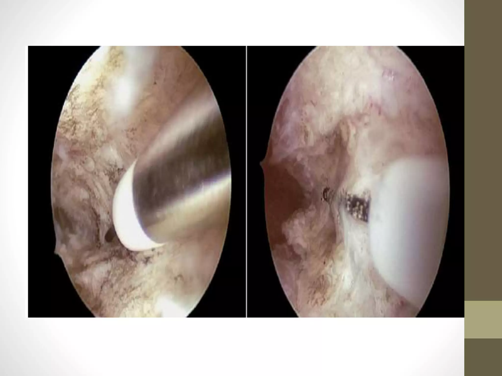
• the hysteroscopic electrocoagulation of internal
orifice of uterine tube: could effectively prevent
hydrosalpinx fluid reflux and induce no
significant complications after surgery.
• a monopolar roller ball electrode (size: 3 mm) is
clung to the internal orifice of the fallopian
tube.. The electrocoagulation is performed with
power of 40-60W for 5~10 sec at each side to
form a deep yellow scabbed plaques.
• The electrical heat energy is utilized to
degenerate the internal orifice tissue of diseased
fallopian so as to scar the electro coagulated
tissue.
• The contracting and even occluding of the
scarred tissue prevented the hydrosalpinx fluid
reflux to uterine cavity.
• Hysteroscopic Tubal Occlusion with the use of Iso-Amyl-
2- Cyanoacrylate:
• Ultrasound-guided aspiration
• This intervention consists of aspirating the
hydrosalpingeal fluid at the time of oocyte
retrieval, after all the oocytes had been
collected, under deep sedation.
• Under ultrasound guidance, the aspiration
needle is inserted into the hydrosalpinx and
suction is applied until no further fluid could be
obtained.
• Recurrence rate of hydrosalpinx after aspiration
is consistent, ranging from 22.2 to 30.8%
• The literature data about the efficacy of this
procedure are controversial.
• Indeed, an RCT did not show a significant
increase in the odds of clinical pregnancy
• Sclerotherapy
• The ultrasound sclerotherapy with 98% ethanol
is another method for the treatment of
hydrosalpinx. An ovum aspiration needle is
inserted under ultrasound guidance into the
hydrosalpinx and suction is applied until no
further fluid could be obtained.
• The tube cavity is washed with gentamicin
sulphate injection and infused with a volume of
98% ethanol equal to a half volume of the
aspirated fluid, that is then left in the tubal
cavity for 5–10 minutes before removal.
• All patients were then monitored for 1 hour after
the procedure and prescribed oral antibiotics for
3 days.
• Two weeks later, ultrasound evaluation is
performed and effectiveness of sclerotherapy is
defined as no visible fluid in the fallopian tube or
remaining fluid less than 10% of original size
before therapy (Jiang et al. 2010).
• A recent study evaluated the effects of this
procedure (Zhang et al. 2014). The rate of
recurrent hydrosalpinx after sclerotherapy was
as high as 21.7%.
• However, the rates of embryo implantation ,
clinical pregnancy and live birth were higher
than non treated patients.
Failed IVF/ RIF
• hysteroscopy will have the following
advantages:
• allow the diagnosis and treatment of
intrauterine pathology,, for those who may have
had a normal hysterosalpingogram before their
IVF cycle.
• Second, uterine instrumentation with
hysteroscopy through the cervical canal may
facilitate future embryo transfers
• as well as improve pregnancy rates through
endometrial injury, provocation of
immunological reaction, subsequent release of
cytokines and growth factors, and increased
implantation rate.
Hysteroscopic Endometrial
Embryo Delivery
• Embryo transfer is traditionally performed by
“blindly” replacing the embryos into the uterine
cavity utilizing a transcervical catheter.
• Many clinicians will transfer the embryos at a
fixed distance (6 cm) from the external os;
however, with varying cervical lengths and
uterine anatomy, this often does not ensure
optimal placement (Brown 2007).
• potential embryo transfer related factors to the
low success rate in pregnancy outcomes such as
uterine contractions, expulsion of embryos,
blood or mucus on the catheter tip, bacterial
contamination of the catheter, and retained
embryos (Schoolcraft 2001).
• Ultrasound guided embryo transfer (UGET) is
currently suggested as the standard clinical
practice and appears to improve the chances of
live/ongoing and clinical pregnancies compared
with clinical touch methods
• a study by Kamrava et al.,2017, utilized a mini-
hysteroscope with a flexible catheter for direct
delivery of embryos under direct visualization.
• ..
Advantages
• procedure increases the reliability of correct
entry into the uterine cavity with direct visual
confirmation.
• placement and subsequent implantation at a
precise location, with minimal volume of transfer
media,
• provides an obvious benefit to patients with
distorted uterine cavities, myomas, and
adenomyosis and uterine adhesions
• performing gas distension of the uterus by an
inert gas (N2), the catheter tip is less likely to
come into contact with the uterine fundus which
has been associated with stimulating uterine
contractions and creating an unfavorable
environment for implantation
• The potential disadvantage and risk of this technique is
disruption of the uterine lining, however the risk is
postulated to be less than “blind” and ultrasound guided
transfers due to the advantage of direct visualization of
the uterine lining and not requiring movement of the
catheter to facilitate identification during ultrasound

More Related Content

What's hot

What is the role of hysteroscopy for the management of women undergoing IVF?
What is the role of hysteroscopy for the management of women undergoing IVF?�What is the role of hysteroscopy for the management of women undergoing IVF?�
What is the role of hysteroscopy for the management of women undergoing IVF?
Ulun Uluğ
 
Diagnosis and classification of tubal factor infertility
Diagnosis and classification of tubal factor infertilityDiagnosis and classification of tubal factor infertility
Diagnosis and classification of tubal factor infertility
Sanjay Makwana
 

What's hot (20)

Role of tubal surgery in era of ivf
Role of tubal surgery in era of ivfRole of tubal surgery in era of ivf
Role of tubal surgery in era of ivf
 
Laparoscopy and fertility
Laparoscopy and fertilityLaparoscopy and fertility
Laparoscopy and fertility
 
Fertility Enhancing Laparoscopic Surgeries Panel Discussion
Fertility Enhancing Laparoscopic Surgeries Panel DiscussionFertility Enhancing Laparoscopic Surgeries Panel Discussion
Fertility Enhancing Laparoscopic Surgeries Panel Discussion
 
Role of hysteroscopy and laparoscopy in ivf
Role of hysteroscopy and laparoscopy in  ivfRole of hysteroscopy and laparoscopy in  ivf
Role of hysteroscopy and laparoscopy in ivf
 
What is the role of hysteroscopy for the management of women undergoing IVF?
What is the role of hysteroscopy for the management of women undergoing IVF?�What is the role of hysteroscopy for the management of women undergoing IVF?�
What is the role of hysteroscopy for the management of women undergoing IVF?
 
Ivf in pcos
Ivf in pcosIvf in pcos
Ivf in pcos
 
Cosmetic and asthetic gynaecology
Cosmetic and asthetic gynaecologyCosmetic and asthetic gynaecology
Cosmetic and asthetic gynaecology
 
CAESAREAN SCAR DEFECT
CAESAREAN SCAR DEFECT  CAESAREAN SCAR DEFECT
CAESAREAN SCAR DEFECT
 
Unexplained Infertility - By Dr Dhorepatil Bharati
Unexplained Infertility - By Dr Dhorepatil BharatiUnexplained Infertility - By Dr Dhorepatil Bharati
Unexplained Infertility - By Dr Dhorepatil Bharati
 
PROTOCOLS Intra Uterine Insemination (sharing personal experience)
PROTOCOLSIntra Uterine Insemination  (sharing personal experience) PROTOCOLSIntra Uterine Insemination  (sharing personal experience)
PROTOCOLS Intra Uterine Insemination (sharing personal experience)
 
Male contraceptive risug
Male contraceptive risugMale contraceptive risug
Male contraceptive risug
 
Office hysteroscopy
Office hysteroscopyOffice hysteroscopy
Office hysteroscopy
 
Management of thin endometrium isar 2019
Management of thin endometrium isar 2019Management of thin endometrium isar 2019
Management of thin endometrium isar 2019
 
Single Embryo Transfer
Single Embryo TransferSingle Embryo Transfer
Single Embryo Transfer
 
Uterus Transplantation Utx (obstetric and gynecology)
Uterus Transplantation Utx (obstetric and gynecology) Uterus Transplantation Utx (obstetric and gynecology)
Uterus Transplantation Utx (obstetric and gynecology)
 
SCAR ECTOPIC
SCAR ECTOPICSCAR ECTOPIC
SCAR ECTOPIC
 
Role of IUI in the era of IVF
Role of IUI in the era of IVFRole of IUI in the era of IVF
Role of IUI in the era of IVF
 
Diagnosis and classification of tubal factor infertility
Diagnosis and classification of tubal factor infertilityDiagnosis and classification of tubal factor infertility
Diagnosis and classification of tubal factor infertility
 
OVULATION INDUCTION FOR IUI BY DR SHASHWAT JANI
OVULATION INDUCTION FOR IUI BY DR SHASHWAT JANIOVULATION INDUCTION FOR IUI BY DR SHASHWAT JANI
OVULATION INDUCTION FOR IUI BY DR SHASHWAT JANI
 
OVARIAN RESERVE
OVARIAN RESERVEOVARIAN RESERVE
OVARIAN RESERVE
 

Similar to Hysteroscopy and infertility

Congenital disorders of female reproductive tract
Congenital disorders of female reproductive tractCongenital disorders of female reproductive tract
Congenital disorders of female reproductive tract
762060
 
Tubal factor and fertility by Dr.Gayathiri
Tubal factor and fertility by Dr.GayathiriTubal factor and fertility by Dr.Gayathiri
Tubal factor and fertility by Dr.Gayathiri
Morris Jawahar
 
Fertility enhancing hysteroscopic surgery
Fertility enhancing hysteroscopic surgeryFertility enhancing hysteroscopic surgery
Fertility enhancing hysteroscopic surgery
DrRokeyaBegum
 
ART Multiple gestation Management.ppt
ART Multiple gestation Management.pptART Multiple gestation Management.ppt
ART Multiple gestation Management.ppt
SriSushmaNagasuri
 
Placenta accreta
Placenta accretaPlacenta accreta
Placenta accreta
Galal Lotfi
 

Similar to Hysteroscopy and infertility (20)

Female infertility.pptx
Female infertility.pptxFemale infertility.pptx
Female infertility.pptx
 
Congenital disorders of female reproductive tract
Congenital disorders of female reproductive tractCongenital disorders of female reproductive tract
Congenital disorders of female reproductive tract
 
Fibroid & infertility
Fibroid & infertilityFibroid & infertility
Fibroid & infertility
 
Subfertility.pptx
Subfertility.pptxSubfertility.pptx
Subfertility.pptx
 
Tubal factor and fertility by Dr.Gayathiri
Tubal factor and fertility by Dr.GayathiriTubal factor and fertility by Dr.Gayathiri
Tubal factor and fertility by Dr.Gayathiri
 
DR SUNITA CHANDRA, LUCKNOW
DR SUNITA CHANDRA, LUCKNOWDR SUNITA CHANDRA, LUCKNOW
DR SUNITA CHANDRA, LUCKNOW
 
Fertility enhancing hysteroscopic surgery
Fertility enhancing hysteroscopic surgeryFertility enhancing hysteroscopic surgery
Fertility enhancing hysteroscopic surgery
 
Mullerian anomalies
Mullerian anomaliesMullerian anomalies
Mullerian anomalies
 
Cervical cancer ppt
Cervical cancer pptCervical cancer ppt
Cervical cancer ppt
 
Adnexal masses in pregnancy
Adnexal masses in pregnancyAdnexal masses in pregnancy
Adnexal masses in pregnancy
 
Basics of tvs color doppler
Basics of tvs color dopplerBasics of tvs color doppler
Basics of tvs color doppler
 
Cervical carcinoma
Cervical carcinomaCervical carcinoma
Cervical carcinoma
 
Salpingectomy for ovarian risk reduction
Salpingectomy for ovarian risk reductionSalpingectomy for ovarian risk reduction
Salpingectomy for ovarian risk reduction
 
Abnormal Invasive Placenta
Abnormal Invasive PlacentaAbnormal Invasive Placenta
Abnormal Invasive Placenta
 
GTD.pptx
GTD.pptxGTD.pptx
GTD.pptx
 
ART Multiple gestation Management.ppt
ART Multiple gestation Management.pptART Multiple gestation Management.ppt
ART Multiple gestation Management.ppt
 
Panel Discussion on Post Menopausal Bleeding
Panel Discussion on Post Menopausal Bleeding Panel Discussion on Post Menopausal Bleeding
Panel Discussion on Post Menopausal Bleeding
 
Presentation on Adherent placenta by Dr. Laxmi Shrikhande
Presentation on Adherent placenta by Dr. Laxmi Shrikhande Presentation on Adherent placenta by Dr. Laxmi Shrikhande
Presentation on Adherent placenta by Dr. Laxmi Shrikhande
 
Placenta accreta
Placenta accretaPlacenta accreta
Placenta accreta
 
Third trimester bleeding
Third trimester bleedingThird trimester bleeding
Third trimester bleeding
 

Recently uploaded

👉 Chennai Sexy Aunty’s WhatsApp Number 👉📞 7427069034 👉📞 Just📲 Call Ruhi Colle...
👉 Chennai Sexy Aunty’s WhatsApp Number 👉📞 7427069034 👉📞 Just📲 Call Ruhi Colle...👉 Chennai Sexy Aunty’s WhatsApp Number 👉📞 7427069034 👉📞 Just📲 Call Ruhi Colle...
👉 Chennai Sexy Aunty’s WhatsApp Number 👉📞 7427069034 👉📞 Just📲 Call Ruhi Colle...
rajnisinghkjn
 
Russian Call Girls In Pune 👉 Just CALL ME: 9352988975 ✅❤️💯low cost unlimited ...
Russian Call Girls In Pune 👉 Just CALL ME: 9352988975 ✅❤️💯low cost unlimited ...Russian Call Girls In Pune 👉 Just CALL ME: 9352988975 ✅❤️💯low cost unlimited ...
Russian Call Girls In Pune 👉 Just CALL ME: 9352988975 ✅❤️💯low cost unlimited ...
chanderprakash5506
 
Guntur Call Girl Service 📞6297126446📞Just Call Divya📲 Call Girl In Guntur No ...
Guntur Call Girl Service 📞6297126446📞Just Call Divya📲 Call Girl In Guntur No ...Guntur Call Girl Service 📞6297126446📞Just Call Divya📲 Call Girl In Guntur No ...
Guntur Call Girl Service 📞6297126446📞Just Call Divya📲 Call Girl In Guntur No ...
Call Girls in Nagpur High Profile Call Girls
 

Recently uploaded (20)

Call Girls Bangalore - 450+ Call Girl Cash Payment 💯Call Us 🔝 6378878445 🔝 💃 ...
Call Girls Bangalore - 450+ Call Girl Cash Payment 💯Call Us 🔝 6378878445 🔝 💃 ...Call Girls Bangalore - 450+ Call Girl Cash Payment 💯Call Us 🔝 6378878445 🔝 💃 ...
Call Girls Bangalore - 450+ Call Girl Cash Payment 💯Call Us 🔝 6378878445 🔝 💃 ...
 
👉 Chennai Sexy Aunty’s WhatsApp Number 👉📞 7427069034 👉📞 Just📲 Call Ruhi Colle...
👉 Chennai Sexy Aunty’s WhatsApp Number 👉📞 7427069034 👉📞 Just📲 Call Ruhi Colle...👉 Chennai Sexy Aunty’s WhatsApp Number 👉📞 7427069034 👉📞 Just📲 Call Ruhi Colle...
👉 Chennai Sexy Aunty’s WhatsApp Number 👉📞 7427069034 👉📞 Just📲 Call Ruhi Colle...
 
💰Call Girl In Bangalore☎️63788-78445💰 Call Girl service in Bangalore☎️Bangalo...
💰Call Girl In Bangalore☎️63788-78445💰 Call Girl service in Bangalore☎️Bangalo...💰Call Girl In Bangalore☎️63788-78445💰 Call Girl service in Bangalore☎️Bangalo...
💰Call Girl In Bangalore☎️63788-78445💰 Call Girl service in Bangalore☎️Bangalo...
 
Bhopal❤CALL GIRL 9352988975 ❤CALL GIRLS IN Bhopal ESCORT SERVICE
Bhopal❤CALL GIRL 9352988975 ❤CALL GIRLS IN Bhopal ESCORT SERVICEBhopal❤CALL GIRL 9352988975 ❤CALL GIRLS IN Bhopal ESCORT SERVICE
Bhopal❤CALL GIRL 9352988975 ❤CALL GIRLS IN Bhopal ESCORT SERVICE
 
Call Girls in Lucknow Just Call 👉👉8630512678 Top Class Call Girl Service Avai...
Call Girls in Lucknow Just Call 👉👉8630512678 Top Class Call Girl Service Avai...Call Girls in Lucknow Just Call 👉👉8630512678 Top Class Call Girl Service Avai...
Call Girls in Lucknow Just Call 👉👉8630512678 Top Class Call Girl Service Avai...
 
Cardiac Output, Venous Return, and Their Regulation
Cardiac Output, Venous Return, and Their RegulationCardiac Output, Venous Return, and Their Regulation
Cardiac Output, Venous Return, and Their Regulation
 
Bhawanipatna Call Girls 📞9332606886 Call Girls in Bhawanipatna Escorts servic...
Bhawanipatna Call Girls 📞9332606886 Call Girls in Bhawanipatna Escorts servic...Bhawanipatna Call Girls 📞9332606886 Call Girls in Bhawanipatna Escorts servic...
Bhawanipatna Call Girls 📞9332606886 Call Girls in Bhawanipatna Escorts servic...
 
Call Girls in Lucknow Just Call 👉👉 8875999948 Top Class Call Girl Service Ava...
Call Girls in Lucknow Just Call 👉👉 8875999948 Top Class Call Girl Service Ava...Call Girls in Lucknow Just Call 👉👉 8875999948 Top Class Call Girl Service Ava...
Call Girls in Lucknow Just Call 👉👉 8875999948 Top Class Call Girl Service Ava...
 
Call Girls Mussoorie Just Call 8854095900 Top Class Call Girl Service Available
Call Girls Mussoorie Just Call 8854095900 Top Class Call Girl Service AvailableCall Girls Mussoorie Just Call 8854095900 Top Class Call Girl Service Available
Call Girls Mussoorie Just Call 8854095900 Top Class Call Girl Service Available
 
ANATOMY AND PHYSIOLOGY OF RESPIRATORY SYSTEM.pptx
ANATOMY AND PHYSIOLOGY OF RESPIRATORY SYSTEM.pptxANATOMY AND PHYSIOLOGY OF RESPIRATORY SYSTEM.pptx
ANATOMY AND PHYSIOLOGY OF RESPIRATORY SYSTEM.pptx
 
Call girls Service Phullen / 9332606886 Genuine Call girls with real Photos a...
Call girls Service Phullen / 9332606886 Genuine Call girls with real Photos a...Call girls Service Phullen / 9332606886 Genuine Call girls with real Photos a...
Call girls Service Phullen / 9332606886 Genuine Call girls with real Photos a...
 
Lucknow Call Girls Service { 9984666624 } ❤️VVIP ROCKY Call Girl in Lucknow U...
Lucknow Call Girls Service { 9984666624 } ❤️VVIP ROCKY Call Girl in Lucknow U...Lucknow Call Girls Service { 9984666624 } ❤️VVIP ROCKY Call Girl in Lucknow U...
Lucknow Call Girls Service { 9984666624 } ❤️VVIP ROCKY Call Girl in Lucknow U...
 
Chennai ❣️ Call Girl 6378878445 Call Girls in Chennai Escort service book now
Chennai ❣️ Call Girl 6378878445 Call Girls in Chennai Escort service book nowChennai ❣️ Call Girl 6378878445 Call Girls in Chennai Escort service book now
Chennai ❣️ Call Girl 6378878445 Call Girls in Chennai Escort service book now
 
Russian Call Girls In Pune 👉 Just CALL ME: 9352988975 ✅❤️💯low cost unlimited ...
Russian Call Girls In Pune 👉 Just CALL ME: 9352988975 ✅❤️💯low cost unlimited ...Russian Call Girls In Pune 👉 Just CALL ME: 9352988975 ✅❤️💯low cost unlimited ...
Russian Call Girls In Pune 👉 Just CALL ME: 9352988975 ✅❤️💯low cost unlimited ...
 
7 steps How to prevent Thalassemia : Dr Sharda Jain & Vandana Gupta
7 steps How to prevent Thalassemia : Dr Sharda Jain & Vandana Gupta7 steps How to prevent Thalassemia : Dr Sharda Jain & Vandana Gupta
7 steps How to prevent Thalassemia : Dr Sharda Jain & Vandana Gupta
 
(RIYA)🎄Airhostess Call Girl Jaipur Call Now 8445551418 Premium Collection Of ...
(RIYA)🎄Airhostess Call Girl Jaipur Call Now 8445551418 Premium Collection Of ...(RIYA)🎄Airhostess Call Girl Jaipur Call Now 8445551418 Premium Collection Of ...
(RIYA)🎄Airhostess Call Girl Jaipur Call Now 8445551418 Premium Collection Of ...
 
Indore Call Girls ❤️🍑7718850664❤️🍑 Call Girl service in Indore ☎️ Indore Call...
Indore Call Girls ❤️🍑7718850664❤️🍑 Call Girl service in Indore ☎️ Indore Call...Indore Call Girls ❤️🍑7718850664❤️🍑 Call Girl service in Indore ☎️ Indore Call...
Indore Call Girls ❤️🍑7718850664❤️🍑 Call Girl service in Indore ☎️ Indore Call...
 
Race Course Road } Book Call Girls in Bangalore | Whatsapp No 6378878445 VIP ...
Race Course Road } Book Call Girls in Bangalore | Whatsapp No 6378878445 VIP ...Race Course Road } Book Call Girls in Bangalore | Whatsapp No 6378878445 VIP ...
Race Course Road } Book Call Girls in Bangalore | Whatsapp No 6378878445 VIP ...
 
Guntur Call Girl Service 📞6297126446📞Just Call Divya📲 Call Girl In Guntur No ...
Guntur Call Girl Service 📞6297126446📞Just Call Divya📲 Call Girl In Guntur No ...Guntur Call Girl Service 📞6297126446📞Just Call Divya📲 Call Girl In Guntur No ...
Guntur Call Girl Service 📞6297126446📞Just Call Divya📲 Call Girl In Guntur No ...
 
ANATOMY AND PHYSIOLOGY OF REPRODUCTIVE SYSTEM.pptx
ANATOMY AND PHYSIOLOGY OF REPRODUCTIVE SYSTEM.pptxANATOMY AND PHYSIOLOGY OF REPRODUCTIVE SYSTEM.pptx
ANATOMY AND PHYSIOLOGY OF REPRODUCTIVE SYSTEM.pptx
 

Hysteroscopy and infertility

  • 2. Fertility .. • Good ovum, Good Sperm • Good embryo • Patent and functioning tube • Good Uterine cavity • Receptive Endometrium
  • 3. Methods to Assess the Uterine Cavity • Evaluation of the uterine cavity is a basic step in female infertility workup. • Out of the four available methods—transvaginal sonography (TVS), hysterosalpingography (HSG), saline contrast sonography (SCS) and hysteroscopy, • HSG is the most frequently used method.
  • 4. Hysterosalpingography (HSG) • Although this test is fairly simple to perform, it does not allow for simultaneous correction of presumed uterine pathology. • Additionally, previous studies have shown that HSG may miss some uterine abnormalities. • HSG has significant limitations when evaluating the endometrial cavity.
  • 5. HSG fulfills the requirements for a good screening test for revealing abnormalities of the uterine cavity, though any abnormalities found will likely need further evaluation to make a definitive diagnosis.
  • 6.
  • 7. Transvaginal ultrasound scan & 3D US • It allows examination of the ovaries to diagnose any ovarian cyst or polycystic ovaries adding valuable information required prior to IVF. • However TVS may not diagnose submucosal fibroids in the presence of multiple fibroids. • Also, it can not distinguish between a hyperplasic endometrium and a large polyp, • or differentiate between an arcuate and a septate uterus. • The advent of transvaginal 3D ultrasonography has enabled the accurate, noninvasive, outpatient diagnosis of congenital uterine anomalies.
  • 8. Hystero-sonography • Hysterosonoggraphy has been proposed as a better diagnostic test of the uterine cavity • However, it also has sensitivity and specificity inferior to that of hysteroscopy in most studies. • Additionally, it does not allow for correction of presumed pathology.
  • 9. Hysteroscopy • Hysteroscopy is the gold standard for the investigation of uterine cavity, particularly when a pathology is suspected. • It is a safe test for the direct and accurate diagnosis of intrauterine abnormalities. • allow for correction of several pathologies.
  • 10. • However, it is the most costly and invasive method so it is reserved for further evaluation and treatment of abnormalities detected by less invasive method as HSG(according to ASRM 2015)
  • 11. The Value of hysteroscopy in infertility
  • 12. Intra cavitary Pathology • Sub mucous Fibroids • Endometrial Polyps • Uterine Septum • Intrauterine Adhesions
  • 13. Other roles • Failed IVF/RIF • Proximal Tubal Occlusion • Chronic Endometritis • Hydrosalpinx.
  • 14. Fibroid and infertility • Leiomyomas are estimated to occur in1 to 2 percent of infertile population particularly those that encroach upon the endometrium. • The location of a fibroid, and not its size, is the key factor regarding the effect on fertility.
  • 15.
  • 16. • Leiomyomas that distort the uterine cavity (submucosal or intramural with an intracavitary component) result in difficulty conceiving a pregnancy and an increased risk of miscarriage. • In contrast, subserosal fibroids do not impact fertility. • The role of intramural fibroids without intracavitary portion in infertility is controversial
  • 17. • Postulated mechanisms by which fibroids cause infertility include the following: • physical impedance to the transport of sperm or embryo • Distortion or obstruction of tubal ostia. • Dysfunctional uterine contractility.
  • 18. • Chronic endometrial inflammation. • Impaired endometrial receptivity. • Implantation failure due to atrophy or venous ectasia over or opposite a submucous fibroid.
  • 19. • in a meta-analysis of 23 studies that compared infertile women with and without leiomyomas. Women with fibroids that were submucosal or intramural with an intracavitary component were less likely to become pregnant and more likely to have a spontaneous abortion (RR 1.7, 95% CI 1.4–2.1).
  • 20. • Consistent with these findings, women with cavity-distorting fibroids who did versus did not undergo myomectomy had a significant increase in conception rate ,but Interestingly, there was no significant decrease in the risk of miscarriage.
  • 21.
  • 22.
  • 23. Whom to treat • For natural conception or pre IVF cycle: • Submucous fibroid: myomectomy for FIGO L0 to L2 of any size. • Intramural fibroid :controversial??? • consider surgery for FIGO L3 to L5 >50mm. • Subserous fibroid :FIGO 6 to L7 only to improve patient symptoms .
  • 24. How to treat?? • Hysteroscopic myomectomy : • only for fibroids that are completely within the endometrial cavity or extend less than 50 percent into the myometrium. • Removal of fibroids with deeper myometrial involvement requires advanced hysteroscopic skills or myomectomy using laparotomy or laparoscopy.
  • 25. • For leiomyomas that are multiple or are >5 cm or deep in myometrium, it is prudent to include in the informed consent the possibility of a two- stage procedure . • Their removal is best accomplished with systematic shaving of sessile leiomyomas that are partially intramural, using the resectoscope and a cutting loop.
  • 26.
  • 27.
  • 28. Endometrial Polyps • Endometrial polyps represent the most common intracavitary finding in the infertile population. • Women undergoing evaluation for infertility may have a finding of an endometrial polyp on ultrasound or hysteroscopy; the reported prevalence in those undergoing in vitro fertilization is 6 to 8 percent.
  • 29. • Postulated mechanisms by which polyps cause infertility include: • may be related to mechanical interference with sperm transportation or as space occupying lesions interfering with embryo implantation.
  • 30. • The glands and stroma in endometrial polyps are unresponsive to progesterone stimulation, leading to defective implantation at the site of the polyp. • Endometrial polyps may also induce local inflammatory changes, which can interfere with normal implantation and embryonic development
  • 31.
  • 32.
  • 33. • Yahaihara et al conducted a retrospective study of 230 women to determine the significance of the location of endometrial polyps and polyp size on infertility. • Site in any area of the uterus did not significantly differ on the impact pregnancy rate; however, the highest pregnancy rate, 50% to 60%, was achieved in those who had polyps removed from the utero-tubal junction.
  • 34. • There is no evidence on which size could impair fertility some suggest only removal of polyps larger than 2 cm whereas others still advocate removal of any space occupying lesion.
  • 35. Uterine Septum • True incidence of congenital uterine anomalies is unknown. • Estimates based on of several studies its incidence around 3% to 4%, and 13% in patients with RPL. • The septate uterus is the most common and has the worst reproductive outcomes of all the congenital uterine malformations. • • It accounts for about 35% of all congenital malformations
  • 36. Classification of Müllerian anomalies according to the American Fertility Society classification system 1988
  • 37. Type I: "Müllerian" agenesis or hypoplasia A. Vaginal (uterus may be normal or exhibit a variety of malformations) B. Cervical C. Fundal D. Tubal E. Combined Type II: Unicornuate uterus A1a. Communicating (endometrial cavity present) A1b. Noncommunicating (endometrial cavity present) A2. Horn without endometrial cavity B. No rudimentary horn Type III: Uterus didelphys
  • 38. Type IV: Uterus bicornuate A. Complete (division down to internal os) B. Partial C. Arcuate Type V: Septate uterus A. Complete (septum to internal os) B. Partial Type VI: Diethylstibestrol-related anomalies A. T-shaped uterus B. T-shaped with dilated horns
  • 39. The European Society of Human Reproduction and Embryology (ESHRE) and the European Society for Gynaecological Endoscopy (ESGE) classification system 2013
  • 40.
  • 41.
  • 42.
  • 43.
  • 44.
  • 45.
  • 46. • HSG and hysteroscopy alone cannot distinguish between septate and bicornuate uteri. • Although laparoscopy to visualize the external fundal contour is the gold standard, three-dimensional (3D) ultrasound and MRI have been shown to have nearly 100% sensitivity and specificity.
  • 47.
  • 48.
  • 49.
  • 50. Effect of septum on fertility • Uterine septa are often diagnosed during an infertility evaluation. The incidence of uterine septa in this population has been noted to be higher than in the general population, suggesting a link with infertility. • Given that infertility can be the result of multiple factors, it is often difficult to determine if the uterine septum is the sole reason for the infertility.
  • 51. • according to ASRM statement 2015 • There is insufficient evidence to conclude that a uterine septum is associated with infertility. • There is fair evidence that a uterine septum contributes to miscarriage and preterm birth. • Some evidence suggests that a uterine septum may increase the risk of other adverse pregnancy outcomes such as malpresentation, intrauterine growth restriction, placental abruption, and perinatal mortality
  • 52. • Proposed mechanisms by which septum can affect fertility: • The septum might contribute to the high rate of early pregnancy loss due to poor blood supply, • deficient estrogen and progesterone receptors, • and inadequate endometrial maturation.
  • 53. Whom to treat?? • Several observational studies indicate that hysteroscopic septoplasty is associated with improved clinical pregnancy rates in women with infertility. • Some limited studies indicate that hysteroscopic septum incision is associated with a reduction in subsequent miscarriage rates and improvement in live-birth rates in patients with a history of recurrent pregnancy loss.
  • 54. • The only fair evidence that hysteroscopic septoplasty is indicated for patients with two or more spontaneous abortions. • However, because hysteroscopic septoplasty is a quick, minimally invasive procedure with negligible risk, surgical correction is reasonable in patients with only one spontaneous abortion, longstanding unexplained infertility, or age greater than 35 years and before undergoing assisted reproductive technology (ART)
  • 55. HOW TO TREAT A UTERINE SEPTUM • Historically, metroplasty for a septum was performed by laparotomy via the Jones or Tompkins procedure, which involved excising a wedge of the uterine fundus containing the septum or opening the fundus and incising the septum, respectively.
  • 56.
  • 57.
  • 58. Hysteroscopic management of a uterine septum • It can be performed in the operating room under anesthesia, or in an office setting. • Commonly used techniques include incision of the septum utilizing cold scissors, unipolar or bipolar cautery, or laser, or resection of the septum. • However, There is insufficient evidence to recommend a specific method for hysteroscopic septum incision.
  • 59. • The procedure is terminated when the hysteroscope can move between the cornua with no intervening septum. • Also, bleeding indicates that the normal myometrium has been reached.
  • 60. Postoperative management • No further treatment is required postoperatively. Intrauterine devices, Foley balloons, high-dose estrogen, and antibiotics are not necessary as formation of intrauterine synechiae is rare, as are postoperative infections. • Endogenous estrogen is sufficient to promote new endometrium within two months of hysteroscopic metroplasty.
  • 61. • A second look hysteroscopy should be performed two months after surgery to assess success. Typically, over 90 percent of the septum is removed during the procedure. Occasionally, further repairs of the septum are required, again in an ambulatory setting • Although the available evidence suggests that the uterine cavity is healed by 2 months postoperatively, there is insufficient evidence to advocate a specific length of time before a woman should conceive
  • 62. Outcome • A summary of 16 retrospective studies reported that 88% of 1062 pregnancies resulted in spontaneously abortion preoperatively versus 14% of 491 pregnancies following hysteroscopic septoplasty. Preterm labor decreased from 9% before septoplasty to 6% after, and term deliveries improved from 3% to 80%
  • 63.
  • 64.
  • 65.
  • 66.
  • 67. Dysmorphic uteri • characterized by a normal outline but with an abnormal lateral wall’s shape of the uterine cavity ( i.e. T-shaped uterus and tubular-shaped/infantile uteri). • These uteri are associated with infertility and pregnancy loss and in the previous American Fertility Society classification were included in class VII and mainly related to (DES) exposure. • However clinical experience has shown that these uteri are more common than expected, mostly diagnosed in young infertile patients with no history of DES exposure.
  • 68. HOME-DU technique • Hysteroscopic Outpatient Metroplasty To Expand Dysmorphic Uteri: the HOME-DU technique). • two incisions of 3–4 mm in depth are made with a 5- Fr bipolar electrode along the lateral walls of the uterine cavity in the isthmic region, followed by additional incisions placed on the anterior and posterior walls of the fundal region up to the isthmus.
  • 69.
  • 70.
  • 71.
  • 72.
  • 73.
  • 74.
  • 75.
  • 76. • Preliminary data on a cohort of 30 infertile patients (i.e. primary infertility, > 2 early abortions or severe preterm delivery) showed a significant increase in the volume of the uterine cavity, with a substantial improvement in uterine morphology. • Moreover, at mean follow-up of 15 months, clinical pregnancy rate was 57% and term delivery rate 65% without any significant obstetrical complications
  • 77. • These positive preliminary data have been confirmed in a larger cohort of patients (64 patients) where together with a clinical pregnancy rate of 55% and a term delivery rate of 69%
  • 78.
  • 79. Chronic endometritis • A common hysteroscopic finding in chronic endometritis is the presence of a thickened edematous endometrium. Also, a thin hyperemic micropolyps layer (less than 1 mm) that appears to float in the endometrial cavity can be seen • Using these criteria, the hysteroscopic diagnosis of chronic endometritis has a sensitivity up to 93%.
  • 80. • However, the gold standard for the diagnosis of CE is histological identification of plasma cells in the endometrial stroma. • The use of immunohistochemistry (IHC) stains for syndecan-1 (CD138), a proteoglycan found on the cell surface of plasma cells and keratinocytes, provides a more accurate diagnosis .
  • 81. • The impact of CE on reproductive capacity is controversial, but reports suggest it may negatively affect fertility outcomes since endometrial receptivity is altered by an abnormal infiltration of plasma cells and secretion of IgM, IgG, and IgA antibodies. • Moreover, an altered endometrial expression of genes encoding for proteins involved in the inflammatory response, proliferation, and apoptosis has been found in women with CE.
  • 82. • Cicinelli and colleagues ,2008 suggest that chronic endometritis was identified in 30.3% of patients with repeated implantation failure at IVF and women diagnosed with CE had lower implantation rates (11.5%) after IVF cycles.
  • 83.
  • 84.
  • 85. • Large area of hyperemic endometrium flushed with white central points, a typical aspect of chronic endometritis called “strawberry aspect.”
  • 86. • 70% of cases of CE demonstrated in EM biopsies were cured by a regimen of 100 mg of doxycycline twice per day for 14 days. • Antibiotic treatment may attenuate the effect of CE on infertility • Cicinelli et al, reported that the clinical pregnancy rate of the group whose hysteroscopic findings normalized 1 year after antibiotic treatment was significantly higher than that of the non-normalized group 74.8% vs. 24.4%.
  • 87. Asherman syndrome • IUAs, or intrauterine synechiae, is a condition in which scar tissue develops within the uterine cavity. Intrauterine adhesions that are accompanied by symptoms (eg, infertility, amenorrhea) are referred to as Asherman syndrome. • Both terms are often used interchangeably.
  • 88. • The manifestations of intrauterine adhesions include menstrual aberrations such as hypomenorrhea or amenorrhea, infertility, pregnancy wastage (including both first and second-trimester abortions), missed abortion, intrauterine fetal demise, and errors of placental implantation (such as placenta accreta, increta, and percreta).
  • 89.
  • 90.
  • 91.
  • 92.
  • 93. • Hysteroscopy is the most accurate method for diagnosis of IUAs and should be the investigation of choice when available. • Classification of IUAs is useful because prognosis is related to the severity of disease. • A number of classification systems have been proposed for IUAs, each of which includes hysteroscopy to determine the characteristics of adhesions.
  • 94. • The ideal classification system should include a comprehensive description of the adhesions which should be graded in terms of severity with correlation with patient symptoms and obstetric performance.
  • 95.
  • 96.
  • 97.
  • 98.
  • 99. • the widely used classification developed on behalf of the American Fertility Society took into account the extent of the disease, menstrual pattern and the morphological feature of the adhesions. • Both hysteroscopy and HSG could be used for this kind of scoring system.
  • 100.
  • 101. • More recently, the classification published in 2000 by Nasr et al. illustrated an innovative way to classify AS • This scoring system included not only the menstrual symptoms but also the obstetric history of the woman. According to this group, clinical history plays a more important role than the extent of the adhesions
  • 102.
  • 103. Management the treatment goals are • to restore uterine architecture to normal, • to prevent re adherence of the uterine walls, • to provide stimulation for endometrial growth over the freshly dissected surfaces,
  • 104. • to verify that the uterine architecture is normal, • and to ascertain that the endometrial development in response to endogenous ovarian steroid production is normal prior to permitting the patient to attempt to conceive
  • 105.
  • 106. Hysteroscopic surgery • When the lesions are filmy, the tip of the hysteroscope and uterine distension may be enough to break down the adhesions. • the treatment of the severe and dense adhesion remains more challenging: in these cases, the cavity may be completely occluded or too narrow to allow the insertion of hysteroscopic sheath inside the cervix.
  • 107. • A wide range of mechanical or electric equipment has been adopted during hysteroscopic adhesiolysis with Ultrasound or laparoscopic guidance was used where ever required(if fundus obliterated) • Adhesions can be removed with sharp scissors or the use of a sharp needle (Touhy needle)
  • 108. • A cold-knife approach is supposed to prevent thermal damage of the residual endometrium and reduce the rate of perforation during the procedure. • The use of powered instruments (electric surgery or laser) has also proven efficient but may be associated with potential damage to the residual endometrium.
  • 109. Outcome of treatment • Infertility — Data are inconsistent about the efficacy of treatment of IUAs in women with infertility. In a systematic review of 28 studies, most studies reported a pregnancy rate of 40 to 80 percent and a live birth rate of 30 to 70 percent. In women with Asherman syndrome who become pregnant following hysteroscopic adhesiolysis, morbidly adherent placenta, including placenta accreta, occurs in approximately 10 percent of cases.
  • 110. • RPL: A study evaluating reproductive outcomes after hysteroscopic adhesiolysis found that out 8 out of 13 women with recurrent pregnancy loss went on to conceive.
  • 111. Recurrence • The recurrence rate following treatment is as high as 33 percent in women with mild to moderate IUAs and 66 percent in women with severe adhesions. • If adhesions recur, a second or third procedure may be necessary to complete the dissection.
  • 112. Prevention of adhesion recurrence • Early second look hysteroscopy: As early as one week rather than after 1-3 months of hormonal treatment(Robinson et al 2008 ). • intrauterine device: to separate the endometrial layers after lysis of IUAs • The best is inert Lippes loop although it is no longer available. • The Levonorgestrel-releasing IUD should not be used for his suppressing effect on the endometrium.
  • 113. • Copper-containing and T-shaped IUDs cannot be recommended because of their inflammation provoking properties and small surface area , respectively. • The use of a Foley catheter for 3 to 10 days following surgical lysis of IUAs is similarly reported to act as a physical intrauterine barrier.
  • 114. • A three-armed pilot RCT assessed fresh amnion versus dried amnion grafts versus intrauterine balloon alone showed that: • Amnion grafts reduced adhesions significantly more than the balloon alone (p < .003), and fresh amnion was superior to dried amnion (p < .05). • Auto-cross-linked hyaluronic acid gel may be suitable for preventing IUAs because of prolonged time on an injured surface such as the postoperative endometrium(the walls of the uterine cavity remained separated for at least 72 h)
  • 115.
  • 116. • Hormonal treatments: Postoperative treatment with estrogen therapy (a daily oral dose of 2.5 mg conjugated equine estrogen with or without opposing progestin for 2 or 3 cycles). • Techniques to increase vascular flow to endometrium: • Various studies have described use of medications such as aspirin, nitroglycerine, and sildenafil citrate to increase vascular perfusion to the endometrium.
  • 117. Tubal causes of infertility • It is estimated that tubal factors account for 14% of the causes of subfertility in women. • Either with • Proximal Tubal Occlusion. • distal tubal occlusion and hydrosalpinges
  • 118. Proximal Tubal Occlusion • Proximal tubal occlusion, leading to obstruction of the anatomic pathway for fertilization, occurs in 10% to 25% of infertile women. • Tubal spasms, mucus plugs, debris, chronic salpingitis, intratubal endometriosis, tubal polyps, and hypoplasia are all known causes of proximal tubal occlusion.
  • 119. • A recent review in 2010 by Allahbadia and Merchant evaluated success and pregnancy rates of tubal recanalization with hysteroscopy. Success rates ranged from 57% to 88%. • Tubal cannulation by using catheter system under combined hysteroscopy and laparoscopy.
  • 120. • outer catheter is directed to uterotubal ostium and selective salpingiogram is performed if block is confirmed the inner flexible catheter with a guide wire is advanced through the proximal tube. • Should only be performed if the distal part of the tube is healthy.
  • 121. Unilateral or bilateral hydrosalpinges • The most severe manifestation of tubal disease is hydrosalpinx, found in 10–30% of couples presenting with infertility from tubal factors. • In addition to its important role in infertility, hydrosalpinx has an adverse effect on the outcome of in vitro fertilization (IVF). • Live birth rate of infertile women with hydrosalpinx following IVF is reduced by 50% in comparison to other tubal factors.
  • 122. • A variety of hypotheses have been proposed to explain the significant decrease in pregnancy and implantation in women with hydrosalpinx. • 1= the accumulation of fluid of any source within the endometrial cavity impairs the embryonic implantation. • 2=hydrosalpinx fluid have direct embryo toxic effect and inhibit the fertilization. By the presence of inflammatory cytokines. • 3= decreased endometrial receptivity.
  • 123. Whom to treat • The United Kingdom’s National Institute of Health and Clinical Excellence guidelines suggest the management of hydrosalpinx by laparoscopic salpingectomy before IVF in order to improve outcomes (O’Flynn 2014). • • Furthermore, a recent Cochrane review found that either laparoscopic salpingectomy or tubal occlusion before IVF increases the odds of pregnancy.
  • 124. Lines of treatment • Therapies include: • Laparscopic approach • Hystrescopic approach • Ultrasound guided aspiration/ sclerotherapy
  • 125. • Laparoscopic procedures (salpingectomy) • Salpingectomy has the advantage that chronically infected tissue is removed totally, thus cancelling the risk of abscess formation or torsion • and increasing the accessibility of the ovary during oocyte retrieval in IVF ,
  • 126. • However ,the laparoscopic approach is not entirely free from complications, such as adhesions, intestinal perforation and injury to the urinary tract. These adverse events may affect fertility even more than the initial condition, especially in a vulnerable group of patients with PID. • Thus, it is worthwhile to search minimally invasive alternative
  • 127. • On the other side, the drawbacks are the invasiveness of the procedure itself and the difficulty of the procedure in case of dense adhesions. • The transection of the oviduct at a site too close to the cornua has been associated with an increased risk of interstitial pregnancy. • Furthermore, it has been suggested that salpingectomy could have a negative effect on the ovarian blood flow and subsequently reduced ovarian response to gonadotrophin stimulation
  • 128. • in their meta‐analysis in 2016 Tsiami and colleagues found that hysteroscopic proximal tubal occlusion appeared to be the most effective intervention, followed by salpingectomy
  • 129. Proximal tubal occlusion • represents an interesting alternative to salpingectomy, especially in cases in which pelvic anatomy is distorted. • It is a less invasive, easier to perform and faster technique that addresses the main demand of elimination of the hydrosalpingeal fluid retrograde flow in the uterine cavity. • Another theoretical advantage of proximal tubal occlusion is the preservation of the ovarian blood supply.
  • 130. • Hysteroscopic insertion of devices: • Two devices, Essure® and Adiana®, are described for hysteroscopic tubal occlusion. Both have been widely used for sterilisation, but only Essure® has been used so far to manage hydrosalpinx. • It is an off label use of the device also hysterscopic evidence that the tail of the device is no longer visible within the endometrial cavity would be necessary considered before ET which typically takes several months to accomplish
  • 131. • The successful placement of Essure® was achieved in 96.5% of women and tubal occlusion was achieved in 98.1%. Subsequent IVF resulted in 38.6% pregnancy rate, 27.9% live birth rate, and 28.6% combined ongoing pregnancy and live birth rate per embryo transfer (Nichols and West 2010).
  • 132.
  • 133.
  • 134. • the hysteroscopic electrocoagulation of internal orifice of uterine tube: could effectively prevent hydrosalpinx fluid reflux and induce no significant complications after surgery. • a monopolar roller ball electrode (size: 3 mm) is clung to the internal orifice of the fallopian tube.. The electrocoagulation is performed with power of 40-60W for 5~10 sec at each side to form a deep yellow scabbed plaques.
  • 135.
  • 136. • The electrical heat energy is utilized to degenerate the internal orifice tissue of diseased fallopian so as to scar the electro coagulated tissue. • The contracting and even occluding of the scarred tissue prevented the hydrosalpinx fluid reflux to uterine cavity.
  • 137. • Hysteroscopic Tubal Occlusion with the use of Iso-Amyl- 2- Cyanoacrylate:
  • 138. • Ultrasound-guided aspiration • This intervention consists of aspirating the hydrosalpingeal fluid at the time of oocyte retrieval, after all the oocytes had been collected, under deep sedation. • Under ultrasound guidance, the aspiration needle is inserted into the hydrosalpinx and suction is applied until no further fluid could be obtained.
  • 139. • Recurrence rate of hydrosalpinx after aspiration is consistent, ranging from 22.2 to 30.8% • The literature data about the efficacy of this procedure are controversial. • Indeed, an RCT did not show a significant increase in the odds of clinical pregnancy
  • 140. • Sclerotherapy • The ultrasound sclerotherapy with 98% ethanol is another method for the treatment of hydrosalpinx. An ovum aspiration needle is inserted under ultrasound guidance into the hydrosalpinx and suction is applied until no further fluid could be obtained. • The tube cavity is washed with gentamicin sulphate injection and infused with a volume of 98% ethanol equal to a half volume of the aspirated fluid, that is then left in the tubal cavity for 5–10 minutes before removal.
  • 141. • All patients were then monitored for 1 hour after the procedure and prescribed oral antibiotics for 3 days. • Two weeks later, ultrasound evaluation is performed and effectiveness of sclerotherapy is defined as no visible fluid in the fallopian tube or remaining fluid less than 10% of original size before therapy (Jiang et al. 2010).
  • 142. • A recent study evaluated the effects of this procedure (Zhang et al. 2014). The rate of recurrent hydrosalpinx after sclerotherapy was as high as 21.7%. • However, the rates of embryo implantation , clinical pregnancy and live birth were higher than non treated patients.
  • 143. Failed IVF/ RIF • hysteroscopy will have the following advantages: • allow the diagnosis and treatment of intrauterine pathology,, for those who may have had a normal hysterosalpingogram before their IVF cycle. • Second, uterine instrumentation with hysteroscopy through the cervical canal may facilitate future embryo transfers
  • 144. • as well as improve pregnancy rates through endometrial injury, provocation of immunological reaction, subsequent release of cytokines and growth factors, and increased implantation rate.
  • 145. Hysteroscopic Endometrial Embryo Delivery • Embryo transfer is traditionally performed by “blindly” replacing the embryos into the uterine cavity utilizing a transcervical catheter. • Many clinicians will transfer the embryos at a fixed distance (6 cm) from the external os; however, with varying cervical lengths and uterine anatomy, this often does not ensure optimal placement (Brown 2007).
  • 146. • potential embryo transfer related factors to the low success rate in pregnancy outcomes such as uterine contractions, expulsion of embryos, blood or mucus on the catheter tip, bacterial contamination of the catheter, and retained embryos (Schoolcraft 2001). • Ultrasound guided embryo transfer (UGET) is currently suggested as the standard clinical practice and appears to improve the chances of live/ongoing and clinical pregnancies compared with clinical touch methods
  • 147. • a study by Kamrava et al.,2017, utilized a mini- hysteroscope with a flexible catheter for direct delivery of embryos under direct visualization.
  • 148. • ..
  • 149. Advantages • procedure increases the reliability of correct entry into the uterine cavity with direct visual confirmation. • placement and subsequent implantation at a precise location, with minimal volume of transfer media, • provides an obvious benefit to patients with distorted uterine cavities, myomas, and adenomyosis and uterine adhesions
  • 150. • performing gas distension of the uterus by an inert gas (N2), the catheter tip is less likely to come into contact with the uterine fundus which has been associated with stimulating uterine contractions and creating an unfavorable environment for implantation
  • 151. • The potential disadvantage and risk of this technique is disruption of the uterine lining, however the risk is postulated to be less than “blind” and ultrasound guided transfers due to the advantage of direct visualization of the uterine lining and not requiring movement of the catheter to facilitate identification during ultrasound