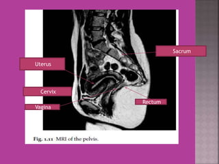
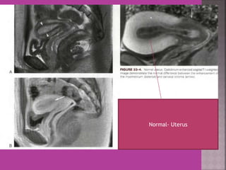
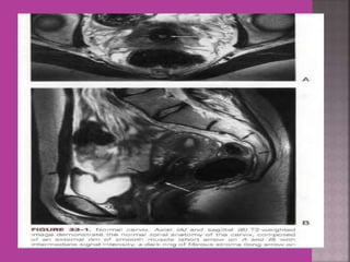
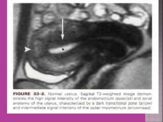
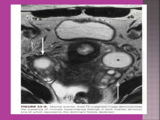
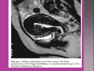
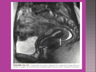
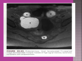
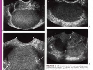
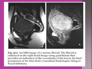
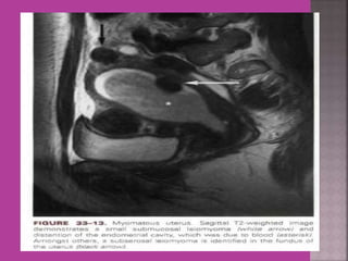
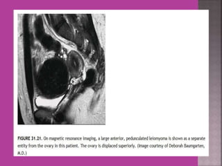
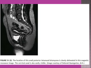
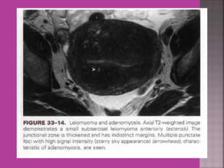
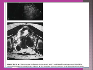
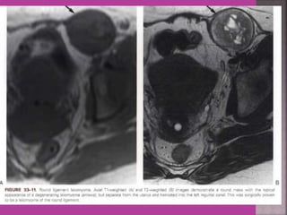
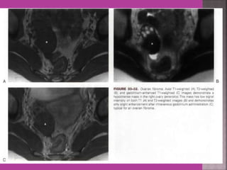
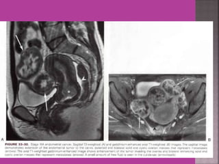
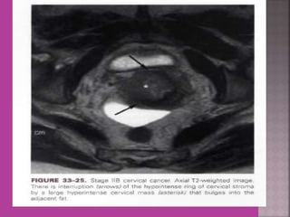
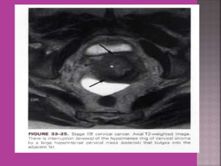
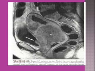
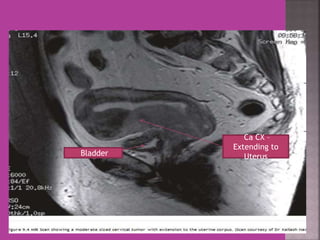
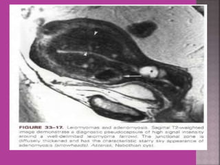
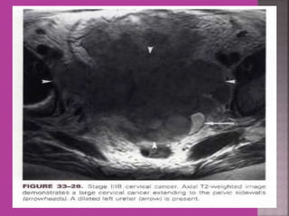
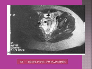
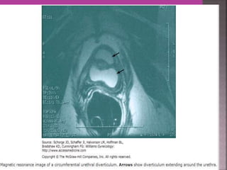
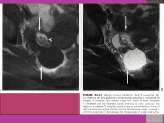
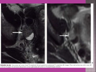
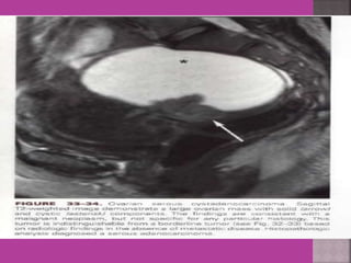
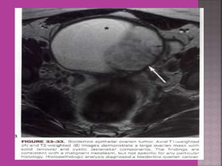
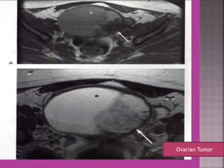
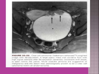
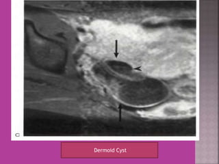
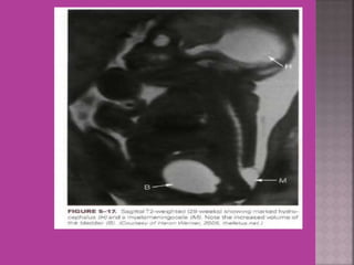
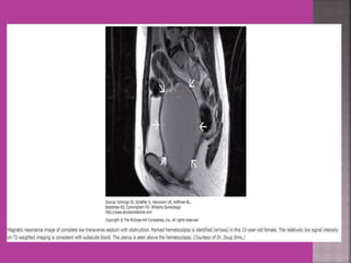
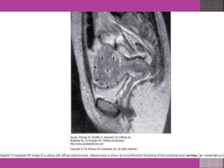
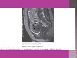
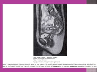
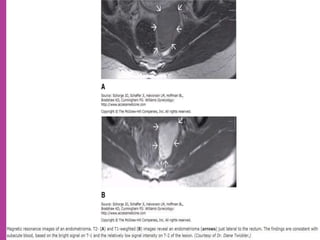
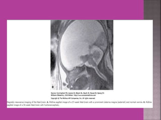
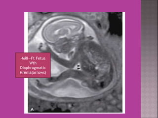
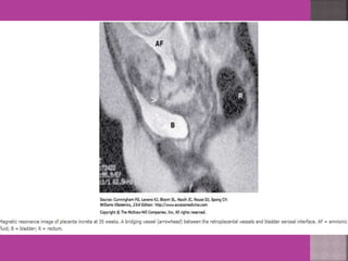
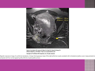
Prof. M.C.Bansal. 
Founder Principal & Controller; 
Jhalawar Medical College And Hospital, Jhalawar. 
Ex . Founder Principal & Controller; 
Mahatma Gandhi Medical College And Hospital, 
Sitapura , jaipur.
 MRI is one of the great , clinically very useful 
modern advances in the field of imaging in 
medical science. 
 Radio frequency pulses are directed at living 
body tissue/ Organ in magnetic field , the tissue 
emits back(reflected )the modulated 
radiofrequency signals which are used to 
construct the image of emitting tissue or organ. 
 By varying the time of application of 
radiofrequency pulses and sampling the emitted 
signals,T1 &T2- weighted images are obtained.
 Different tissues appear with different 
intensity and brightness in T1 and T2 images. 
 Depending upon their molecular morphology 
different tissue emit variable radiofrequency 
signals—hence different intensity and 
brightness. 
 Paramagnetic contrast agents like 
Gadolinium- DTPA are used for better 
delineation ( demarcation ) of adjoining 
tissue / organs.
 Study of normal uterus and adnexa – with 
aim to have clear image of normal 
myometrium, endometrium , ovary and 
follicles within the ovary. 
 Myoma—to differentiate myoma from 
adenomyoma.localization of myoma—sub 
serous/ intra mural or sub mucous. 
 Adenomyosis---accurate diagnosis. 
 Congenital uterovaginal anomalies— 
bicornuate, septate,subseptate , 
unicornuate, didelphys ,rudimentary horn 
and vaginal atrasia etc
 Gynaecological cancers--- 
1. Cervical cancer –extent of parametrium 
and pelvic organs , pelvic L.N., Endocervical. 
2.Endometrial cancer –Myometrial 
invasion, downward extension to cervix. 
3. ovarian cancer—malignant versus 
benign mass, ascites , retroperitoneal node > 
1cm , Bowel bladder , ano rectal . Parietal 
peritoneum , liver and or omental 
metastasis. 
4. Pregnancy --- complicated by genital 
cancers
 MRI is most sensitive for diagnosis of Adenomyosis. 
 MRI Is more accurate and informative in diagnosis of 
congenital malformation of genito-urinary tract. 
 Retroparitoneal lymph node > 1cm can be identified 
so also such small deposits on bowel / bladder liver 
can be identified in cases of cancers. 
 It is more sensitive and accurate tool to assess 
infiltration of myometrial/ cervical wall, adjoining 
organs and parametrium --thus helps in pre 
operative staging of genital carcinomas. 
 As it is safe in pregnancy –it is useful in diagnosis of 
obstetrical conditions as well s differentiation of 
tumors (benign / malignant ) complicating pregnancy.
Uterus 
Cervix 
Vagina 
Rectum 
Sacrum
Normal- Uterus
A 
B
UTERUS ----DIDELPHYS
Ovarian Endometriomaa
Degeneration Of Fibroid
Cystic Degeneration of 
Fibroid
Endometrial 
Ca 
Endo-ca— 
extending in 
cervical canal 
Pelvic Nodes
Ca CX – 
Extending to 
Bladder Uterus
MRI-----Bilateral ovaries with PCOD changes
Ovarian Tumor
Dermoid Cyst
MRI---Pituitary adenoma (A) MRI– Pituitary fosa after surgery of 
macroadenoma 
empty 
Fosa
MRI Normal Fetal Brain at Term
-MRI—Ft Fetus 
Wth 
Diaphragmatic 
Hrenia(arrows)
Mri in ob  gy practice

Mri in ob gy practice

  • 1.
    Prof. M.C.Bansal. FounderPrincipal & Controller; Jhalawar Medical College And Hospital, Jhalawar. Ex . Founder Principal & Controller; Mahatma Gandhi Medical College And Hospital, Sitapura , jaipur.
  • 2.
     MRI isone of the great , clinically very useful modern advances in the field of imaging in medical science.  Radio frequency pulses are directed at living body tissue/ Organ in magnetic field , the tissue emits back(reflected )the modulated radiofrequency signals which are used to construct the image of emitting tissue or organ.  By varying the time of application of radiofrequency pulses and sampling the emitted signals,T1 &T2- weighted images are obtained.
  • 3.
     Different tissuesappear with different intensity and brightness in T1 and T2 images.  Depending upon their molecular morphology different tissue emit variable radiofrequency signals—hence different intensity and brightness.  Paramagnetic contrast agents like Gadolinium- DTPA are used for better delineation ( demarcation ) of adjoining tissue / organs.
  • 4.
     Study ofnormal uterus and adnexa – with aim to have clear image of normal myometrium, endometrium , ovary and follicles within the ovary.  Myoma—to differentiate myoma from adenomyoma.localization of myoma—sub serous/ intra mural or sub mucous.  Adenomyosis---accurate diagnosis.  Congenital uterovaginal anomalies— bicornuate, septate,subseptate , unicornuate, didelphys ,rudimentary horn and vaginal atrasia etc
  • 5.
     Gynaecological cancers--- 1. Cervical cancer –extent of parametrium and pelvic organs , pelvic L.N., Endocervical. 2.Endometrial cancer –Myometrial invasion, downward extension to cervix. 3. ovarian cancer—malignant versus benign mass, ascites , retroperitoneal node > 1cm , Bowel bladder , ano rectal . Parietal peritoneum , liver and or omental metastasis. 4. Pregnancy --- complicated by genital cancers
  • 6.
     MRI ismost sensitive for diagnosis of Adenomyosis.  MRI Is more accurate and informative in diagnosis of congenital malformation of genito-urinary tract.  Retroparitoneal lymph node > 1cm can be identified so also such small deposits on bowel / bladder liver can be identified in cases of cancers.  It is more sensitive and accurate tool to assess infiltration of myometrial/ cervical wall, adjoining organs and parametrium --thus helps in pre operative staging of genital carcinomas.  As it is safe in pregnancy –it is useful in diagnosis of obstetrical conditions as well s differentiation of tumors (benign / malignant ) complicating pregnancy.
  • 7.
    Uterus Cervix Vagina Rectum Sacrum
  • 8.
  • 13.
  • 16.
  • 24.
  • 36.
  • 38.
  • 42.
    Endometrial Ca Endo-ca— extending in cervical canal Pelvic Nodes
  • 48.
    Ca CX – Extending to Bladder Uterus
  • 51.
  • 57.
  • 60.
  • 68.
    MRI---Pituitary adenoma (A)MRI– Pituitary fosa after surgery of macroadenoma empty Fosa
  • 71.
    MRI Normal FetalBrain at Term
  • 73.
    -MRI—Ft Fetus Wth Diaphragmatic Hrenia(arrows)