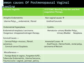
1. POSTMENOPAUSAL VAGINAL
BLEEDING
PROF. M.C.Bansal.
Founder Principal & Controller
Jhalwar Medical College and Hospital Jhalwar.
Ex . Principal& Controller Mahatma Gandhi Medical College and
Hospital ., Sitapura , Jaipur.
3. Atrophic Vaginitis
Senile Vaginitis is incorrect term . It is due to menopausla
deficiency of estrogen ; resulting in thinning of vaginal
epithelium (basal and 2-3 layered Para basal layer ) lacking in
glycogen content hence defiance mechanism provided by
lacto bacilli and acidic pH make this atrophic vaginal
epithelium vulnerable for inflammation and infection by
opportunistic common inhabitant bacteria . Slightest trauma
even coital dabbing oneself dry may result in bleeding due to
atrophic changes. P/S examination will reveal blood stained
unhealthy vaginal discharge and puncted hemorrhagic spots
on vaginal wall.
Removal of any foreign body, local estriole vaginal cream(1%)
and anti biotic cream containing soframycin / povidine and
metronidazole therapy for a week will be sufficient . Oral
estrogen can be given but carry the risk of endometral
stimulation. Local estradiole is poorly absorbed systemically.
Exclusion of neoplastic causes of RT& UT are necessary.
4. Atrophic Endometritis
Endometrial thinning<4mm and inflammation that
occurs as a result of estrogen deficiency.. Is called
atrophic Endometritis.
It may result in spotting or even bleeding more so in
hypertensive women .
This diagnosis is done after exclusion of more serious
causes of postmenopausal bleeding from uterus ; being
excluded by hysteroscopy and biopsy .
Other extra uterine cause also need their exclusion by
thorough history , local and syststemic examination and
pelvic USG.
Oral estrogen therapy and broad spectrum antibiotics
are given with a caution of endometrial hyperplasia and
other side effects of HRT.
5. Uterine Polyp
Uterine polyps are common cause of
postmenopausal bleeding .Polyps maybe
endometrial , myomatous , carcinomatous or
sarcomatous .
TVS / saline sonohysterography and
Hysteroscopy , their removal and HP Reporting
is must .
Blind D7C may miss its removal specially when it
is pedunculated and mobile .
6. Endometrial Hyperplasia
Hyperplasia means thickened endometrial lining
>4mm in post menopausal women .
HPR –classification
1. simple hyperplasia (risk of malignancy <1 %)
2. Complex Hyperplasia (3% risk of malignancy
3. Simple Hyperplasia with Atypia ( 8% risk )
4. Complex Hyperplasia with atypia (30% of
malignancy)
7. Endometrial Hyperplasia---
These hyperplasia without atypia; can safely treated by
oral progesterone (medroxy, didrogesterone 10-30mg
/day ) therapy for 3months ; followed by repeat D&c
and HPR .
If hyperplasia has reverted to normal , progesterone
therapy is continued till next 9months ---TVs
examination if needed D&c should be done in follow up
at the interval of 3months .
If hyperplasia persist / worsen in its grade/ atypia is
present hysterectomy is the best option .
In absence of H/O un opposed estrogen therapy
given, endogenous source like small grannulosa cell
tumor in ovary must be searched. Assessing estrogen
and inhibit –A level ,TVS and palpable ovary in
menopausal women will help in its detection .
8. Endometrial Neoplasisa.
Endometrial neoplasia ; its type and grade is
diagnosed by HP Reporting of endometrial
tissue obtained by fractional D&c.
It has to be managed according to its grading
, staging done and appropriate investigations.
Follow up , rehabilitation after surgery , chemo /
radiotherapy is equally important.
9. Exogenous Intake of
Estrogen(ERT) of “ women's Health Initiative
After2003 , with release
“and “ Million Women Study “ results , the use of HRT
has decrease significantly .
Before this exogenous estrogen therapy was
commonest cause of postmenopausal bleeding .
Missed dose of drug and failure to follow the advised
schedule for HRT resulted in increased i9ncidences of
Endometrial hyperplasia and neoplasia .
Tomoxifen therapy With its paradoxical estrogen like
effect on endometrium can also cause bleeding episode.
Such women need regular follow up by TVS ,
hysteroscopy and D&C (HPR ) ; incase the patient report
with bleeding P/V.
10. Miscellaneous causes bleeding
from Genital tract
Intrauterine foreign body like forgotten IUCD or its
broken , retained piece may cause pyometra.
Cervical lesion such as infected ectropion, cervicitis,
erosion, cervical polyp , post coital trauma and
carcinoma may result in postmenopausal bleeding . the
lesions are visible and easily identifiable on speculum
examination . If there is no active bleeding Pap smear
should be taken cervicoscopy to visualize cervical canal
and colposcopy will also be done.
Obvious infection and contact bleeding should be
treated by local antibiotic creams and later followed by
Colposcopy, Papsmear is to be done to exclude
carcinoma situ or dysplasia.
11. Miscellaneous causes ------
Adnexal mass arising from ovary / tube may be
benign / malignant –can also present as
postmenopausal bleeding by virtue of functional
ovarian tumor producing estrogen , associated with
pelvic congestion and increased uterine vascularity in
cases of large nonfunctional ovarian tumors.
Endometrial tuberculosis is also not uncommon in this
age group., in Indian subcontinent.
Rarely uterine sarcoma/ mixed mullerian tumors may
present with postmenopausal bleeding .
Trauma to private parts– fall on sharp object , bull
horn injury and offense on females may cause PMB
12. Systemic bleeding disorders
It may be superimposed over atrophic vaginitis /
endometritis.
Common varieties of these disorders -
Thrombocytopenia
Leukaemia.
Pancytopenia from immunosuppression ,
chemotherapy marrow suppression
Anti coagulant ( iatrogenic ) therapy specially when
high International normalized Ratio (INR ) is required .
Secondary Coagulopathy due to liver dysfunction .
High degree of suspicion is needed to diagnose these
conditions as a cause of PMB.
13. Non Vaginal Bleeding
Non vaginal bleeding often may be mistaken by
women to be of vaginal origin .
Surrounding structures and problems that need
to be considered are from urogenital part of
perineum ---
Urethral carbuncle urethral prolapse Hematuria ,
bleeding is usually painless , noticed by women
on toilet seat.
Similarly rectal bleeding may also be mistaken
for vaginal bleeding .
14. Initial Evaluation And
Stabilization
Assessment of blood loss
In some cases the blood loss may be excessive ,rapid
and possibly life threatening .
Rapid restoration of blood volume ,vital parameters is
followed by local examination to find out the site and
source of bleeding .
Tears need to be sutured.
bleeding from cervical malignancy can effectively stopped
by tight vaginal packing. Bleeding from uterine cavity
need therapeutic D&C and quick ,bed side USG ; tissue
obtained is collected in saline for HPR.
Haemostatic agents like extracts of miocronised
flavonoids , Tranexemic acid / anti PGs agents ---N-saids (
mefanic acid ) should be initiated.
15. Initial Evaluation And
Stabilization----
In some circumstances when bleeding from uterus is
not controlled , large doses of androgenic
progestogens are needed. , intra uterine temponade
may also be put in; by using foley’s catheter and
blowing its balloon to appropriate volume .
Uncontrolled life threatening bleeding unresponsive
to the above measures ; may require uterine artery
embolisation / internal artery ligation on either side.
There is risk of DIC.
16. Initial evaluation and Stabilizatio.
Acute life threatening Non life Threatening
blood loss Blood loss
History
Initiate resuscitation General examination
Ascertain source of bleeding Abd. Examination.
Associated DIC, check FDC Pelvic examination
Control bleeding and Rx Bimanual Examination
TVS and color Doppler ,
SalineHysteroscopy , Biopsy Cx , D&C,
HPReporting
Suture tear , Vaginal packing If end –thickness < 4mm -
Discharge
Papsmear , colposcopy
hysteroscopy.
Androgenic Progestogens, Counseling
Uterine Temponade, Uterine Cause of Bleeding
artery embolisation , Bilateral Management Options
Internal artery ligation , emergency Hysterectomy Explain Prognosis
outline follow up plan
17. Diagnostic Approach
History Go0d and detailed history will
reveal details of pattern of bleeding , is it post
coital or other precipitating factor present?
The premenopausal menstrual history gives
useful back ground information.
H/o pre existing fibroids. CIN , HRT ,
systemic bleeding disorders , tamoxifen
therapy or local estrogen cream application .
It is necessary to establish whether bleedi9ng
is from vagina / rectum or urethra.
18. Diagnostic Approach----
Examination General physical examination ;
whether patient is Haemodynamically stable or
not . Is it a case of acute bleeding ? Immediate
resuscitation is to be started .
Vircow’s Lymphadenopathy , palpable liver with
irregular margin and hard nodular surface
indicating distance metastasis , palpable lump in
supra pubic area may be noted .
Pelvic examination starts with speculum
examination in good spot less bright light . A pap
smear to be taken , colposcopy , cervical biopsy
, office hysteroscopy , TVS and If required D7C at
the same sitting will help nit only in reaching the
diagnosis but help in control of bleeding .
19. Diagnostic Approach-----
On bimanual examination uterine fibroids,
adnexal mass , enlarged uterus < bulky
without fibroids and adenomyosis is seldom
present in menopausal woman ; indicating
the possibility of existing pyometra / uterine
body carcinoma .
Cervical cancer is easily picked up on
speculum examination , probe test and punch
biopsy.
20. Diagnostic Approach---
Investigations
1. TVS– measure the endometrial thickness / presence
of homogeneity, polyps , sub mucus fibroid , adnexal
mass –provide very useful clue.
2. Saline hysterosonography --- useful when intra cavity
polyps , fibroid s are suspected. Endometrial
calcification indicates tuberculosis .
3. Full blood count – necessary to assess blood loss and
if operative intervention is required .
4. Hysteroscopy—Intrauterine pathology is directly
visualized .
5. Pap smear , colposcopy , Cx Biopsy , Fractional –
curette ---tissue --- HPR
21. Future Management
After initial assessing Patient and her
relatives are counseled to alley anxiety and
fear , explain about possible cause , out lining
management plan , explain prognosis and
help the women to take informed decision
about her health .
This help in long term treatment and follow
ups.
Those women with malignancy , it forms the
basis of further treatment plan
, management, support as suitable to
individual woman .