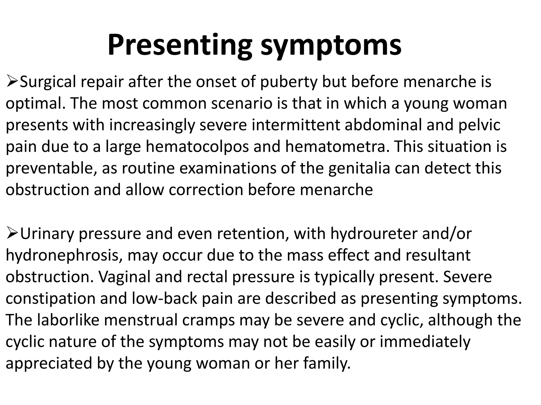
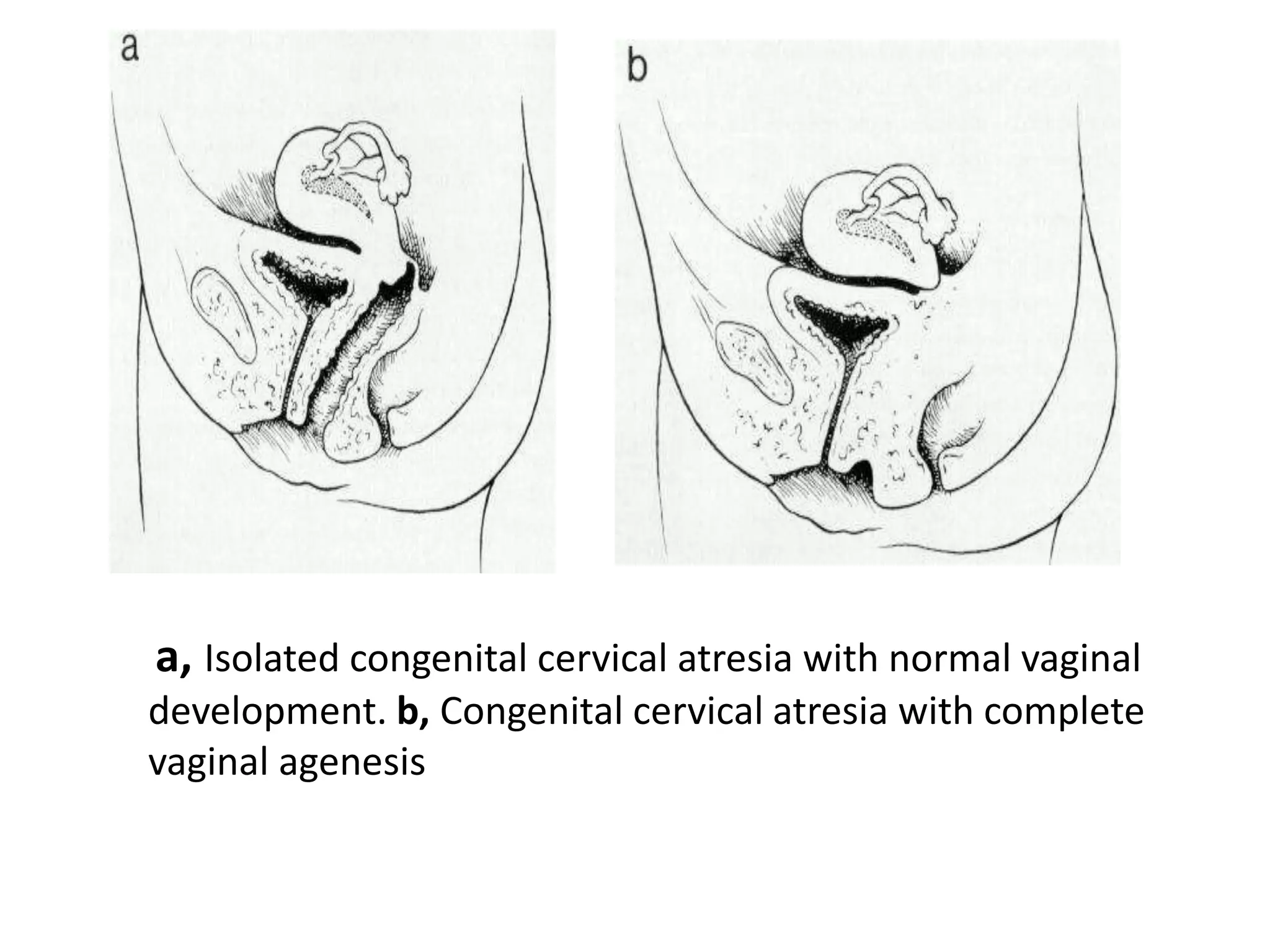
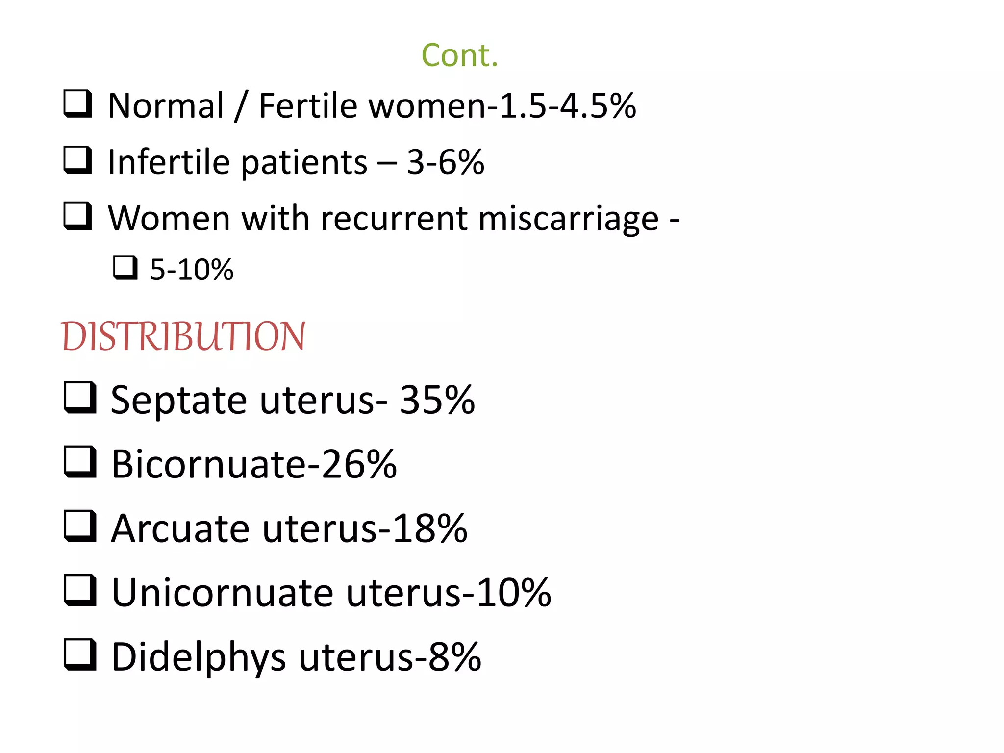
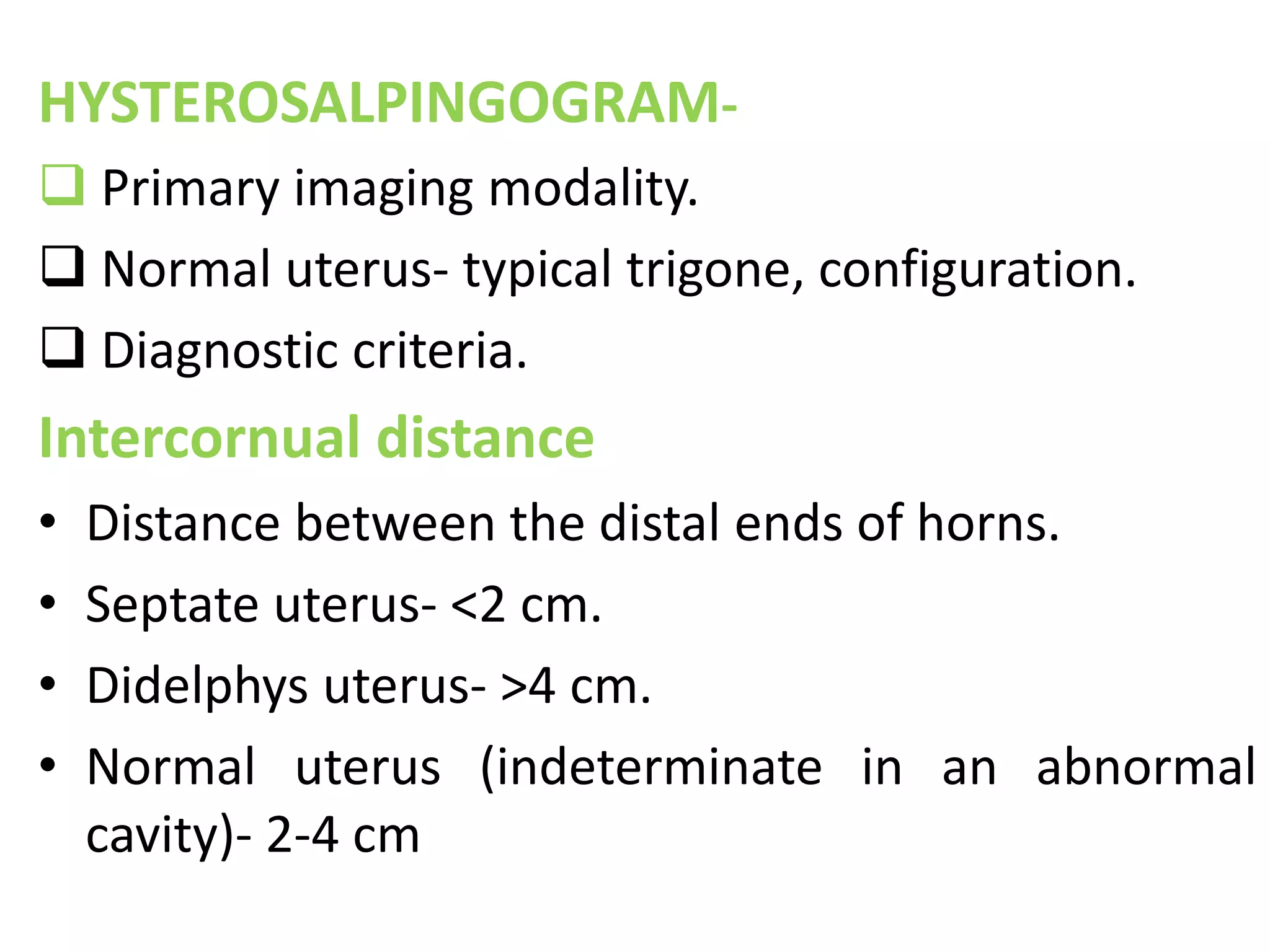
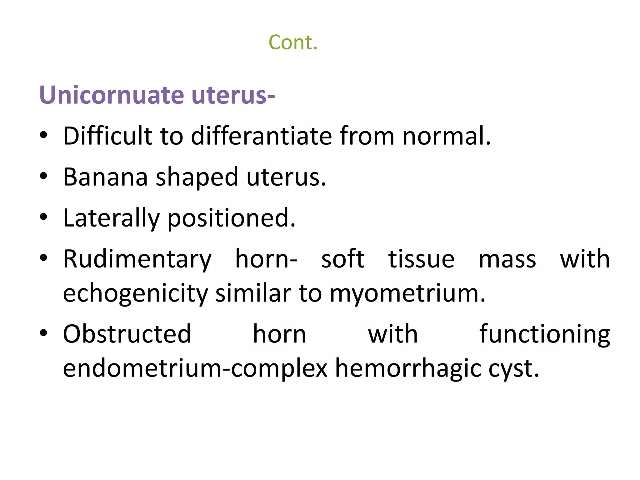
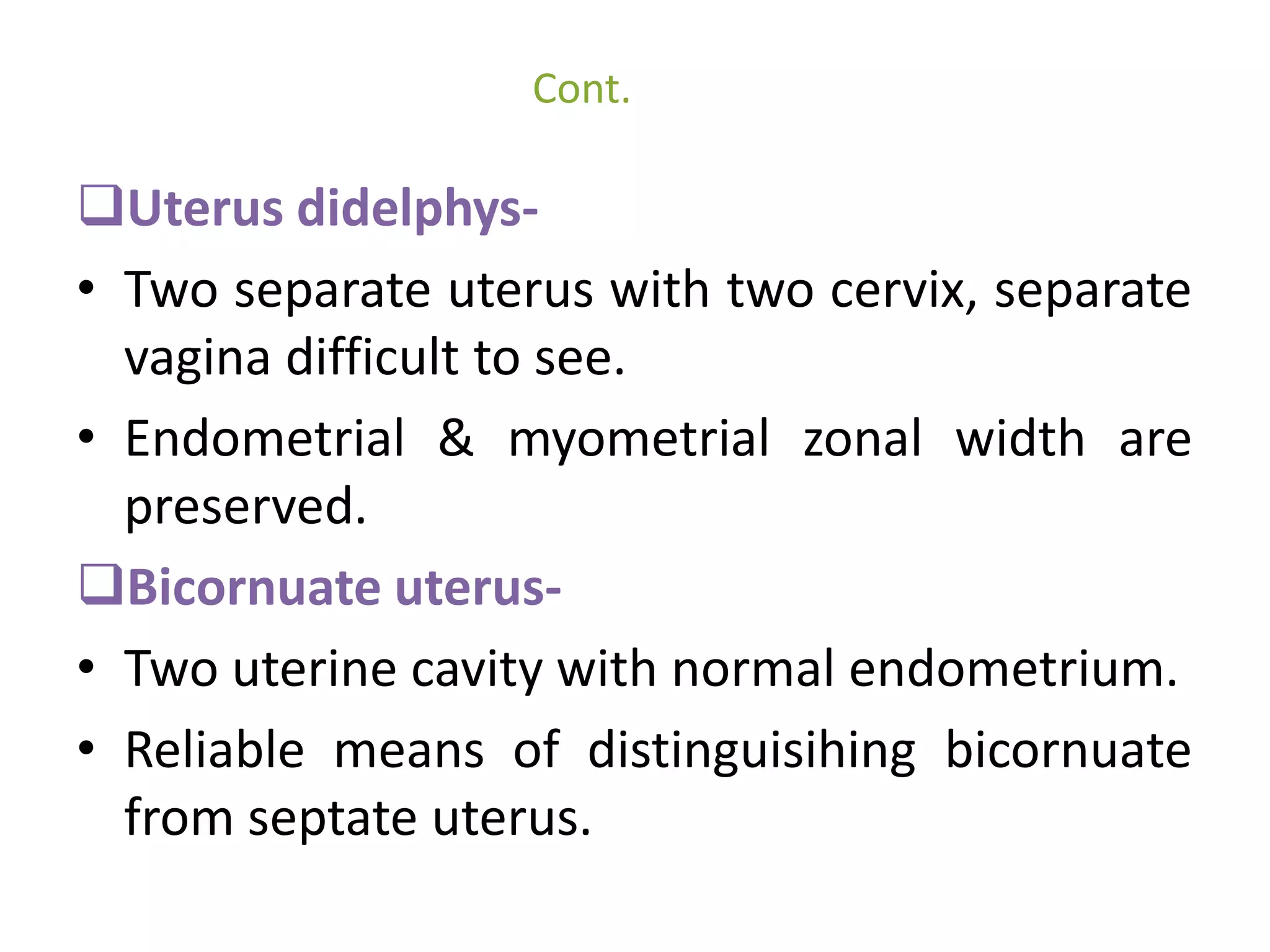
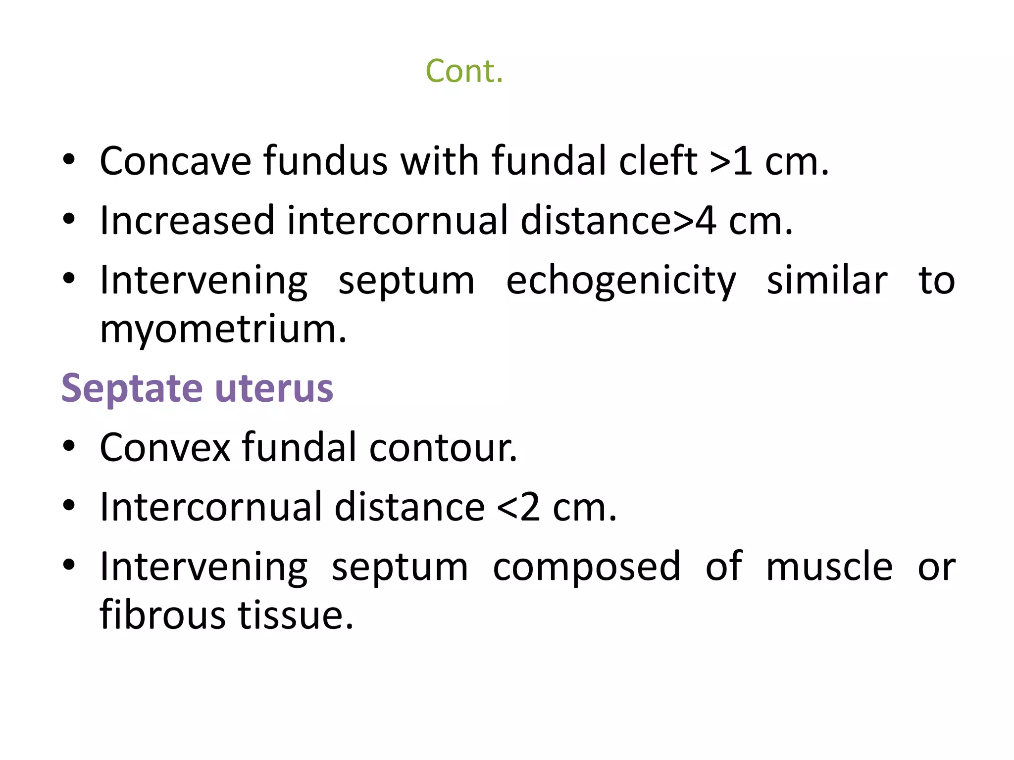
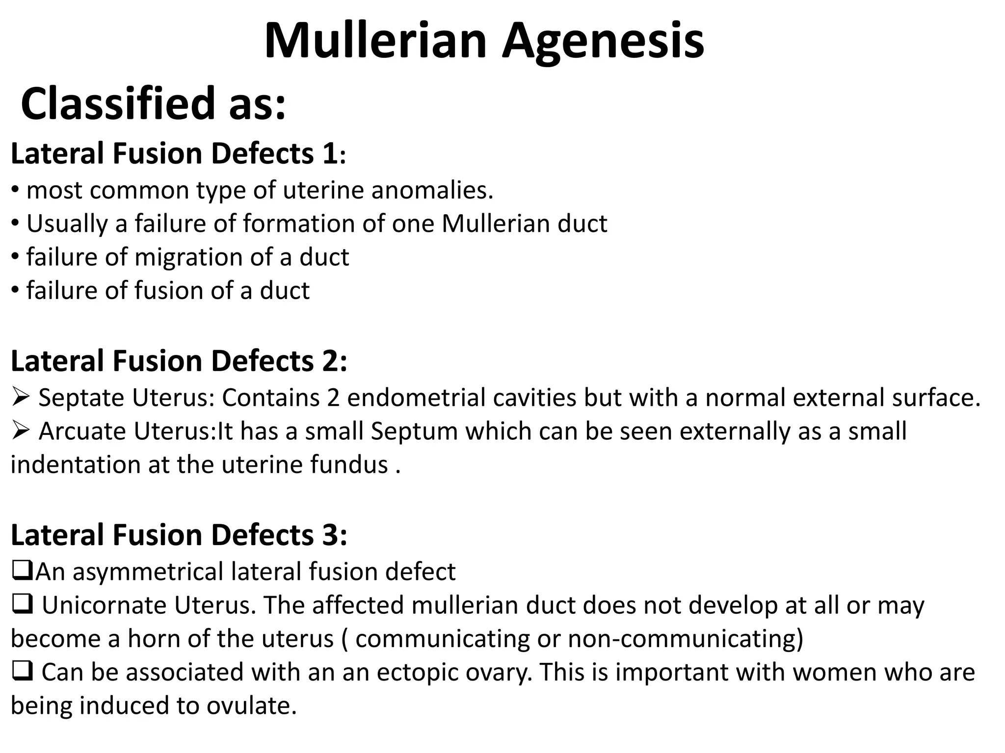
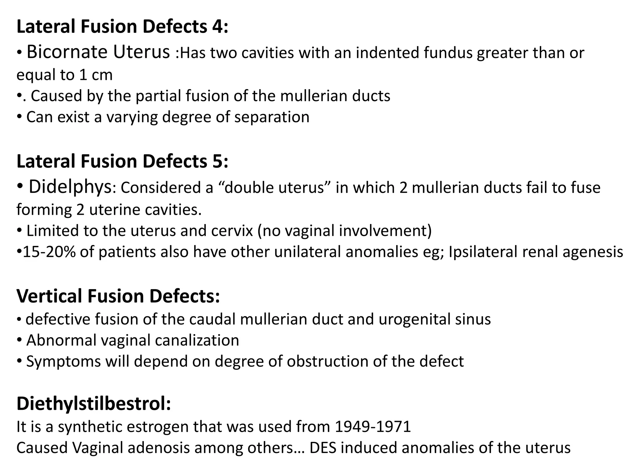
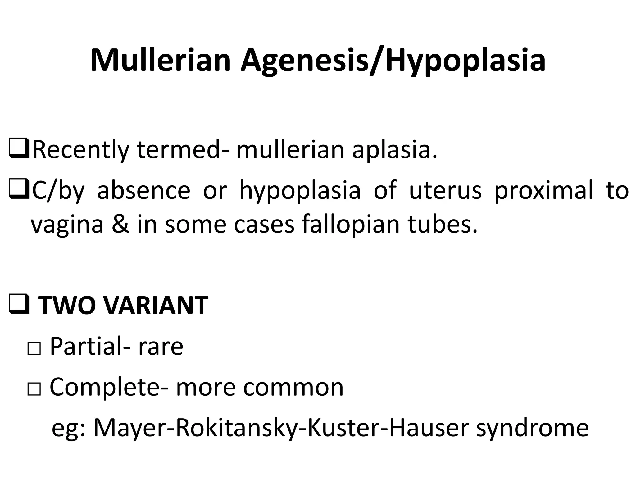
Mullerian duct anomalies occur due to abnormal development of the paired mullerian ducts in females during embryological development. The three main phases of mullerian duct development are organogenesis, fusion, and septal resorption. When one or more of these phases are disrupted, it can lead to mullerian duct anomalies such as a bicornuate or septate uterus. Mullerian duct anomalies are diagnosed using imaging modalities like ultrasound, MRI, and hysterosalpingography which allow visualization of the uterine cavity and identification of the specific anomaly present. The most common anomalies include septate uterus, bicornuate uterus, and arcuate uterus.


![Imperforate hymen
Imperforate hymen is embryologically not of
mullerian origin although clinically have a similar
presentation
Imperforate:[8][9] hymenal opening nonexistent;
will require minor surgery if it has not corrected
itself by puberty to allow menstrual fluids to
escape.
Cribriform, or microperforate: sometimes
confused for imperforate, the hymenal opening
appears to be nonexistent, but has, under close
examination, small openings.
Septate: the hymenal opening has one or more
bands of tissue extending across the opening](https://image.slidesharecdn.com/mulleriananomalies-141019023155-conversion-gate02/75/Mullerian-anomalies-80-2048.jpg)