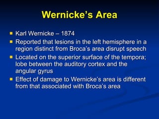
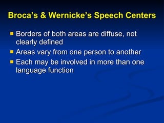
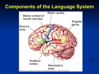
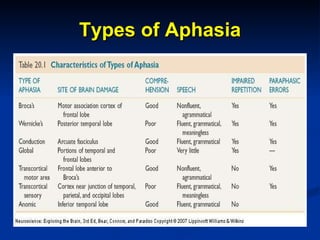
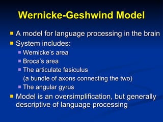
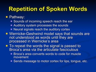
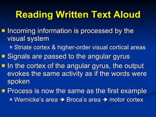
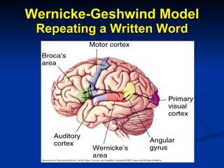
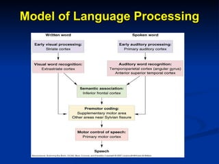
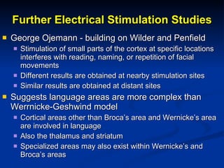
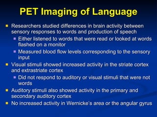
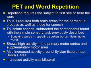
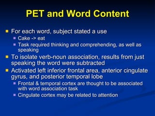
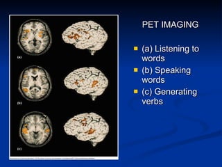
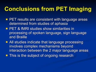
Language & the Brain
What is Language? A system by which sounds, symbols, and gestures are used for communication Sensory/Motor Convergence Language enters the brain through sensory inputs (vision, hearing – touch?) Produces motor responses    speech, writing Processing between sensory and motor systems is the essence of language
For Discussion Is language universal in human society? Is language universal among animals? Can animals be taught human language? How we define language effects our answers     Language = communication Language (human) is a complex system that employs predictable rules (grammar)
Animals & Language Many animals use specific vocalizations to communicate    claim territory, warn of danger, attract a mate Communication is limited, specific, stereotypical Human language is a creative process The possibilities are effectively endless, limited only by the rules of grammar
Animals & Language Nim Chimpsky Washoe Chimps raised in human families; taught American sign language Koko A lowland gorilla, learned more than 1000 words in American sign language and understood at least 2000 Demonstrated complex grammar use Can animals “learn” human language, or rules of grammar?
Koko
Historical Perspective Greek & Roman Empires Thought that the  tongue controlled speech Sixteenth century:  Discovered that speech impairment did not reflect tongue paralysis Johann Gesner – 1770 Inability to associate images or abstract ideas with their expressive verbal symbols Attributed to brain damage from disease Cognitive ability may be intact but verbal expression is lost The first steps toward modern understanding of aphasia
Historical Perspective Franz Joseph Gall & phrenology Speech loss while other mental faculties are retained suggests that there is a specific brain area devoted to speech Jean Baptiste Bouillard – 1825 Speech is controlled by the frontal lobes Simon Alexandre Aubertin – 1861 Described a man who had shot away his frontal bone in a failed suicide attempt Pressing a spatula against the exposed brain tissue halted speech, which resumed immediately when the pressure was released
Broca’s Area Paul Broca – 1861 With Aubertin, examined the brain of a man unable to speak Found lesion in the frontal lobes  Published a study of 8 similar patients in 1863 Showed that language expression is controlled by a portion of the frontal lobe in one hemisphere, usually the left First clear demonstration that brain function can be localized Now called Broca’s area
Broca’s Area
The Wada Procedure A simple procedure to study the function of a single cerebral hemisphere Fast acting barbiturate is injected into the carotid artery on one side Preferentially carried to the ipsilateral hemisphere Acts as a short-term anesthetic Limbs on the contralateral side become paralyzed Can then assess patients’ ability to speak If the injection is on the side dominant for speech, patient is completely unable to talk Opposite side retains complete speech ability
Handedness Handedness is an obvious functional asymmetry 90% of people are right handed Left hemisphere specialized for fine motor control Other species show about equal numbers of left and right handers The left hemisphere is dominant in speech in 96% of right handed people and 70% of left-handed people  Thus the left hemisphere is dominant for language in 93% of people Bilateral representation of speech occurs only in left-handers
Wernicke’s Area Karl Wernicke – 1874 Reported that lesions in the left hemisphere in a region distinct from Broca’s area disrupt speech Located on the superior surface of the tempora; lobe between the auditory cortex and the angular gyrus Effect of damage to Wernicke’s area is different from that associated with Broca’s area
Broca’s & Wernicke’s Speech Centers Borders of both areas are diffuse, not clearly defined Areas vary from one person to another Each may be involved in more than one language function
Components of the Language System
Aphasia Partial or complete loss of language function following brain damage Much of what we know about language and the brain comes from studying patients with specific deficits The occurrence of distinct types of aphasia suggests that language is processed in several stages at several locations in the brain.
Types of Aphasia
Broca’s Aphasia Also known as motor or nonfluent aphasia Person has difficulty speaking, although they can understand both spoken and written language People with Broca’s aphasia often pause, searching for the right word Anomia – inability to find the right word Telegraphic speech using only content words  agrammatism use nouns verbs & adjectives omit function words – articles, pronouns conjunctions
Wernicke’s Aphasia Clearly different from Broca’s aphasia Speech is fluent but comprehension is poor Content does not make sense Mixtures of clarity & gibberish More paraphasic errors Substitution of incorrect sounds, sound-alike words Seem undisturbed by their own speech Comprehension, such as following directions, is lost Written language, music are similarly affected
Implication’s of Wernicke’s Aphasia Wernicke’s area is located on the superior temporal gyrus near the primary auditory cortex May play a critical role in relating incoming sounds to their meaning Stores memories of the sounds that make up words
Wernicke-Geshwind Model A model for language processing in the brain System includes: Wernicke’s area  Broca’s area The articulate fasiculus  (a bundle of axons connecting the two) The angular gyrus Model is an oversimplification, but generally descriptive of language processing
Repetition of Spoken Words Pathway: Sounds of incoming speech reach the ear Auditory system processes the sounds Neural signals reach the auditory cortex Wernicke-Geshwind model says that sounds are not understood as words until they are processed in Wernicke’s area To repeat the words the signal is passed to Broca’s area via the articulate fasciculous Broca’s area converts words to code for muscle movement Sends message to motor cortex for lips, tongue, etc.
Wernicke-Geshwind Model Repeating a Spoken Word
Reading Written Text Aloud Incoming information is processed by the visual system Striate cortex & higher-order visual cortical areas Signals are passed to the angular gyrus In the cortex of the angular gyrus, the output evokes the same activity as if the words were spoken Process is now the same as the first example Wernicke’s area    Broca’s area    motor cortex
Wernicke-Geshwind Model Repeating a Written Word
Conduction Aphasia A lesion disconnects Wernicke’s area from Broca’s area, but leaves both intact Usually involves damage to the parietal cortex and the arcuate fasciculus Comprehension is good and speech is fluent Difficulty is in repeating words Greatest difficulty with function words, nonsense sounds Comprehends sentences they read aloud, though spoken words contain errors
Aphasia in Bilinguals If an individual is bilingual, does a stroke produce aphasia for both languages? Depends on order learned and relative fluency If languages were learned to same level of fluency at about the same time, both are equally affected Language is better preserved in the language learned more fluently and/or earlier in life If learned at different times, one will be affected more than the other Suggests that a second language may use a different population of neurons
Aphasia in the Deaf Left hemispheric lesions cause a language deficit in deaf subjects who use American Sign Language, similar to verbal aphasics Conditions analogous to both Broca’s and Wernicke’s aphasia exist Suggests universality of language processing in the brain
Model of Language Processing
Split Brain Studies Corpus callosum is severed to treat seizures Hemispheres may retain some communication via the brain stem and smaller commisures No significant impact observed in animal studies Humans are different Asymmetry in ability to verbalize answers to questions perceived by the different hemispheres Demonstrated by presenting visual stimuli to one hemisphere
Visual Stimulation of One Hemisphere
Asymmetrical Visual Stimulation   Only the right hemisphere sees objects to the left of the point of fixation and vice versa True as long as the eyes can’t move to bring images together in the fovea Pictures or words are flashed for a fraction of a second Shorter than time needed to move the eyes Numbers, words, & pictures presented to the right visual field are repeated/described easily; on the left they cannot be described Objects manipulated with the right hand can be described; objects in the left hand cannot
Language on the Right Side of the Brain
Functional Asymmetry Deficit is speech specific, not language specific Can read and understand letters and numbers with the right hemisphere if the response is nonverbal. Patient “says” he sees nothing - but successfully picks matching card Some patients can write with the right hemisphere Right brain can understand complex pictures Right brain can be shown to dominate certain tasks such as perspective and complex puzzles The 2 hemispheres can function as independent brains that have different language abilities
Anatomical Asymmetry If function differs, does structure differ between the hemispheres? Most significant difference is in the  planum temporale  on the superior surface of the temporal lobe Larger on the left side than on the right in about 65% of brains In some cases 5x larger Larger even in prenatal development, so not the result of use Relationship to language asymmetry  uncertain
Asymmetry of the Sylvian Fissure In most right-handed people, Sylvian fissure is longer on the left hemisphere than on the right Runs at a more shallow angle
Brain Stimulation and Language Wilder & Penfield Electrical stimulation at different cortical sites without general anesthesia (patients are conscious) Stimulation of motor cortex in area that controls mouth and lips caused speech arrest Occurred with stimulation on either side of the brain Stimulation of Broca’s area (left hemisphere only) Speech stopped, or with weaker stimulation, was hesitant Mild transient anomia Stimulation of 2 other sites also affected speech In the area of Wernicke’s area and the arcuate fasciculus
Further Electrical Stimulation Studies George Ojemann - building on Wilder and Penfield Stimulation of small parts of the cortex at specific locations interferes with reading, naming, or repetition of facial movements Different results are obtained at nearby stimulation sites Similar results are obtained at distant sites Suggests language areas are more complex than Werrnicke-Geshwind model Cortical areas other than Broca’s area and Wernicke’s area are involved in language Also the thalamus and striatum Specialized areas may also exist within Wernicke’s and Broca’s areas
PET Imaging of Language Researchers studied differences in brain activity between sensory responses to words and production of speech Either listened to words that were read or looked at words flashed on a monitor Measured blood flow levels corresponding to the sensory input Visual stimuli showed increased activity in the striate cortex and extrastriate cortex Did not respond to auditory or visual stimuli that were not words Auditory stimuli also showed activity in the primary and secondary auditory cortex No increased activity in Wernicke’s area or the angular gyrus
PET and Word Repetition Repetition requires the subject to first see or hear the word Thus it requires both brain areas for the perceptual process as well as those for speech To isolate speech, subtracted the components found with the simple sensory task previously described Speaking words = repeating spoken words - listening to words Shows high activity in the primary motor cortex and supplementary motor area Also increased activity around Sylvian fissure near Broca’s area Increased activity was bilateral
PET and Word Content For each word, subject stated a use Cake -> eat Task required thinking and comprehending, as well as speaking To isolate verb-noun association, results from just speaking the word were subtracted Activated left inferior frontal area, anterior cingulate gyrus, and posterior temporal lobe Frontal & temporal cortex are thought to be associated with word association task Cingulate cortex may be related to attention
PET IMAGING (a) Listening to words (b) Speaking words (c) Generating verbs
Conclusions from PET Imaging PET results are consistent with language areas determined from studies of aphasia PET & fMRI studies show similarities in processing of spoken language, sign language and Braille All studies indicate that language processing involves complex mechanisms beyond interaction between the 2 major language areas This is the subject of ongoing research

Language

  • 1.
  • 2.
    What is Language?A system by which sounds, symbols, and gestures are used for communication Sensory/Motor Convergence Language enters the brain through sensory inputs (vision, hearing – touch?) Produces motor responses  speech, writing Processing between sensory and motor systems is the essence of language
  • 3.
    For Discussion Islanguage universal in human society? Is language universal among animals? Can animals be taught human language? How we define language effects our answers  Language = communication Language (human) is a complex system that employs predictable rules (grammar)
  • 4.
    Animals & LanguageMany animals use specific vocalizations to communicate  claim territory, warn of danger, attract a mate Communication is limited, specific, stereotypical Human language is a creative process The possibilities are effectively endless, limited only by the rules of grammar
  • 5.
    Animals & LanguageNim Chimpsky Washoe Chimps raised in human families; taught American sign language Koko A lowland gorilla, learned more than 1000 words in American sign language and understood at least 2000 Demonstrated complex grammar use Can animals “learn” human language, or rules of grammar?
  • 6.
  • 7.
    Historical Perspective Greek& Roman Empires Thought that the tongue controlled speech Sixteenth century: Discovered that speech impairment did not reflect tongue paralysis Johann Gesner – 1770 Inability to associate images or abstract ideas with their expressive verbal symbols Attributed to brain damage from disease Cognitive ability may be intact but verbal expression is lost The first steps toward modern understanding of aphasia
  • 8.
    Historical Perspective FranzJoseph Gall & phrenology Speech loss while other mental faculties are retained suggests that there is a specific brain area devoted to speech Jean Baptiste Bouillard – 1825 Speech is controlled by the frontal lobes Simon Alexandre Aubertin – 1861 Described a man who had shot away his frontal bone in a failed suicide attempt Pressing a spatula against the exposed brain tissue halted speech, which resumed immediately when the pressure was released
  • 9.
    Broca’s Area PaulBroca – 1861 With Aubertin, examined the brain of a man unable to speak Found lesion in the frontal lobes Published a study of 8 similar patients in 1863 Showed that language expression is controlled by a portion of the frontal lobe in one hemisphere, usually the left First clear demonstration that brain function can be localized Now called Broca’s area
  • 10.
  • 11.
    The Wada ProcedureA simple procedure to study the function of a single cerebral hemisphere Fast acting barbiturate is injected into the carotid artery on one side Preferentially carried to the ipsilateral hemisphere Acts as a short-term anesthetic Limbs on the contralateral side become paralyzed Can then assess patients’ ability to speak If the injection is on the side dominant for speech, patient is completely unable to talk Opposite side retains complete speech ability
  • 12.
    Handedness Handedness isan obvious functional asymmetry 90% of people are right handed Left hemisphere specialized for fine motor control Other species show about equal numbers of left and right handers The left hemisphere is dominant in speech in 96% of right handed people and 70% of left-handed people Thus the left hemisphere is dominant for language in 93% of people Bilateral representation of speech occurs only in left-handers
  • 13.
    Wernicke’s Area KarlWernicke – 1874 Reported that lesions in the left hemisphere in a region distinct from Broca’s area disrupt speech Located on the superior surface of the tempora; lobe between the auditory cortex and the angular gyrus Effect of damage to Wernicke’s area is different from that associated with Broca’s area
  • 14.
    Broca’s & Wernicke’sSpeech Centers Borders of both areas are diffuse, not clearly defined Areas vary from one person to another Each may be involved in more than one language function
  • 15.
    Components of theLanguage System
  • 16.
    Aphasia Partial orcomplete loss of language function following brain damage Much of what we know about language and the brain comes from studying patients with specific deficits The occurrence of distinct types of aphasia suggests that language is processed in several stages at several locations in the brain.
  • 17.
  • 18.
    Broca’s Aphasia Alsoknown as motor or nonfluent aphasia Person has difficulty speaking, although they can understand both spoken and written language People with Broca’s aphasia often pause, searching for the right word Anomia – inability to find the right word Telegraphic speech using only content words agrammatism use nouns verbs & adjectives omit function words – articles, pronouns conjunctions
  • 19.
    Wernicke’s Aphasia Clearlydifferent from Broca’s aphasia Speech is fluent but comprehension is poor Content does not make sense Mixtures of clarity & gibberish More paraphasic errors Substitution of incorrect sounds, sound-alike words Seem undisturbed by their own speech Comprehension, such as following directions, is lost Written language, music are similarly affected
  • 20.
    Implication’s of Wernicke’sAphasia Wernicke’s area is located on the superior temporal gyrus near the primary auditory cortex May play a critical role in relating incoming sounds to their meaning Stores memories of the sounds that make up words
  • 21.
    Wernicke-Geshwind Model Amodel for language processing in the brain System includes: Wernicke’s area Broca’s area The articulate fasiculus (a bundle of axons connecting the two) The angular gyrus Model is an oversimplification, but generally descriptive of language processing
  • 22.
    Repetition of SpokenWords Pathway: Sounds of incoming speech reach the ear Auditory system processes the sounds Neural signals reach the auditory cortex Wernicke-Geshwind model says that sounds are not understood as words until they are processed in Wernicke’s area To repeat the words the signal is passed to Broca’s area via the articulate fasciculous Broca’s area converts words to code for muscle movement Sends message to motor cortex for lips, tongue, etc.
  • 23.
  • 24.
    Reading Written TextAloud Incoming information is processed by the visual system Striate cortex & higher-order visual cortical areas Signals are passed to the angular gyrus In the cortex of the angular gyrus, the output evokes the same activity as if the words were spoken Process is now the same as the first example Wernicke’s area  Broca’s area  motor cortex
  • 25.
  • 26.
    Conduction Aphasia Alesion disconnects Wernicke’s area from Broca’s area, but leaves both intact Usually involves damage to the parietal cortex and the arcuate fasciculus Comprehension is good and speech is fluent Difficulty is in repeating words Greatest difficulty with function words, nonsense sounds Comprehends sentences they read aloud, though spoken words contain errors
  • 27.
    Aphasia in BilingualsIf an individual is bilingual, does a stroke produce aphasia for both languages? Depends on order learned and relative fluency If languages were learned to same level of fluency at about the same time, both are equally affected Language is better preserved in the language learned more fluently and/or earlier in life If learned at different times, one will be affected more than the other Suggests that a second language may use a different population of neurons
  • 28.
    Aphasia in theDeaf Left hemispheric lesions cause a language deficit in deaf subjects who use American Sign Language, similar to verbal aphasics Conditions analogous to both Broca’s and Wernicke’s aphasia exist Suggests universality of language processing in the brain
  • 29.
  • 30.
    Split Brain StudiesCorpus callosum is severed to treat seizures Hemispheres may retain some communication via the brain stem and smaller commisures No significant impact observed in animal studies Humans are different Asymmetry in ability to verbalize answers to questions perceived by the different hemispheres Demonstrated by presenting visual stimuli to one hemisphere
  • 31.
    Visual Stimulation ofOne Hemisphere
  • 32.
    Asymmetrical Visual Stimulation Only the right hemisphere sees objects to the left of the point of fixation and vice versa True as long as the eyes can’t move to bring images together in the fovea Pictures or words are flashed for a fraction of a second Shorter than time needed to move the eyes Numbers, words, & pictures presented to the right visual field are repeated/described easily; on the left they cannot be described Objects manipulated with the right hand can be described; objects in the left hand cannot
  • 33.
    Language on theRight Side of the Brain
  • 34.
    Functional Asymmetry Deficitis speech specific, not language specific Can read and understand letters and numbers with the right hemisphere if the response is nonverbal. Patient “says” he sees nothing - but successfully picks matching card Some patients can write with the right hemisphere Right brain can understand complex pictures Right brain can be shown to dominate certain tasks such as perspective and complex puzzles The 2 hemispheres can function as independent brains that have different language abilities
  • 35.
    Anatomical Asymmetry Iffunction differs, does structure differ between the hemispheres? Most significant difference is in the planum temporale on the superior surface of the temporal lobe Larger on the left side than on the right in about 65% of brains In some cases 5x larger Larger even in prenatal development, so not the result of use Relationship to language asymmetry uncertain
  • 36.
    Asymmetry of theSylvian Fissure In most right-handed people, Sylvian fissure is longer on the left hemisphere than on the right Runs at a more shallow angle
  • 37.
    Brain Stimulation andLanguage Wilder & Penfield Electrical stimulation at different cortical sites without general anesthesia (patients are conscious) Stimulation of motor cortex in area that controls mouth and lips caused speech arrest Occurred with stimulation on either side of the brain Stimulation of Broca’s area (left hemisphere only) Speech stopped, or with weaker stimulation, was hesitant Mild transient anomia Stimulation of 2 other sites also affected speech In the area of Wernicke’s area and the arcuate fasciculus
  • 38.
    Further Electrical StimulationStudies George Ojemann - building on Wilder and Penfield Stimulation of small parts of the cortex at specific locations interferes with reading, naming, or repetition of facial movements Different results are obtained at nearby stimulation sites Similar results are obtained at distant sites Suggests language areas are more complex than Werrnicke-Geshwind model Cortical areas other than Broca’s area and Wernicke’s area are involved in language Also the thalamus and striatum Specialized areas may also exist within Wernicke’s and Broca’s areas
  • 39.
    PET Imaging ofLanguage Researchers studied differences in brain activity between sensory responses to words and production of speech Either listened to words that were read or looked at words flashed on a monitor Measured blood flow levels corresponding to the sensory input Visual stimuli showed increased activity in the striate cortex and extrastriate cortex Did not respond to auditory or visual stimuli that were not words Auditory stimuli also showed activity in the primary and secondary auditory cortex No increased activity in Wernicke’s area or the angular gyrus
  • 40.
    PET and WordRepetition Repetition requires the subject to first see or hear the word Thus it requires both brain areas for the perceptual process as well as those for speech To isolate speech, subtracted the components found with the simple sensory task previously described Speaking words = repeating spoken words - listening to words Shows high activity in the primary motor cortex and supplementary motor area Also increased activity around Sylvian fissure near Broca’s area Increased activity was bilateral
  • 41.
    PET and WordContent For each word, subject stated a use Cake -> eat Task required thinking and comprehending, as well as speaking To isolate verb-noun association, results from just speaking the word were subtracted Activated left inferior frontal area, anterior cingulate gyrus, and posterior temporal lobe Frontal & temporal cortex are thought to be associated with word association task Cingulate cortex may be related to attention
  • 42.
    PET IMAGING (a)Listening to words (b) Speaking words (c) Generating verbs
  • 43.
    Conclusions from PETImaging PET results are consistent with language areas determined from studies of aphasia PET & fMRI studies show similarities in processing of spoken language, sign language and Braille All studies indicate that language processing involves complex mechanisms beyond interaction between the 2 major language areas This is the subject of ongoing research