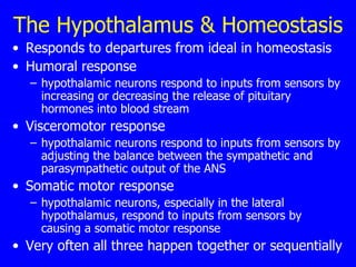
Motivation and Homeostasis The Role of the Hypothalamus
Motivation  Motivation is the driving force in behavior  The stronger the motivation, the more likely the behavior  Control of behavior involves decisions among different motivated actions with different outcomes  Motivation for behaviors basic to survival is controlled by the hypothalamus
The Hypothalamus & Homeostasis Responds to departures from ideal in homeostasis  Humoral response  hypothalamic neurons respond to inputs from sensors by increasing or decreasing the release of pituitary hormones into blood stream  Visceromotor response  hypothalamic neurons respond to inputs from sensors by adjusting the balance between the sympathetic and parasympathetic output of the ANS  Somatic motor response  hypothalamic neurons, especially in the lateral hypothalamus, respond to inputs from sensors by causing a somatic motor response  Very often all three happen together or sequentially
Regulation of Feeding Behavior   Energy balance regulates feeding Hormonal and hypothalamic regulation of body fat and feeding
Feeding & Energy Balance   Prandial state  after eating a meal  Postabsorptive state  fasting times between meals  Obesity occurs when there is consistently more intake than usage and adipose cells enlarge and increase in number  Starvation occurs when there is consistently less intake than usage and loss of fat tissue occurs  In virtually all mammals except humans and their pets, homeostatic mechanisms act to avoid both obesity and starvation
The Prandial State Occurs after eating a meal  Glucose, fatty acids, and ketones are produced for all cells; neurons only use glucose  Excess energy is stored, either as glycogen in the liver and skeletal muscles, or as triglycerides in adipose tissue = fat cells  Anabolism is the assembly of glycogen and triglycerides from what you eat  Because the storage capacity for glycogen is limited, excess energy is stored as fat
The Postabsorptive State  Fasting times between meals  Energy is produced from glycogen and triglycerides  Catabolism is the process of breaking down glycogen and triglycerides and making glucose, fatty acids, and ketones
Hormonal Regulation of Feeding   Lipostatic  hypothesis  The brain monitors the amount of body fat and acts to keep it constant  Basically true  Adipocytes release leptin into blood  Neurons in the periventricular zone of hypothalamus detect a drop in leptin levels  Neurons in the lateral hypothalamus are then excited and drive feeding behavior
Elevated Leptin & Feeding From 'holiday feasting' for example Leptin receptors on neurons in the arcuate nucleus are activated  These neurons use two peptide transmitters: aMSH and CART  Response is three-part, triggered by nerve fibers leaving the arcuate nucleus
The Response Humoral response is through the paraventricular nucleus  Leads to increased release of TSH and ACTH both of which serve to raise the metabolic rate  Visceromotor response is to activate the sympathetic division of ANS  increases the metabolic rate, partly by raising body temperature.  occurs both directly because of activity of arcuate nucleus outputs and through the paraventricular nucleus outputs to the spinal cord  Somatic motor response is to inhibit feeding behavior through the neurons of the lateral hypothalamic area
Lowered Leptin & Feeding Turns off arcuate nucleus neurons that use aMSH and CART as transmitters  Turns on other arcuate nucleus neurons that use NPY and AgRP as transmitters  These neurons have connections with the paraventricular nucleus and the lateral hypothalamic area  Inhibit secretion of TSH and ACTH  Activate the parasympathetic division of ANS  Stimulate feeding behavior
Key Neurotransmitters aMSH and CART  depress feeding behavior  therefore called  anorectic  peptides  NPY and AgRP  stimulate feeding behavior  therefore called  orexigenic  peptides
Role of the Lateral Hypothalamus Some neurons in the lateral hypothalamic area receive direct input from the arcuate nucleus neurons These use the peptide transmitter, MCH  MCH neurons innervate most of the cerebral cortex  Other neurons in the lateral hypothalamic area also receive direct input from the arcuate nucleus neurons  Use the peptide transmitter, orexin.  Orexin neurons also innervate most of the cerebral cortex  So, when leptin levels fall, there is a major system to drive feeding behavior!
Regulation of Feeding Behavior Starting a meal:  Cephalic phase  sight and smell of food activates the parasympathetic and enteric divisions of the ANS saliva is secreted in the mouth and digestive juices, in the stomach  Gastric phase  the cephalic phase responses are intensified by chewing, swallowing, and filling the stomach with food  Intestinal phase (or substrate phase)  As the filling stomach begins to empty in to the intestines, satiety signals arise to stop feeding
Satiety Ending a meal Gastric distention  mechanoreceptors in the stomach wall connect to the nucleus of the solitary tract via the vagus nerve and inhibit eating  The gustatory nucleus is part of the nucleus of the solitary tract, which is why great food can override signals from a full stomach and lead to a totally bloated stomach
Cholecystokinin (CCK) Released from neurons in the enteric nervous system and other cells lining the intestines  Triggered in response to stimulation of intestines by fatty foods  Acts on sensory receptors connected to the vagus nerve and signals to stop eating
Insulin Pancreatic cells produce insulin necessary to transport glucose into all cells except neurons  Directly affects neurons in arcuate nucleus and ventromedial nucleus of the hypothalamus  similar to leptin  Late in the eating stage, rising insulin is a satiety signal
The Role of Dopamine Liking  food and  wanting  food are mediated by two different systems  Wanting  food is driven by the mesocorticolimbic dopamine system  Animals with lesions in this system still have the hedonic experience (like food), but seem unmotivated to want it  Cravings that lead to addiction involve the same dopamine system
Drinking Water, not alcohol Two different drives for drinking  use two different mechanisms  Decrease in blood volume drives drinking  Increase in the concentration of solutes in the blood, blood osmolarity, drives drinking
Volumetric Thirst Decreased blood volume drives magnocellular neurons in the hypothalamus  release ADH  leads to concentrated urine  humoral response  Decreased blood volume stimulates the sympathetic division of the ANS helps to correct low blood pressure visceromotor response  Decreased blood volume stimulates neurons in the lateral hypothalamus  drives seeking and consuming water  somatic motor response
Osmometric Thirst Hypertonicity of the blood is sensed by vascular organ of the lamina terminalis (OVLT)  outside the blood brain barrier just like the subfornical organ  OVLT cells when excited, directly drive magnocellular neurons to release ADH  humoral response  Hypertonicity drives the OVLT to stimulate the motivation to drink water through the lateral hypothalamic area  somatic motor response
Diabetes Insipidus One type of diabetes includes failure to release ADH  Caused by loss of magnocellular neurosecretory neurons in hypothalamus, or viral infection of hypothalamus  ADH is not secreted, urine is very dilute  Osmometric thirst drives patients to drink vast amounts of water  Vast amounts of urine are passed  Patients become sorely sleep deprived - dangerous  Synthetic ADH can be administered nasally
Temperature Regulation In addition to the somatosensory thermal receptors, there are lots of neurons in the brain sensitive to temperature.  Most important cluster is in the anterior hypothalamus monitors blood temperature  Humoral and visceromotor responses are then initiated by the medial preoptic area of the hypothalamus  Somatic motor responses are initiated by the lateral area of the hypothalamus
Decreasing Temperature A fall in temperature in the blood leads to: Humoral response  TSH is released by the anterior pituitary causing: thyroxin release by the thyroid gland increases cellular metabolism and produces heat  Visceromotor response  constricted blood vessels near the skin surface and piloerection (goose bumps)  Somatic motor response  involuntary shivering  voluntary action to seek warmth
Increasing Temperature A rise in temperature in the blood leads to:  Reduction of TSH release humoral response  Blood is shunted toward surface capillary beds visceromotor response  Animal seeks shade  voluntary somatic motor response  Animal pants or humans sweat  involuntary somatic motor response

Motivation

  • 1.
    Motivation and HomeostasisThe Role of the Hypothalamus
  • 2.
    Motivation Motivationis the driving force in behavior The stronger the motivation, the more likely the behavior Control of behavior involves decisions among different motivated actions with different outcomes Motivation for behaviors basic to survival is controlled by the hypothalamus
  • 3.
    The Hypothalamus &Homeostasis Responds to departures from ideal in homeostasis Humoral response hypothalamic neurons respond to inputs from sensors by increasing or decreasing the release of pituitary hormones into blood stream Visceromotor response hypothalamic neurons respond to inputs from sensors by adjusting the balance between the sympathetic and parasympathetic output of the ANS Somatic motor response hypothalamic neurons, especially in the lateral hypothalamus, respond to inputs from sensors by causing a somatic motor response Very often all three happen together or sequentially
  • 4.
    Regulation of FeedingBehavior Energy balance regulates feeding Hormonal and hypothalamic regulation of body fat and feeding
  • 5.
    Feeding & EnergyBalance Prandial state after eating a meal Postabsorptive state fasting times between meals Obesity occurs when there is consistently more intake than usage and adipose cells enlarge and increase in number Starvation occurs when there is consistently less intake than usage and loss of fat tissue occurs In virtually all mammals except humans and their pets, homeostatic mechanisms act to avoid both obesity and starvation
  • 6.
    The Prandial StateOccurs after eating a meal Glucose, fatty acids, and ketones are produced for all cells; neurons only use glucose Excess energy is stored, either as glycogen in the liver and skeletal muscles, or as triglycerides in adipose tissue = fat cells Anabolism is the assembly of glycogen and triglycerides from what you eat Because the storage capacity for glycogen is limited, excess energy is stored as fat
  • 7.
    The Postabsorptive State Fasting times between meals Energy is produced from glycogen and triglycerides Catabolism is the process of breaking down glycogen and triglycerides and making glucose, fatty acids, and ketones
  • 8.
    Hormonal Regulation ofFeeding Lipostatic hypothesis The brain monitors the amount of body fat and acts to keep it constant Basically true Adipocytes release leptin into blood Neurons in the periventricular zone of hypothalamus detect a drop in leptin levels Neurons in the lateral hypothalamus are then excited and drive feeding behavior
  • 9.
    Elevated Leptin &Feeding From 'holiday feasting' for example Leptin receptors on neurons in the arcuate nucleus are activated These neurons use two peptide transmitters: aMSH and CART Response is three-part, triggered by nerve fibers leaving the arcuate nucleus
  • 10.
    The Response Humoralresponse is through the paraventricular nucleus Leads to increased release of TSH and ACTH both of which serve to raise the metabolic rate Visceromotor response is to activate the sympathetic division of ANS increases the metabolic rate, partly by raising body temperature. occurs both directly because of activity of arcuate nucleus outputs and through the paraventricular nucleus outputs to the spinal cord Somatic motor response is to inhibit feeding behavior through the neurons of the lateral hypothalamic area
  • 11.
    Lowered Leptin &Feeding Turns off arcuate nucleus neurons that use aMSH and CART as transmitters Turns on other arcuate nucleus neurons that use NPY and AgRP as transmitters These neurons have connections with the paraventricular nucleus and the lateral hypothalamic area Inhibit secretion of TSH and ACTH Activate the parasympathetic division of ANS Stimulate feeding behavior
  • 12.
    Key Neurotransmitters aMSHand CART depress feeding behavior therefore called anorectic peptides NPY and AgRP stimulate feeding behavior therefore called orexigenic peptides
  • 13.
    Role of theLateral Hypothalamus Some neurons in the lateral hypothalamic area receive direct input from the arcuate nucleus neurons These use the peptide transmitter, MCH MCH neurons innervate most of the cerebral cortex Other neurons in the lateral hypothalamic area also receive direct input from the arcuate nucleus neurons Use the peptide transmitter, orexin. Orexin neurons also innervate most of the cerebral cortex So, when leptin levels fall, there is a major system to drive feeding behavior!
  • 14.
    Regulation of FeedingBehavior Starting a meal: Cephalic phase sight and smell of food activates the parasympathetic and enteric divisions of the ANS saliva is secreted in the mouth and digestive juices, in the stomach Gastric phase the cephalic phase responses are intensified by chewing, swallowing, and filling the stomach with food Intestinal phase (or substrate phase) As the filling stomach begins to empty in to the intestines, satiety signals arise to stop feeding
  • 15.
    Satiety Ending ameal Gastric distention mechanoreceptors in the stomach wall connect to the nucleus of the solitary tract via the vagus nerve and inhibit eating The gustatory nucleus is part of the nucleus of the solitary tract, which is why great food can override signals from a full stomach and lead to a totally bloated stomach
  • 16.
    Cholecystokinin (CCK) Releasedfrom neurons in the enteric nervous system and other cells lining the intestines Triggered in response to stimulation of intestines by fatty foods Acts on sensory receptors connected to the vagus nerve and signals to stop eating
  • 17.
    Insulin Pancreatic cellsproduce insulin necessary to transport glucose into all cells except neurons Directly affects neurons in arcuate nucleus and ventromedial nucleus of the hypothalamus similar to leptin Late in the eating stage, rising insulin is a satiety signal
  • 18.
    The Role ofDopamine Liking food and wanting food are mediated by two different systems Wanting food is driven by the mesocorticolimbic dopamine system Animals with lesions in this system still have the hedonic experience (like food), but seem unmotivated to want it Cravings that lead to addiction involve the same dopamine system
  • 19.
    Drinking Water, notalcohol Two different drives for drinking use two different mechanisms Decrease in blood volume drives drinking Increase in the concentration of solutes in the blood, blood osmolarity, drives drinking
  • 20.
    Volumetric Thirst Decreasedblood volume drives magnocellular neurons in the hypothalamus release ADH leads to concentrated urine humoral response Decreased blood volume stimulates the sympathetic division of the ANS helps to correct low blood pressure visceromotor response Decreased blood volume stimulates neurons in the lateral hypothalamus drives seeking and consuming water somatic motor response
  • 21.
    Osmometric Thirst Hypertonicityof the blood is sensed by vascular organ of the lamina terminalis (OVLT) outside the blood brain barrier just like the subfornical organ OVLT cells when excited, directly drive magnocellular neurons to release ADH humoral response Hypertonicity drives the OVLT to stimulate the motivation to drink water through the lateral hypothalamic area somatic motor response
  • 22.
    Diabetes Insipidus Onetype of diabetes includes failure to release ADH Caused by loss of magnocellular neurosecretory neurons in hypothalamus, or viral infection of hypothalamus ADH is not secreted, urine is very dilute Osmometric thirst drives patients to drink vast amounts of water Vast amounts of urine are passed Patients become sorely sleep deprived - dangerous Synthetic ADH can be administered nasally
  • 23.
    Temperature Regulation Inaddition to the somatosensory thermal receptors, there are lots of neurons in the brain sensitive to temperature. Most important cluster is in the anterior hypothalamus monitors blood temperature Humoral and visceromotor responses are then initiated by the medial preoptic area of the hypothalamus Somatic motor responses are initiated by the lateral area of the hypothalamus
  • 24.
    Decreasing Temperature Afall in temperature in the blood leads to: Humoral response TSH is released by the anterior pituitary causing: thyroxin release by the thyroid gland increases cellular metabolism and produces heat Visceromotor response constricted blood vessels near the skin surface and piloerection (goose bumps) Somatic motor response involuntary shivering voluntary action to seek warmth
  • 25.
    Increasing Temperature Arise in temperature in the blood leads to: Reduction of TSH release humoral response Blood is shunted toward surface capillary beds visceromotor response Animal seeks shade voluntary somatic motor response Animal pants or humans sweat involuntary somatic motor response