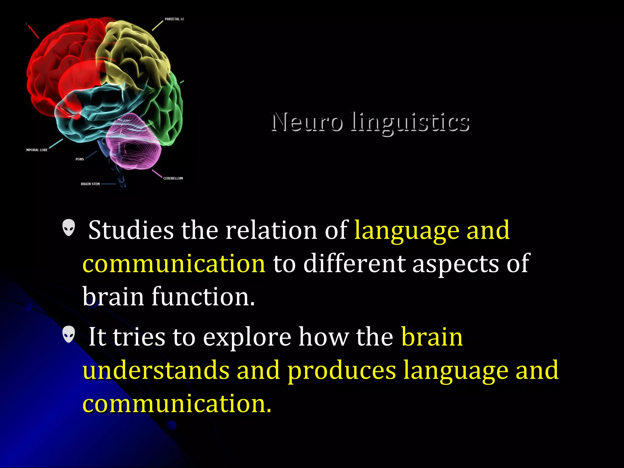
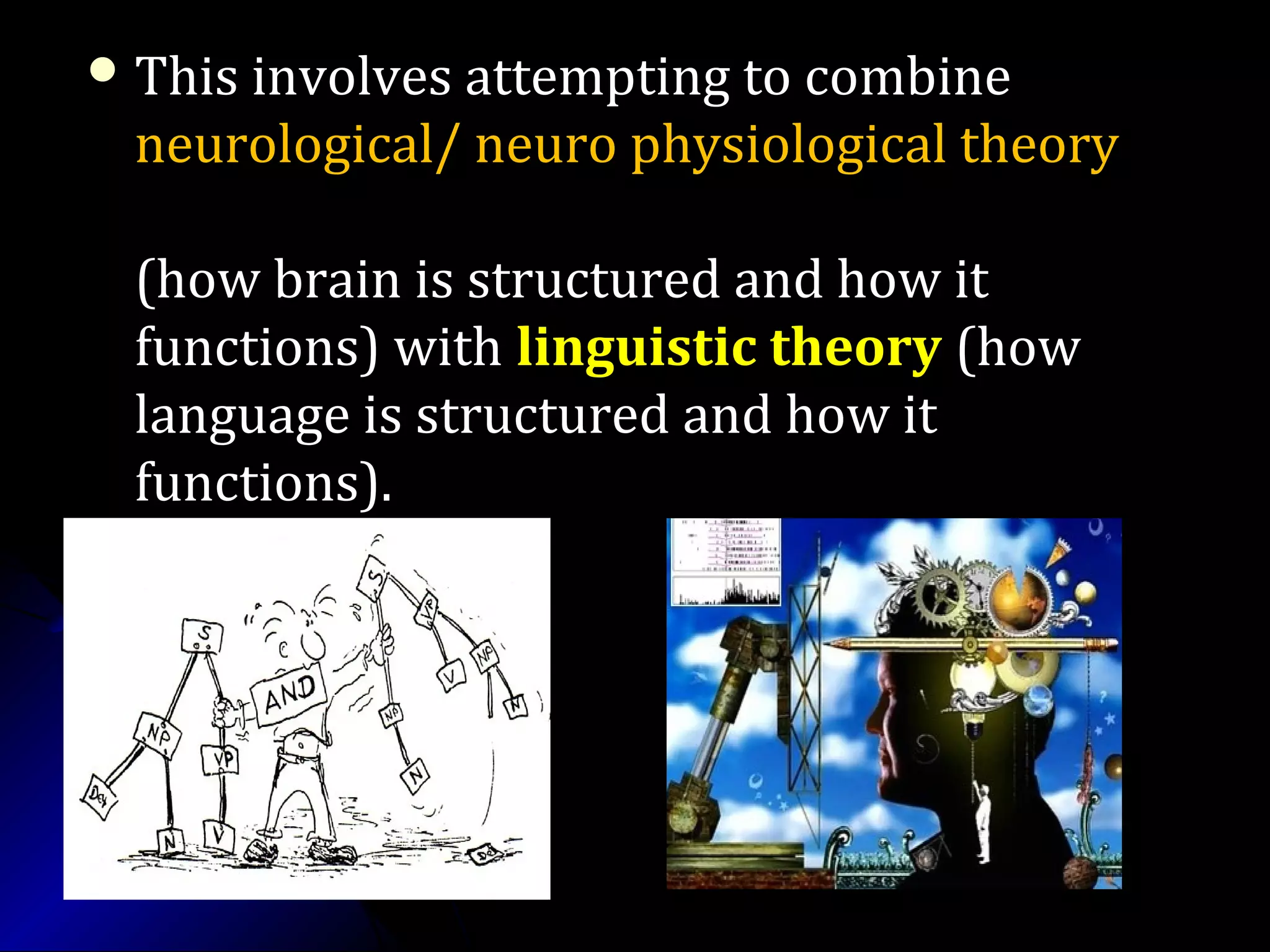
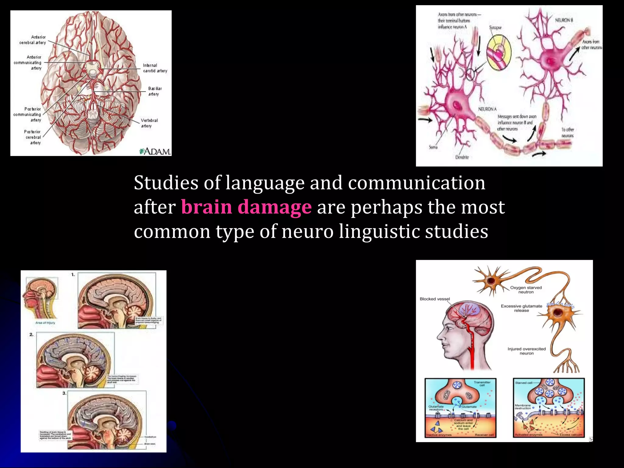
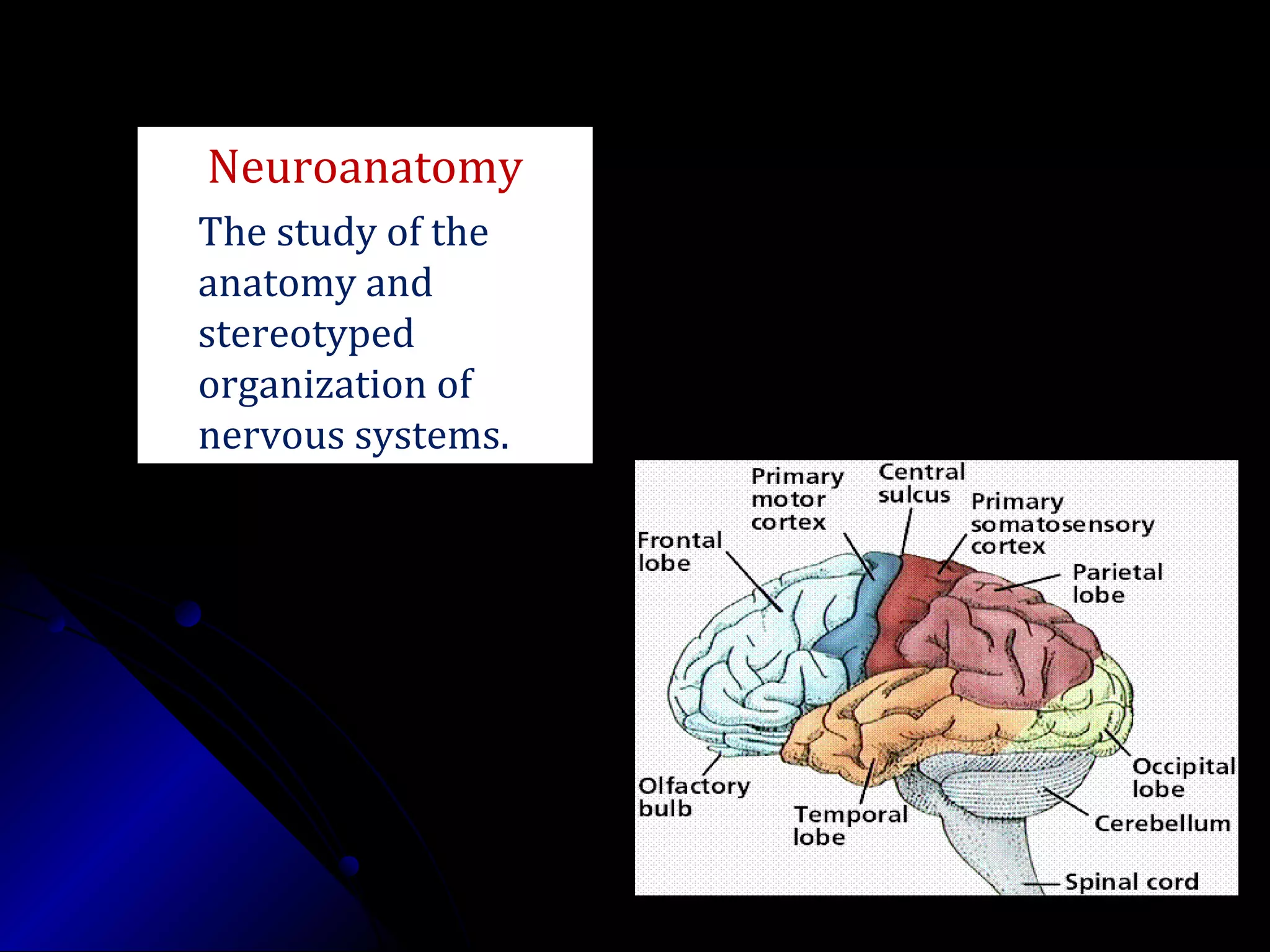
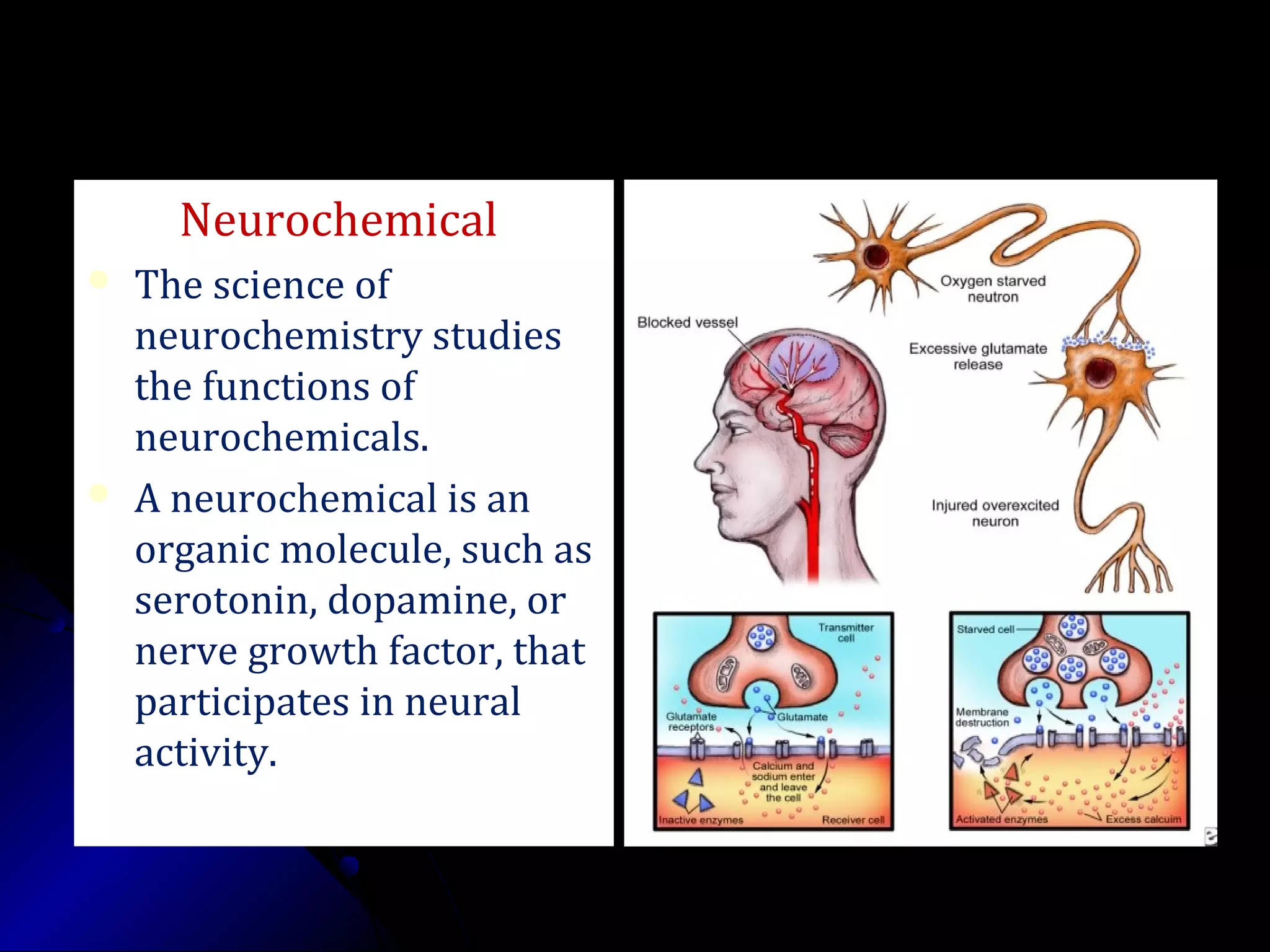
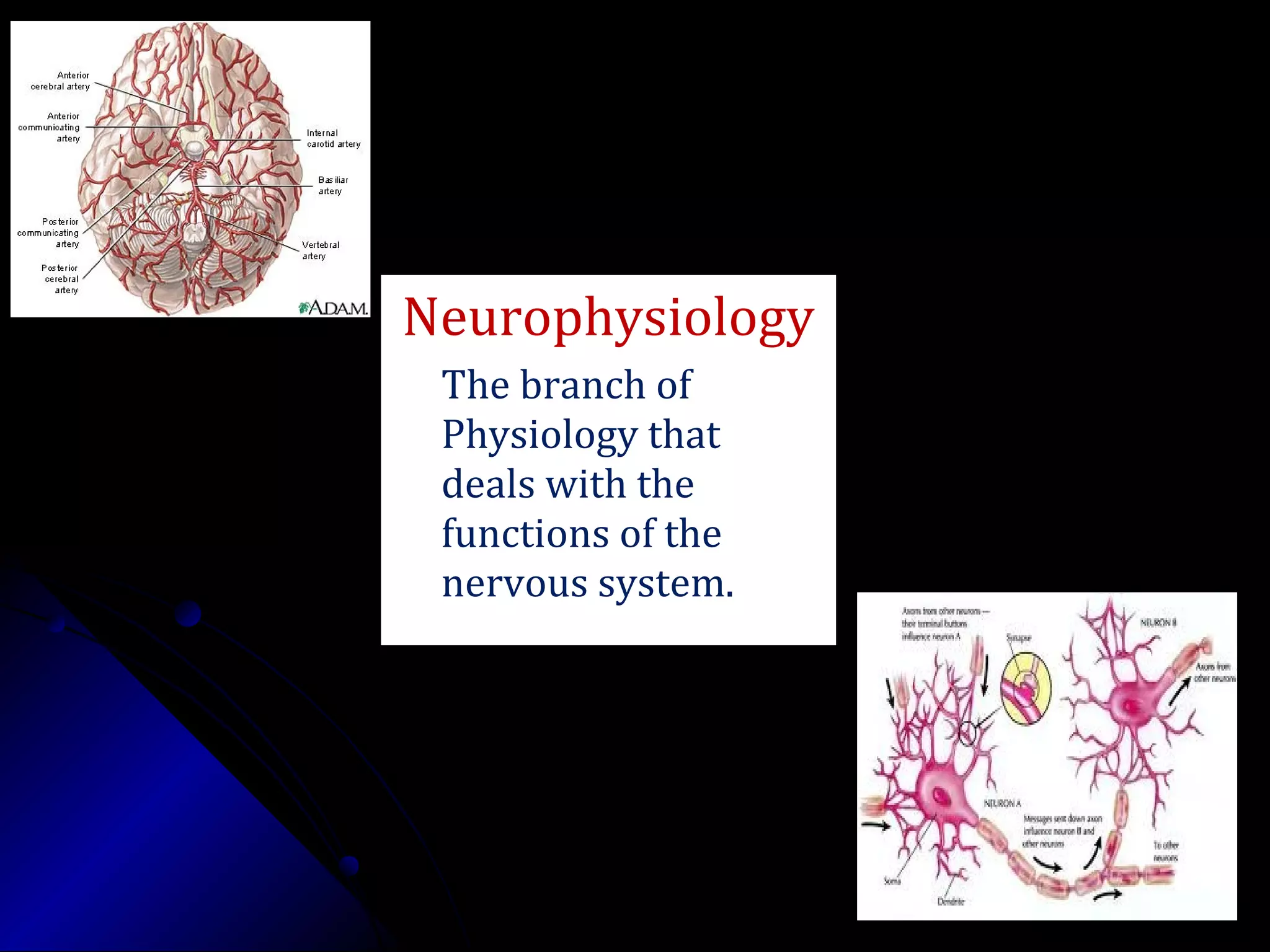
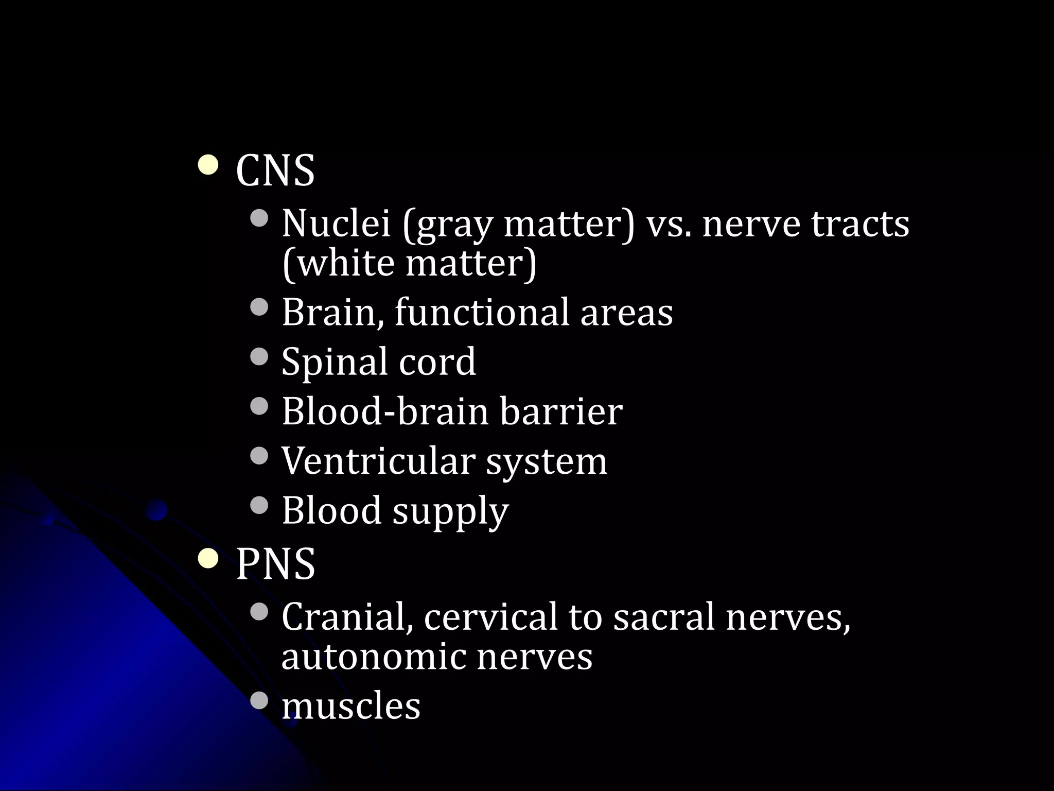
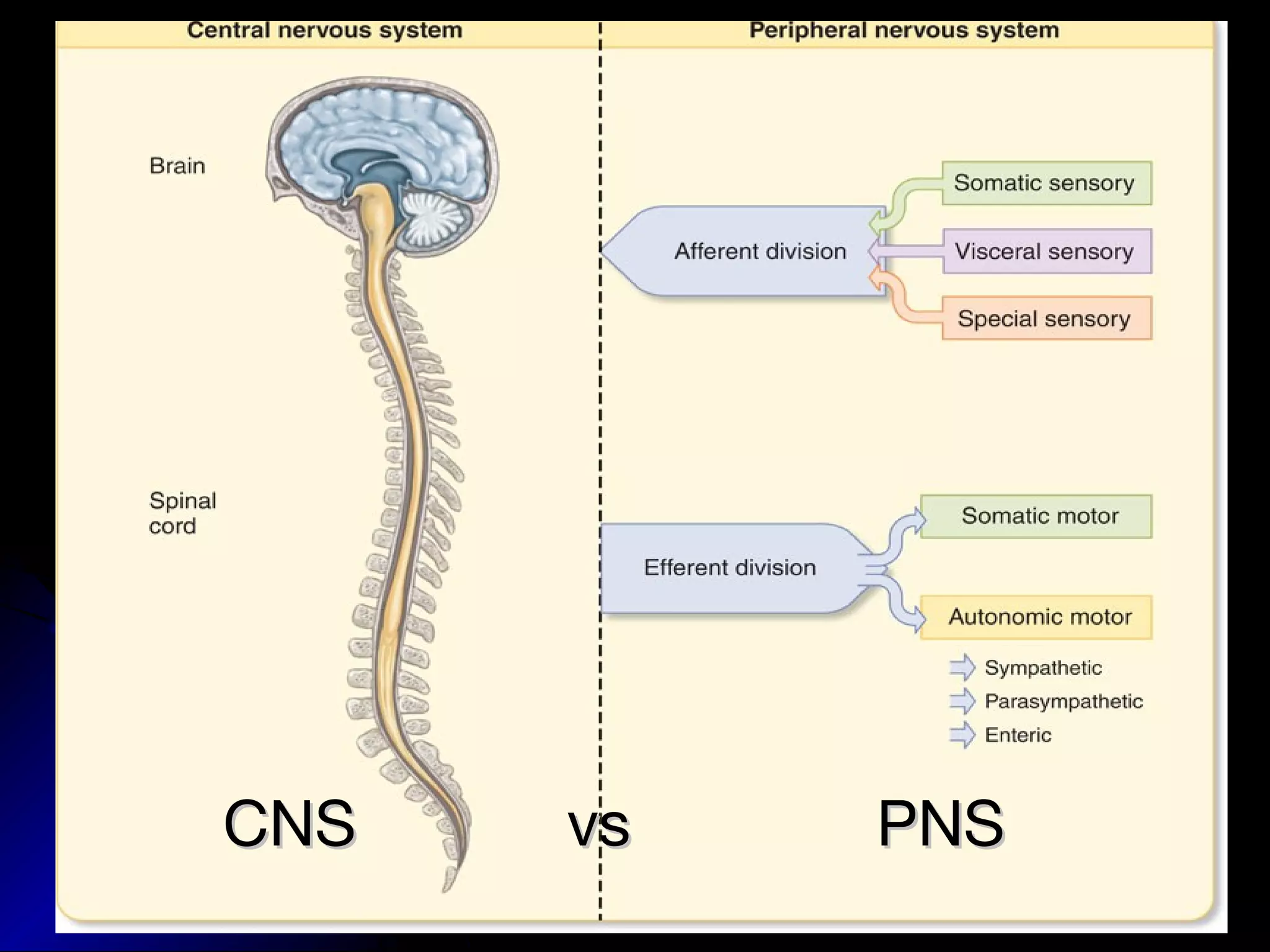
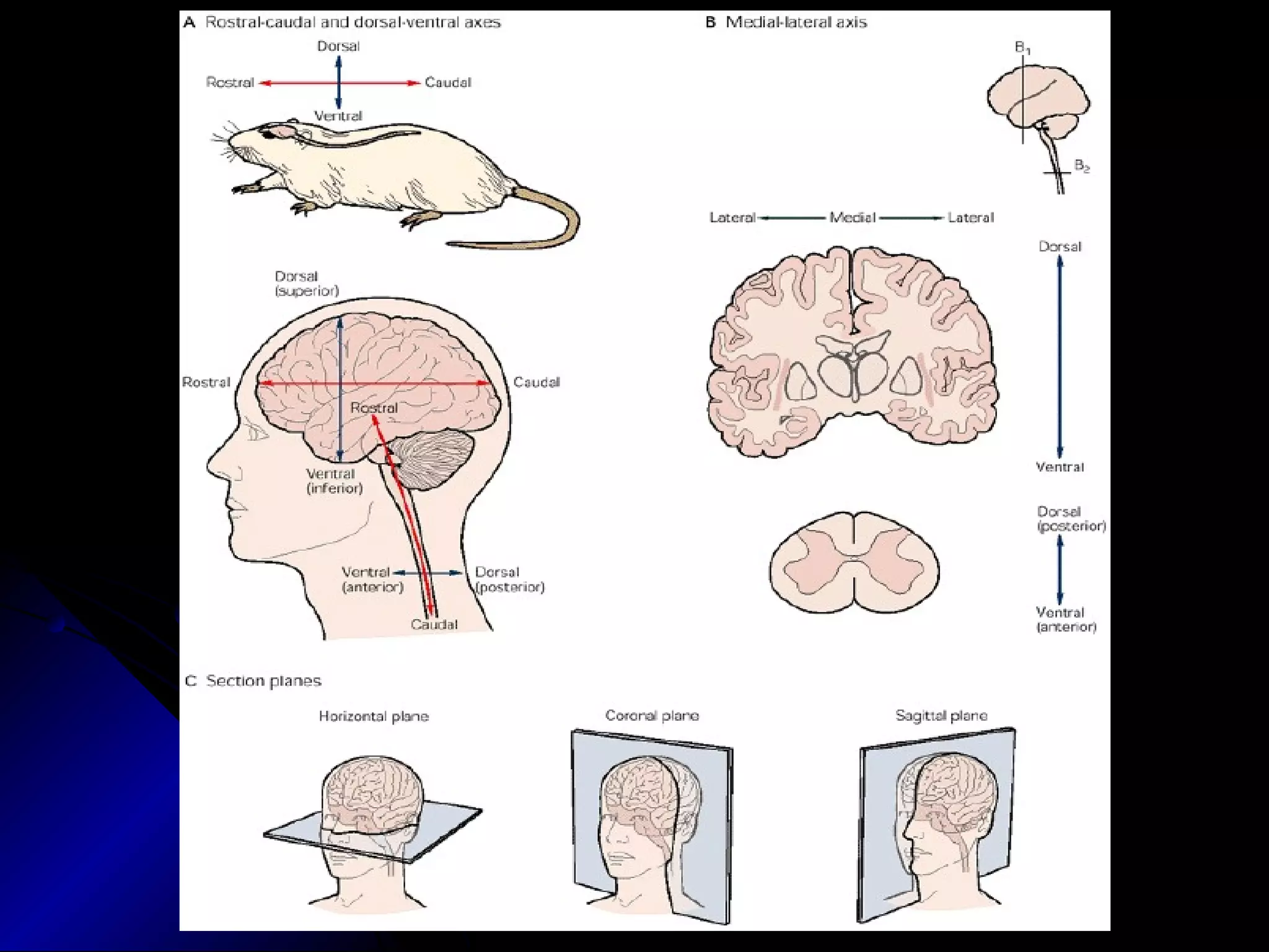
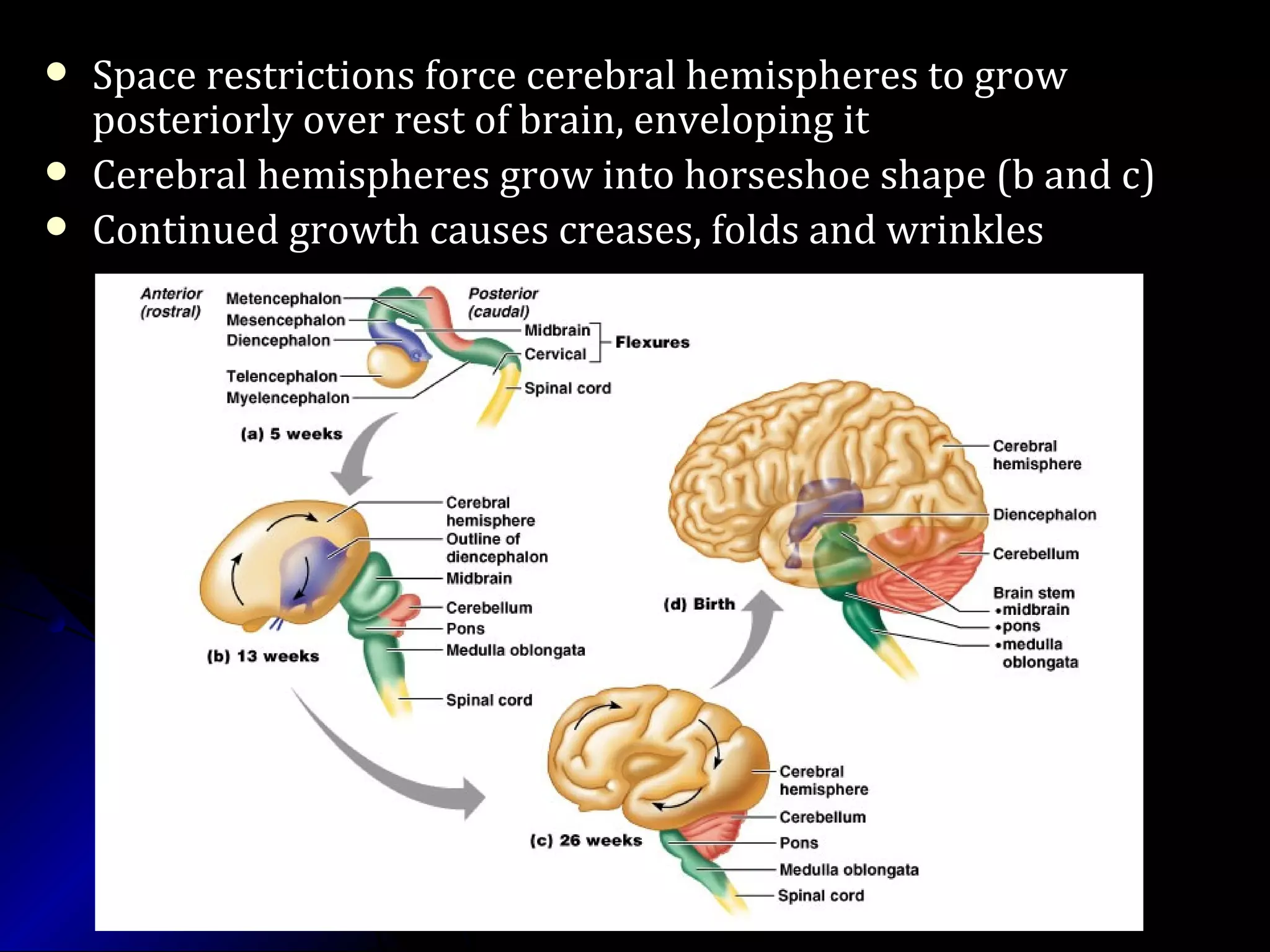
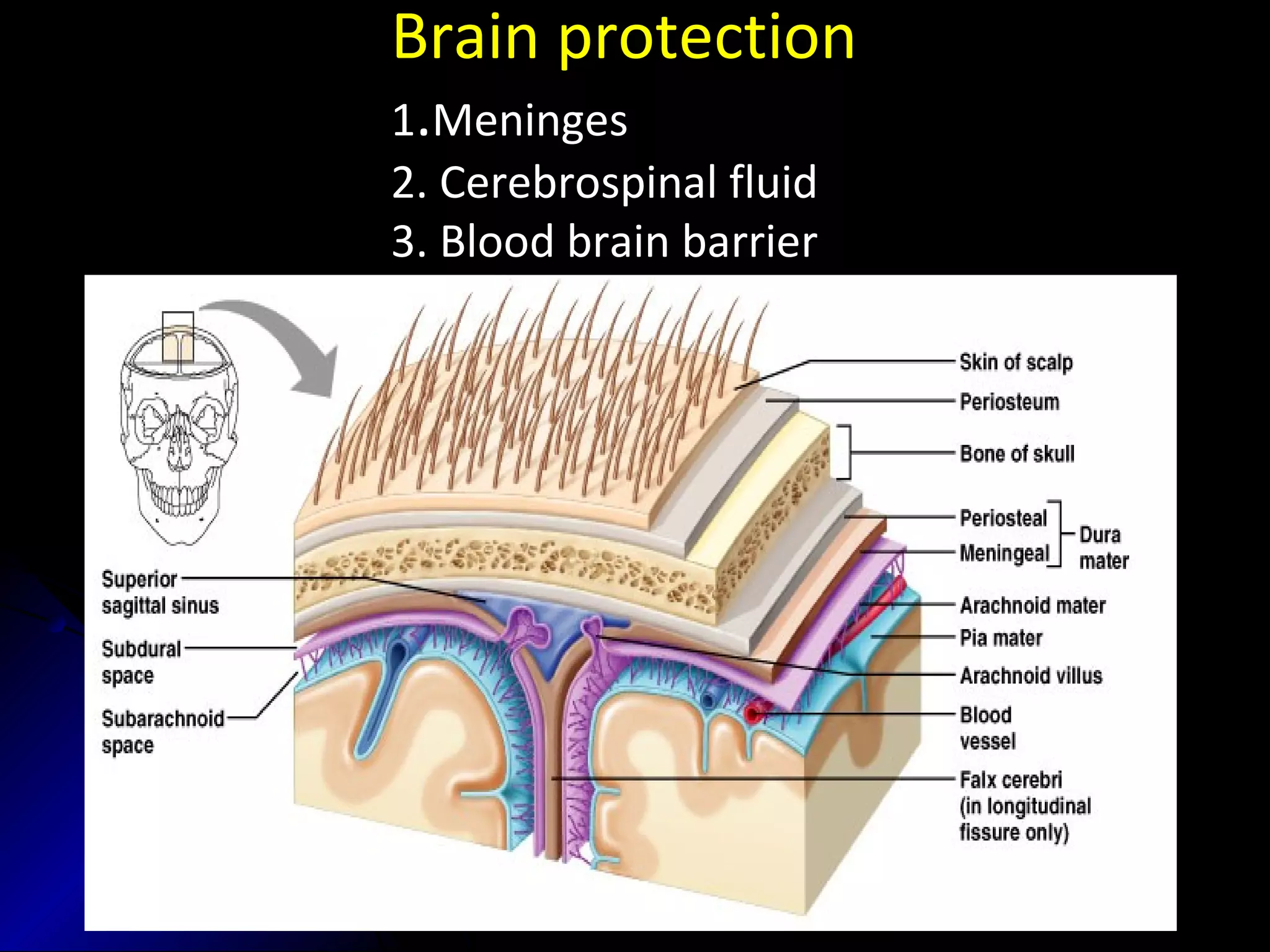
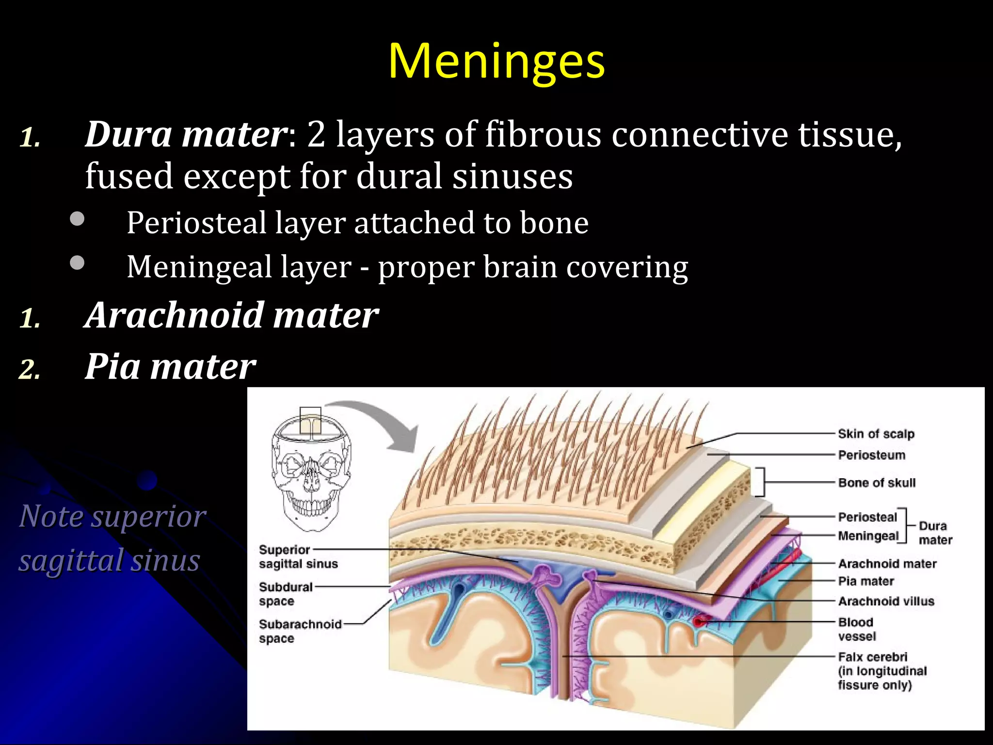
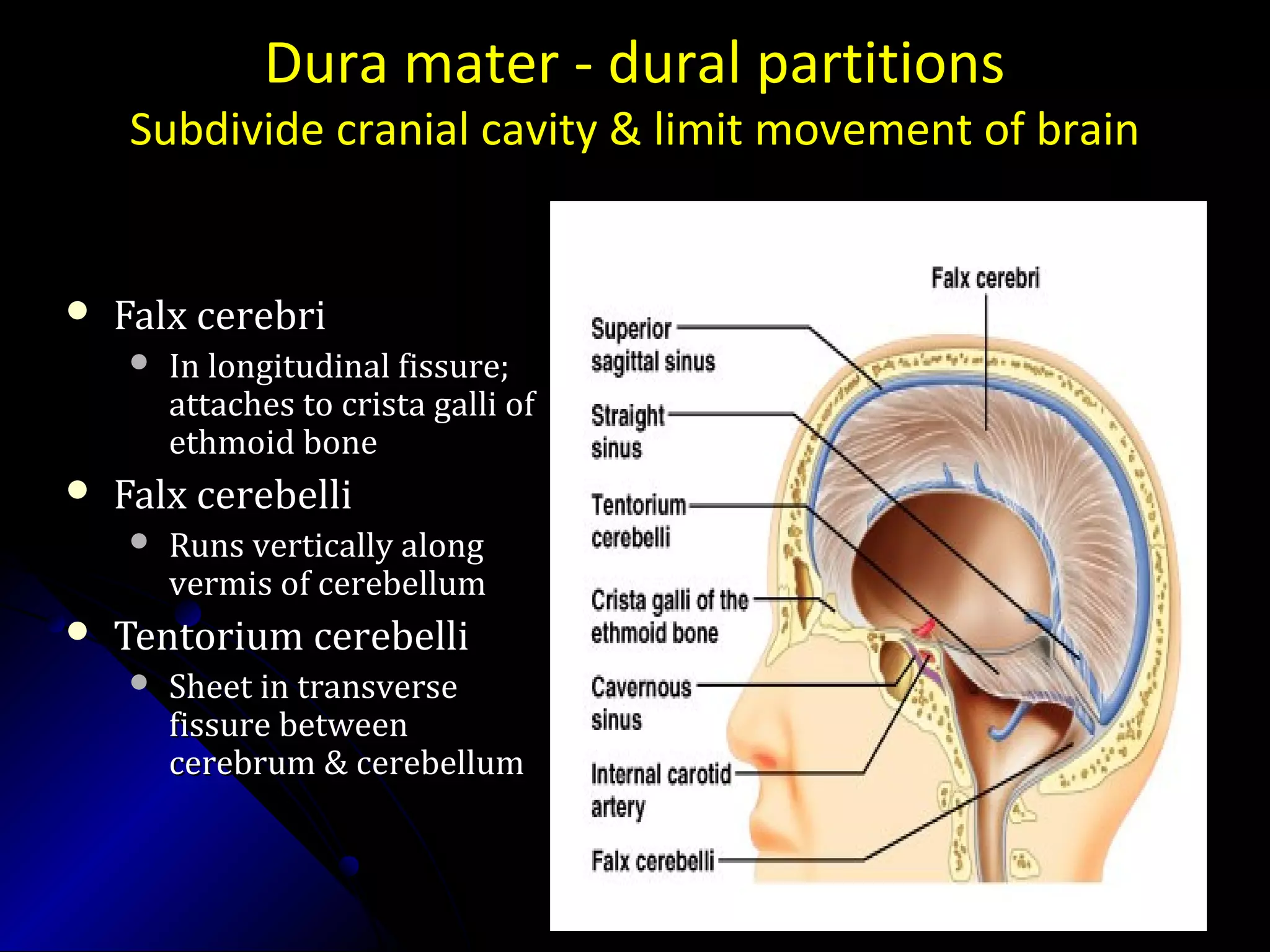
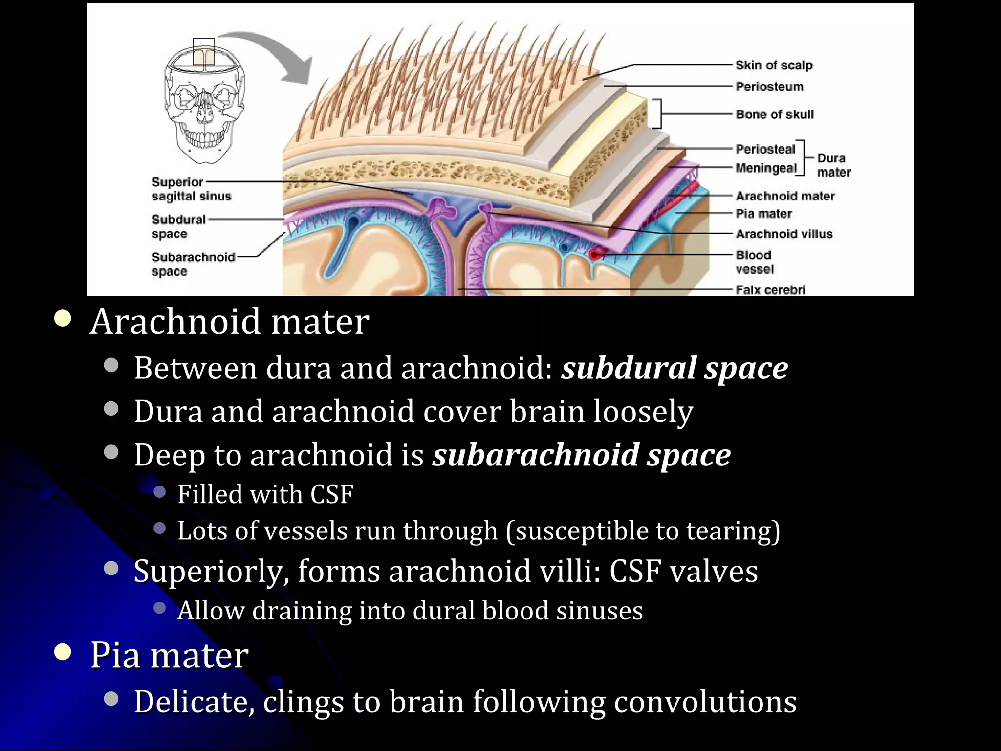
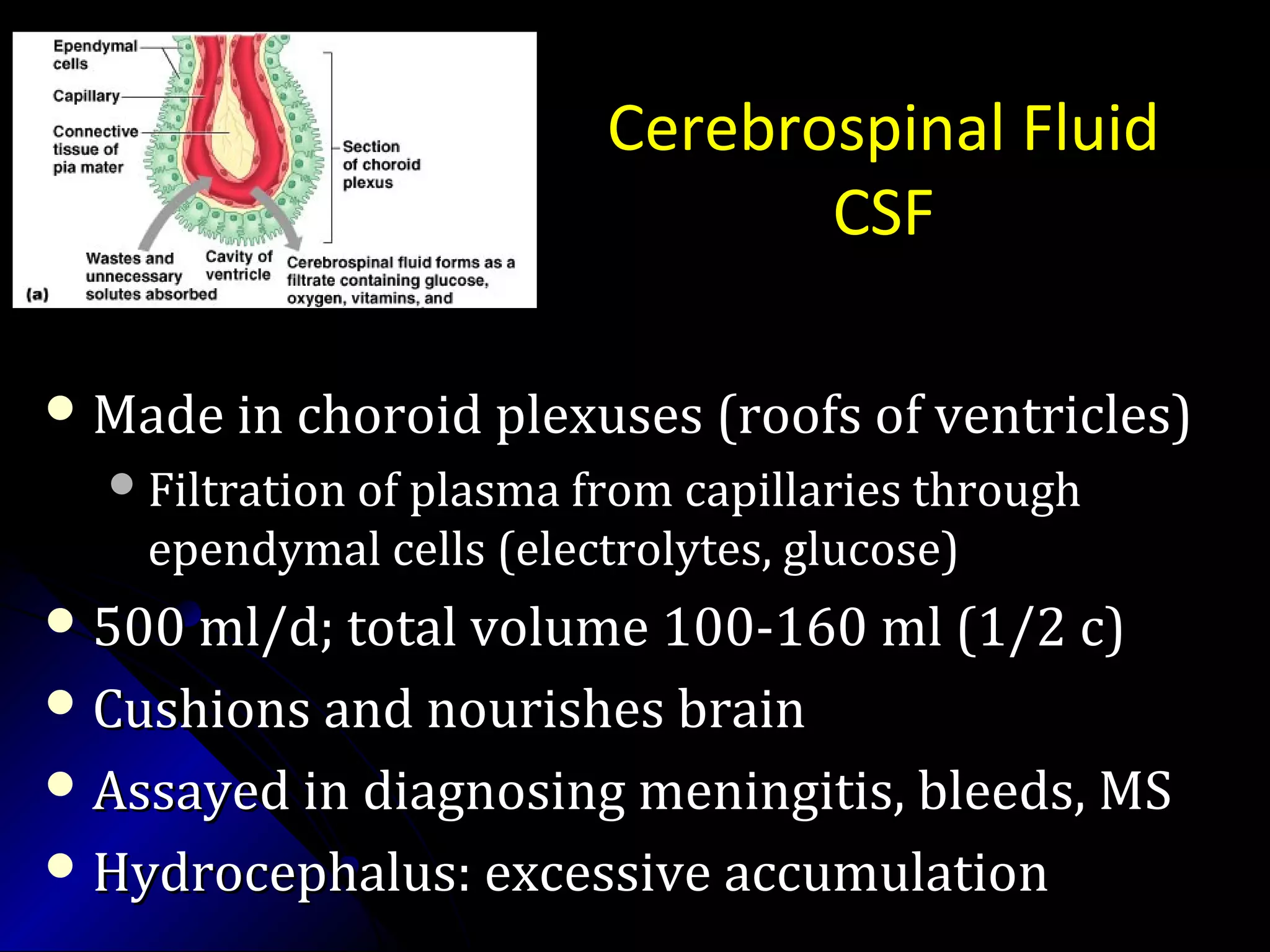
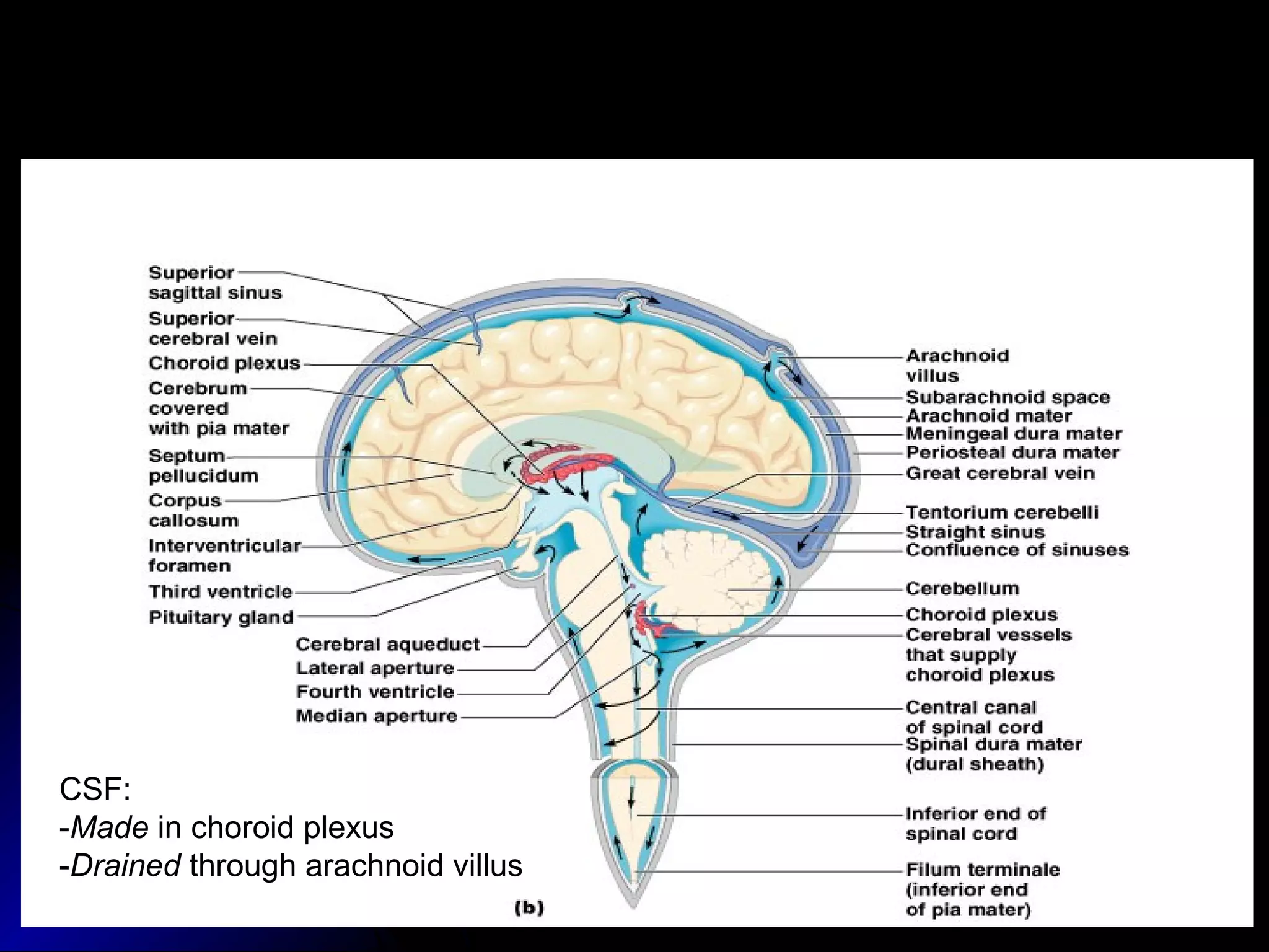
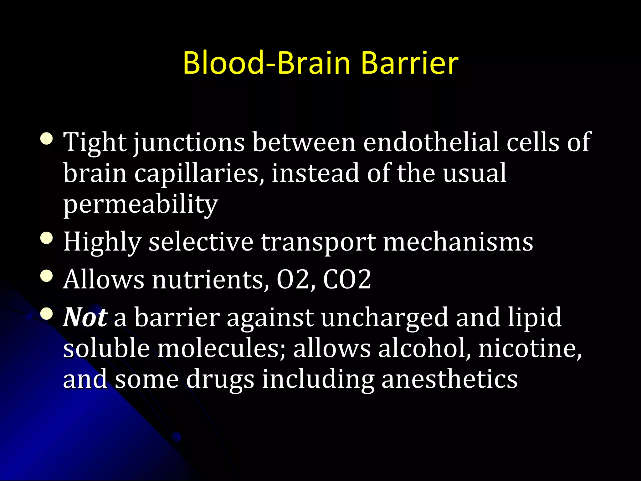
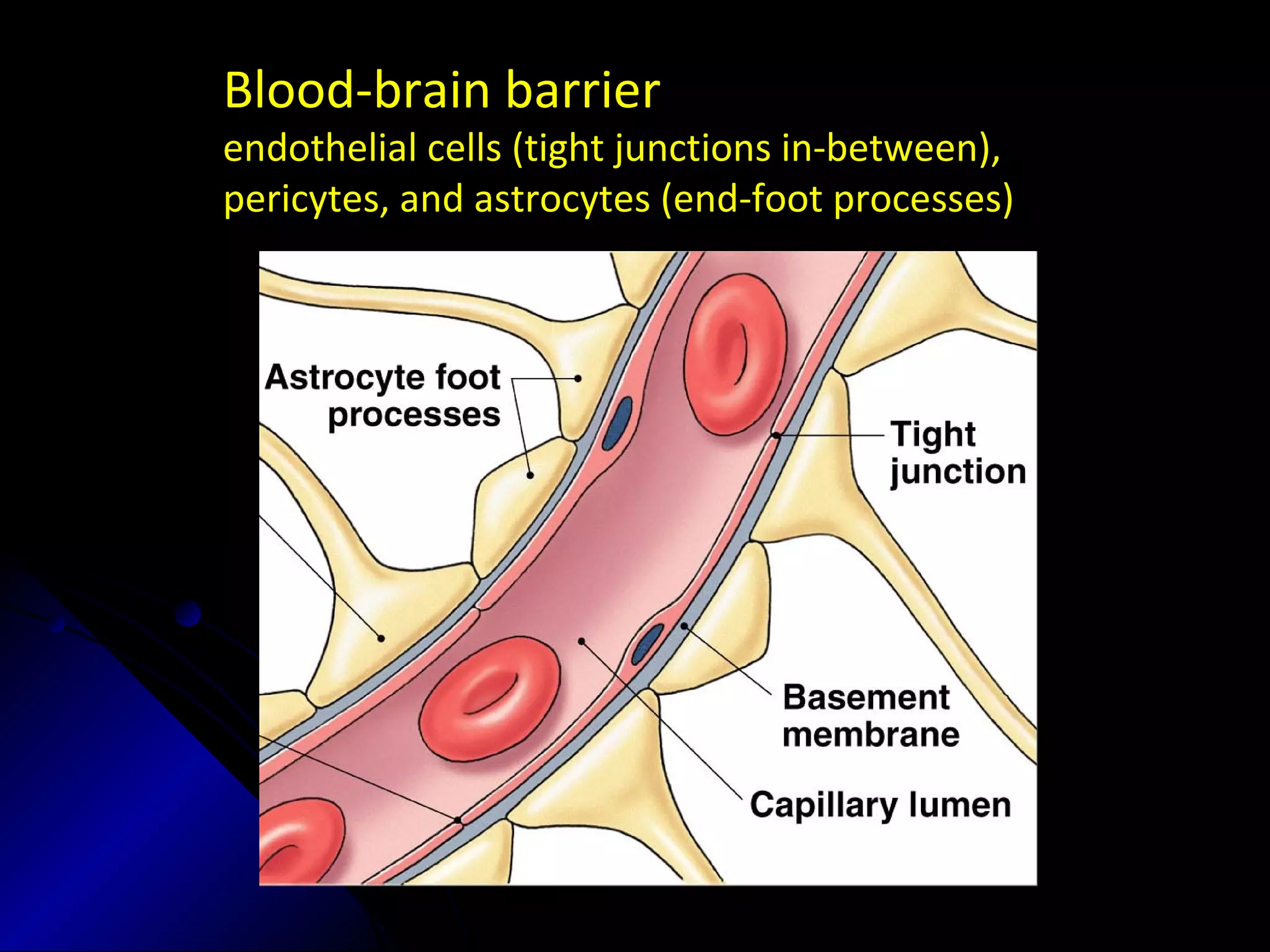
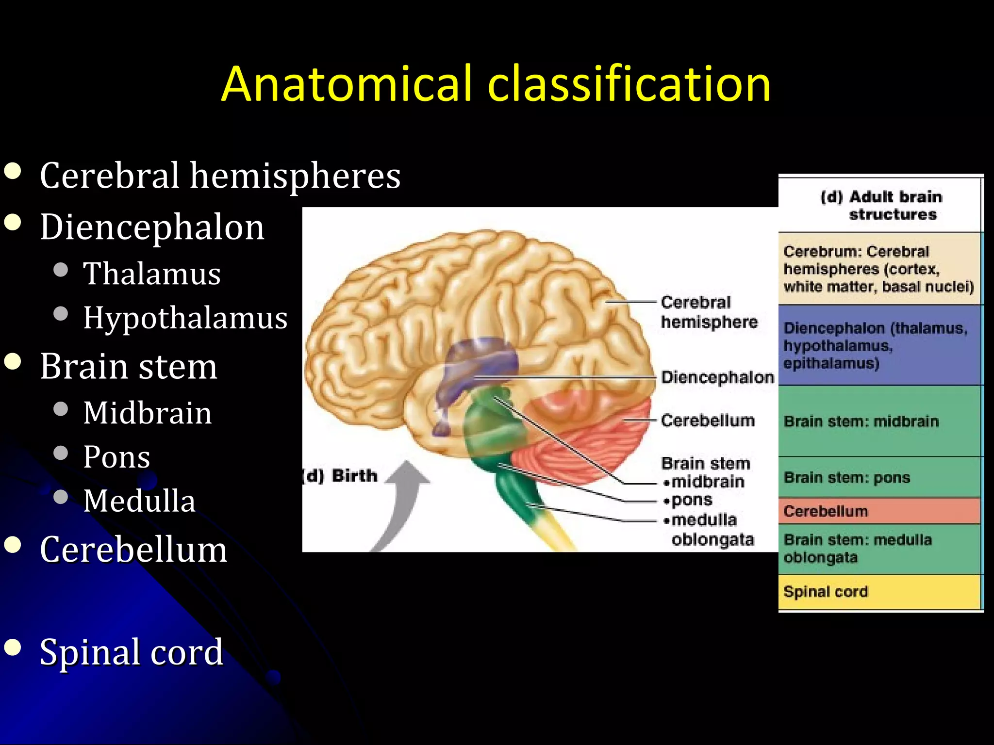
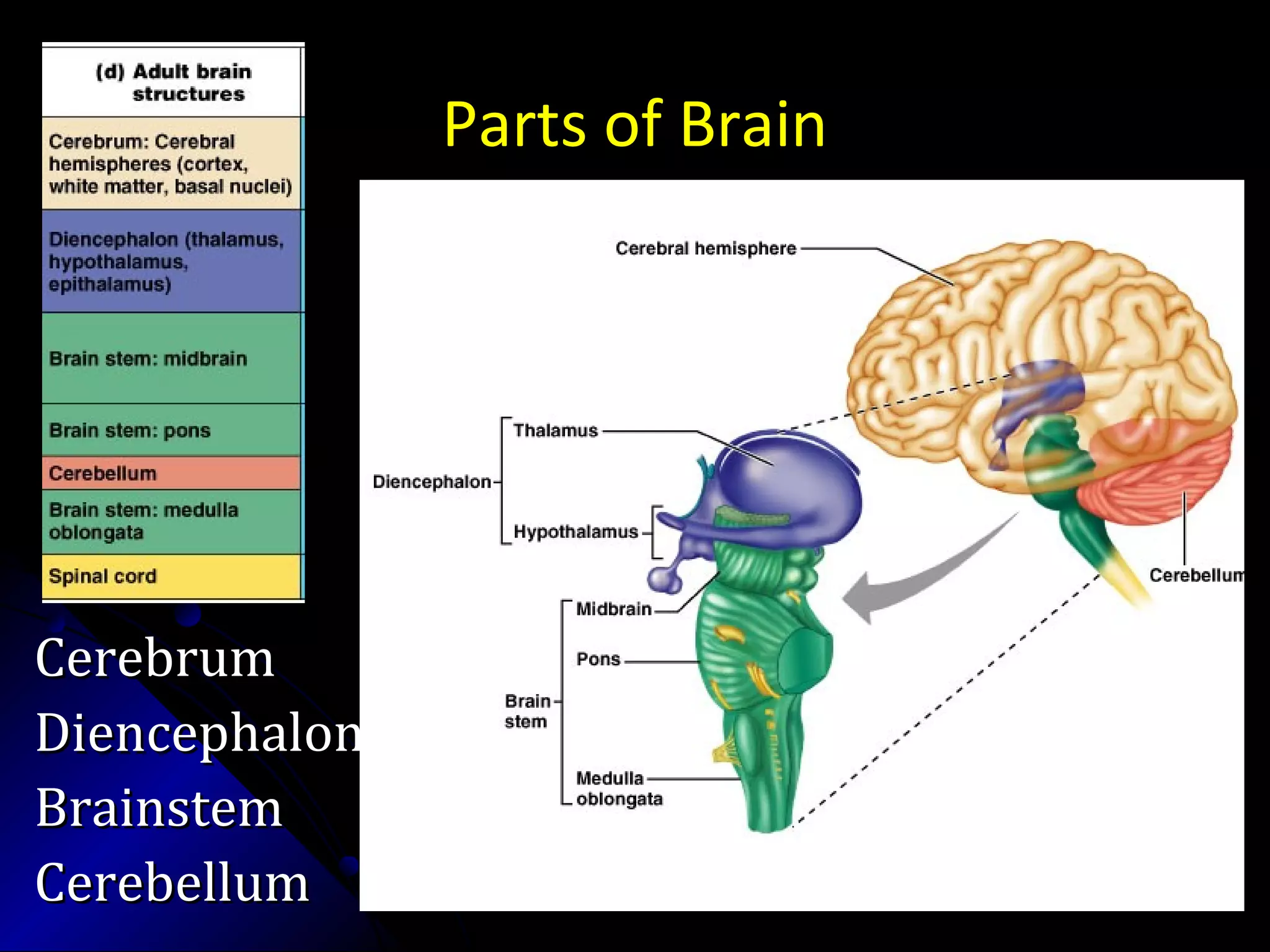
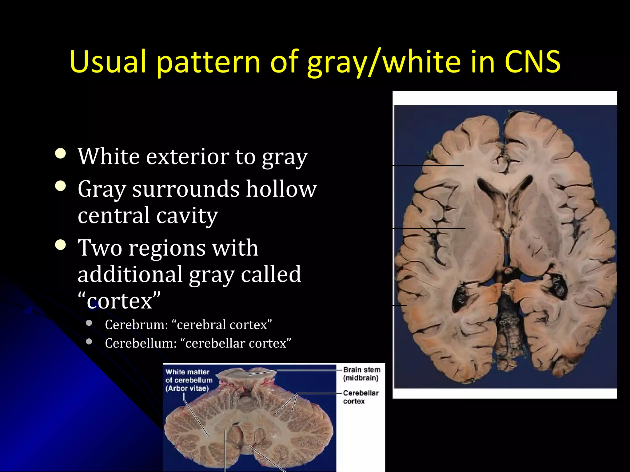
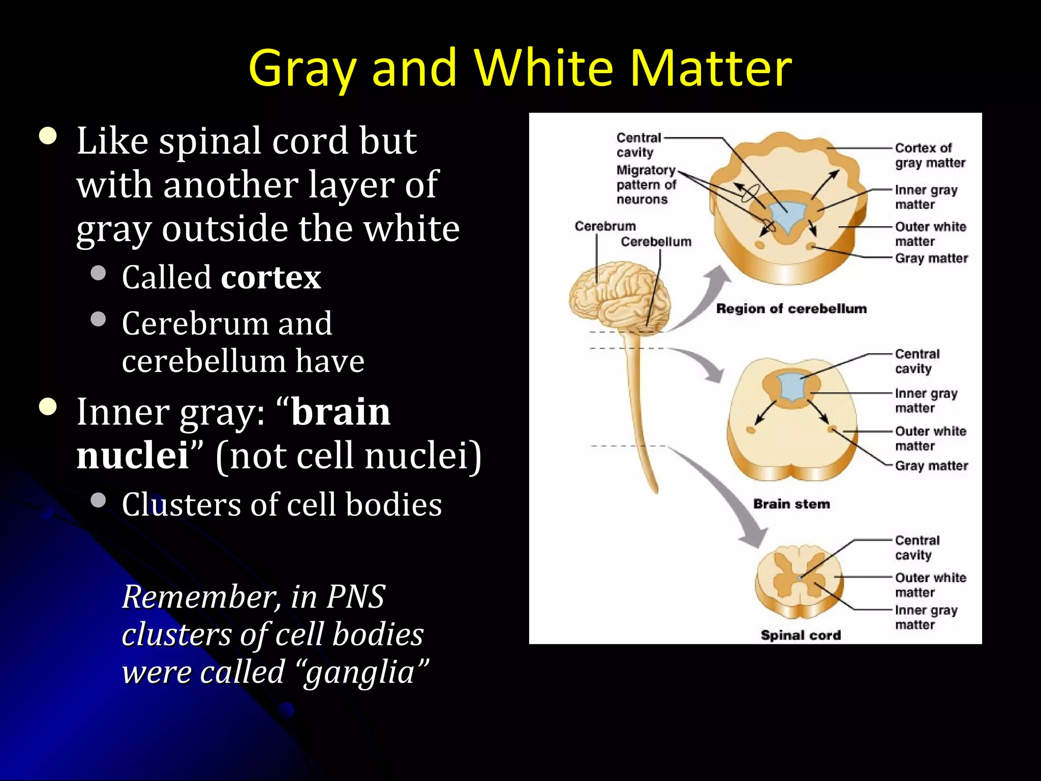
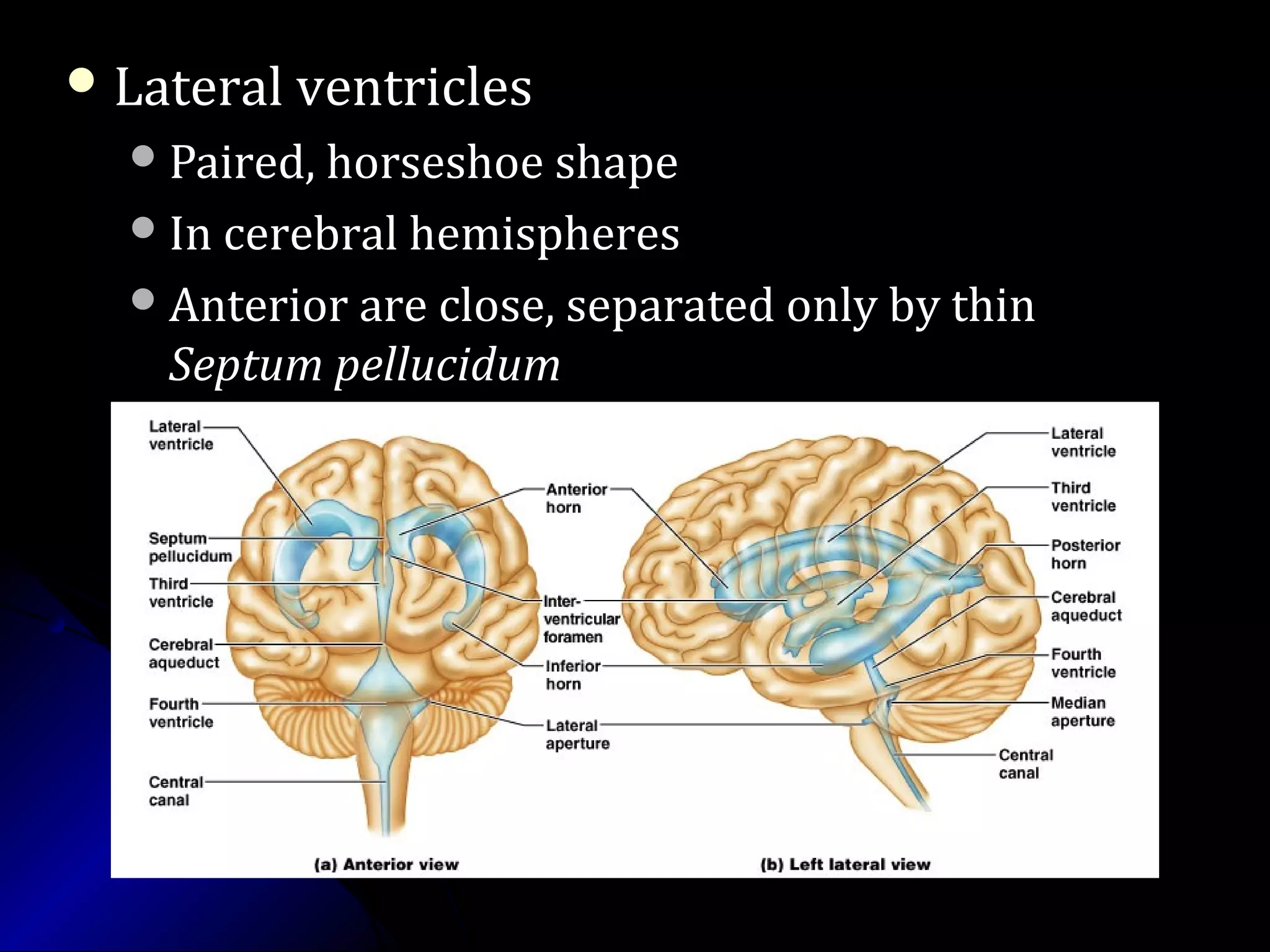
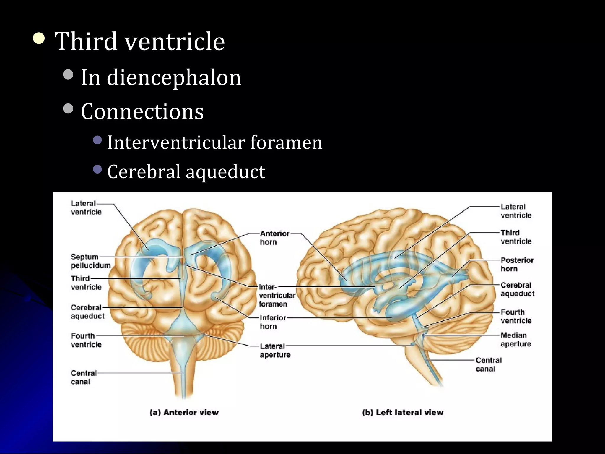
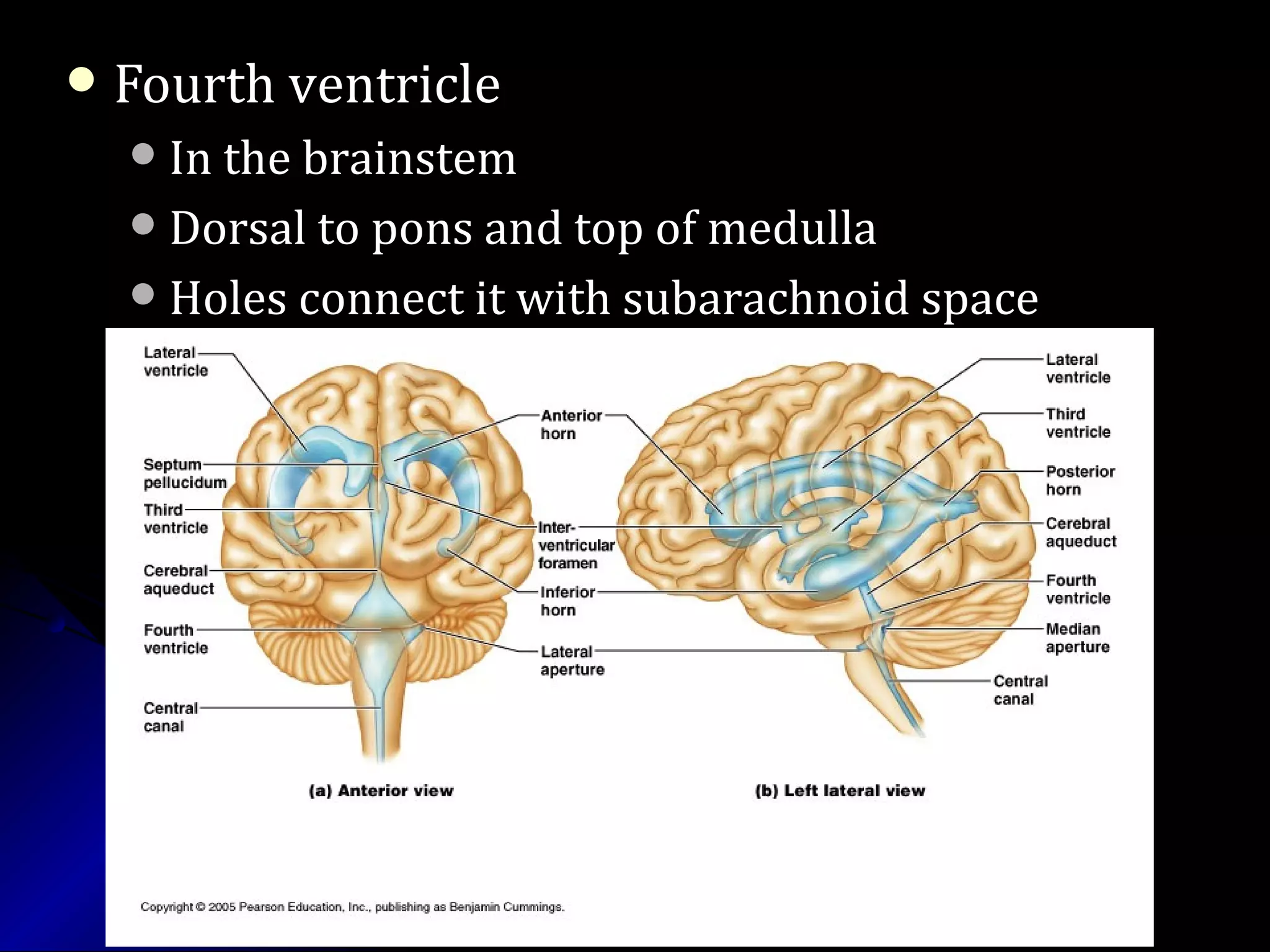
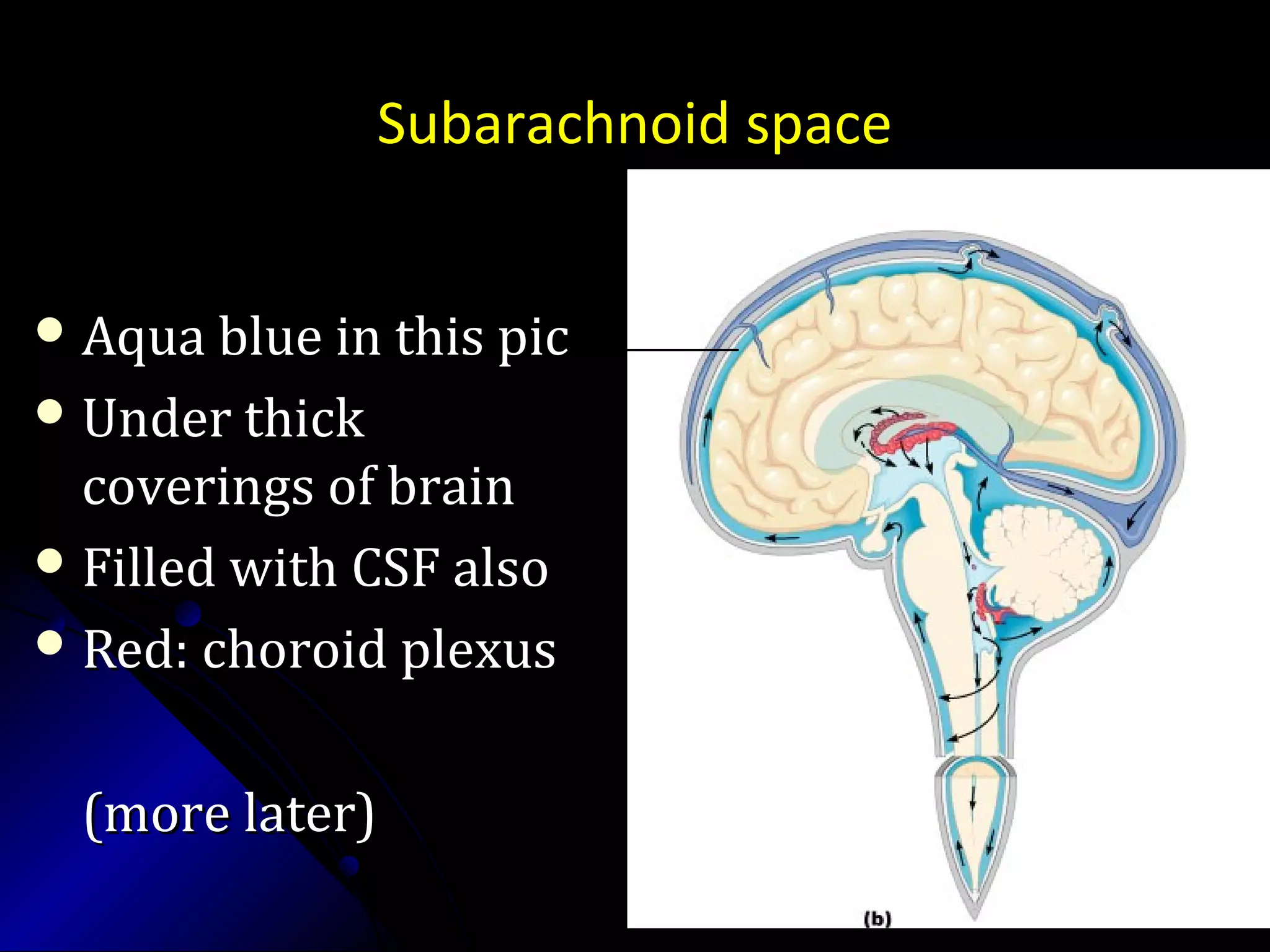
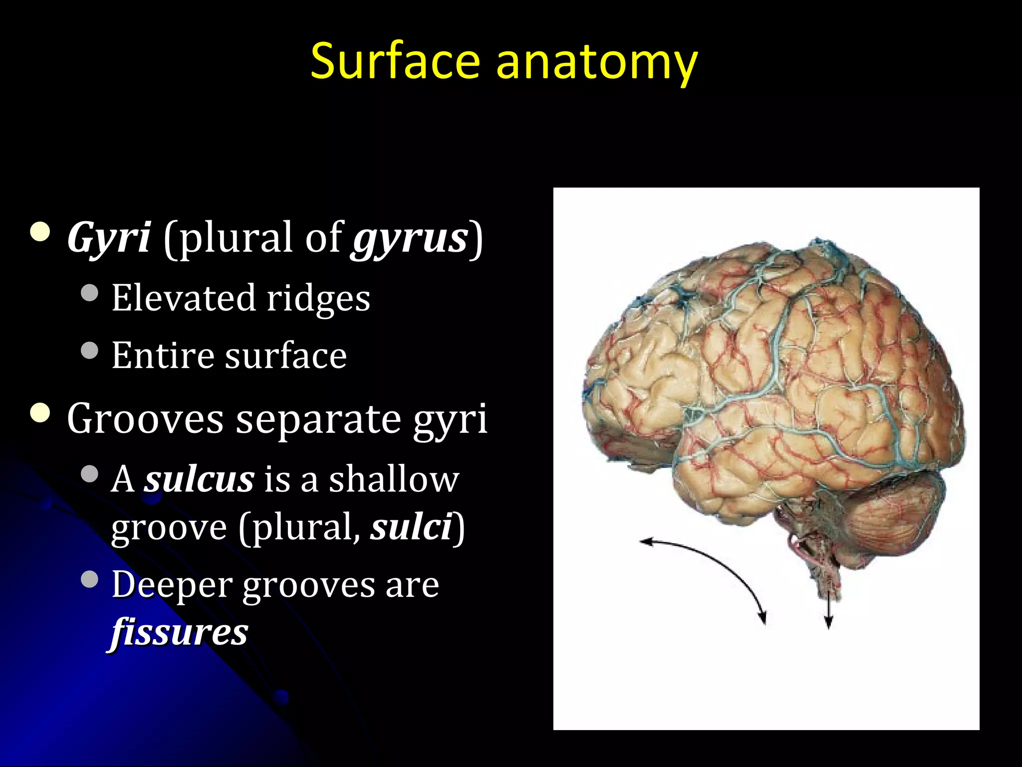
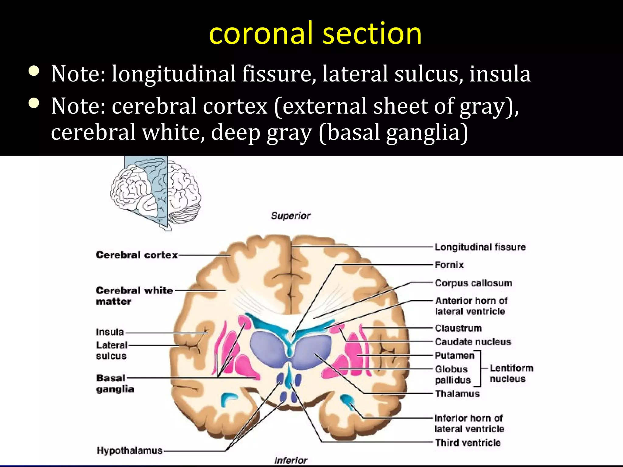
The document discusses the neural bases of language. It describes how post-mortem studies have found no consistent results on the localization of language functions in the brain, as different types of aphasia can cause similar behavioral disturbances. Neurolinguistics studies the relationship between language, communication, and brain function by attempting to combine neurological and linguistic theories. Common neurolinguistic studies examine language and communication after brain damage. The document then provides details on the anatomy and structures of the central and peripheral nervous systems, including the meninges, cerebrospinal fluid, and blood-brain barrier that protect the brain.
Due to damage of the CNS or PNS or both. There is some involvement of the basic motor processes used in speech and this results in a movement disorder...
Auditory brainstem response (ABR)
Approximately 1 of every 1000 children is born deaf. Many more are born with less severe degrees of hearing impairment, while others may acquire hearing loss during early childhood.
combination of technological advances in ABR and otoacoustic emissions (OAE) testing methods are used for evaluation of hearing in newborns.
An introduction to the biology and neurophysiology of human speech. The target audience is researchers and engineers working on speech recognition technology.
Nervous sytem and its divisions: Neuro AnatomyPriyanka Pundir
Neuro Anatomy Introduction, Nervous System, Classification of Nervous System, Cellular Architecture, Neuron Structure, Classification of Neuron, Skull: Osteology, Bones of skull, Skull Joints, Anatomical Position of Skull, Methods of Study of skull.
USMLE NEUROANATOMY 016 White matter of the brain corpus calloum.pdfAHMED ASHOUR
Neurosurgery involving the white matter of the brain typically focuses on addressing specific conditions or abnormalities within this tissue. The white matter comprises nerve fibers, or axons, which are responsible for transmitting signals between different regions of the brain and connecting various parts of the central nervous system. Surgery involving the white matter of the brain is highly specialized and requires a thorough understanding of the brain's anatomy, neuroimaging, and advanced surgical techniques. Neurosurgeons carefully plan interventions to achieve therapeutic goals while minimizing damage to critical white matter tracts that play a crucial role in neural communication.
at a glance
Introduction
Terminologies used in the nervous system
Division of nervous system
Types of nerves- structure and functions
Brain
Cranial nerves
Spinal cord
Motor and sensory pathways of the spinal cord
Autonomic nervous system
Prix Galien International 2024 Forum ProgramLevi Shapiro
June 20, 2024, Prix Galien International and Jerusalem Ethics Forum in ROME. Detailed agenda including panels:
- ADVANCES IN CARDIOLOGY: A NEW PARADIGM IS COMING
- WOMEN’S HEALTH: FERTILITY PRESERVATION
- WHAT’S NEW IN THE TREATMENT OF INFECTIOUS,
ONCOLOGICAL AND INFLAMMATORY SKIN DISEASES?
- ARTIFICIAL INTELLIGENCE AND ETHICS
- GENE THERAPY
- BEYOND BORDERS: GLOBAL INITIATIVES FOR DEMOCRATIZING LIFE SCIENCE TECHNOLOGIES AND PROMOTING ACCESS TO HEALTHCARE
- ETHICAL CHALLENGES IN LIFE SCIENCES
- Prix Galien International Awards Ceremony
micro teaching on communication m.sc nursing.pdfAnurag Sharma
Microteaching is a unique model of practice teaching. It is a viable instrument for the. desired change in the teaching behavior or the behavior potential which, in specified types of real. classroom situations, tends to facilitate the achievement of specified types of objectives.
Title: Sense of Taste
Presenter: Dr. Faiza, Assistant Professor of Physiology
Qualifications:
MBBS (Best Graduate, AIMC Lahore)
FCPS Physiology
ICMT, CHPE, DHPE (STMU)
MPH (GC University, Faisalabad)
MBA (Virtual University of Pakistan)
Learning Objectives:
Describe the structure and function of taste buds.
Describe the relationship between the taste threshold and taste index of common substances.
Explain the chemical basis and signal transduction of taste perception for each type of primary taste sensation.
Recognize different abnormalities of taste perception and their causes.
Key Topics:
Significance of Taste Sensation:
Differentiation between pleasant and harmful food
Influence on behavior
Selection of food based on metabolic needs
Receptors of Taste:
Taste buds on the tongue
Influence of sense of smell, texture of food, and pain stimulation (e.g., by pepper)
Primary and Secondary Taste Sensations:
Primary taste sensations: Sweet, Sour, Salty, Bitter, Umami
Chemical basis and signal transduction mechanisms for each taste
Taste Threshold and Index:
Taste threshold values for Sweet (sucrose), Salty (NaCl), Sour (HCl), and Bitter (Quinine)
Taste index relationship: Inversely proportional to taste threshold
Taste Blindness:
Inability to taste certain substances, particularly thiourea compounds
Example: Phenylthiocarbamide
Structure and Function of Taste Buds:
Composition: Epithelial cells, Sustentacular/Supporting cells, Taste cells, Basal cells
Features: Taste pores, Taste hairs/microvilli, and Taste nerve fibers
Location of Taste Buds:
Found in papillae of the tongue (Fungiform, Circumvallate, Foliate)
Also present on the palate, tonsillar pillars, epiglottis, and proximal esophagus
Mechanism of Taste Stimulation:
Interaction of taste substances with receptors on microvilli
Signal transduction pathways for Umami, Sweet, Bitter, Sour, and Salty tastes
Taste Sensitivity and Adaptation:
Decrease in sensitivity with age
Rapid adaptation of taste sensation
Role of Saliva in Taste:
Dissolution of tastants to reach receptors
Washing away the stimulus
Taste Preferences and Aversions:
Mechanisms behind taste preference and aversion
Influence of receptors and neural pathways
Impact of Sensory Nerve Damage:
Degeneration of taste buds if the sensory nerve fiber is cut
Abnormalities of Taste Detection:
Conditions: Ageusia, Hypogeusia, Dysgeusia (parageusia)
Causes: Nerve damage, neurological disorders, infections, poor oral hygiene, adverse drug effects, deficiencies, aging, tobacco use, altered neurotransmitter levels
Neurotransmitters and Taste Threshold:
Effects of serotonin (5-HT) and norepinephrine (NE) on taste sensitivity
Supertasters:
25% of the population with heightened sensitivity to taste, especially bitterness
Increased number of fungiform papillae
These lecture slides, by Dr Sidra Arshad, offer a quick overview of physiological basis of a normal electrocardiogram.
Learning objectives:
1. Define an electrocardiogram (ECG) and electrocardiography
2. Describe how dipoles generated by the heart produce the waveforms of the ECG
3. Describe the components of a normal electrocardiogram of a typical bipolar leads (limb II)
4. Differentiate between intervals and segments
5. Enlist some common indications for obtaining an ECG
Study Resources:
1. Chapter 11, Guyton and Hall Textbook of Medical Physiology, 14th edition
2. Chapter 9, Human Physiology - From Cells to Systems, Lauralee Sherwood, 9th edition
3. Chapter 29, Ganong’s Review of Medical Physiology, 26th edition
4. Electrocardiogram, StatPearls - https://www.ncbi.nlm.nih.gov/books/NBK549803/
5. ECG in Medical Practice by ABM Abdullah, 4th edition
6. ECG Basics, http://www.nataliescasebook.com/tag/e-c-g-basics
Anti ulcer drugs and their Advance pharmacology ||
Anti-ulcer drugs are medications used to prevent and treat ulcers in the stomach and upper part of the small intestine (duodenal ulcers). These ulcers are often caused by an imbalance between stomach acid and the mucosal lining, which protects the stomach lining.
||Scope: Overview of various classes of anti-ulcer drugs, their mechanisms of action, indications, side effects, and clinical considerations.
Couples presenting to the infertility clinic- Do they really have infertility...Sujoy Dasgupta
Dr Sujoy Dasgupta presented the study on "Couples presenting to the infertility clinic- Do they really have infertility? – The unexplored stories of non-consummation" in the 13th Congress of the Asia Pacific Initiative on Reproduction (ASPIRE 2024) at Manila on 24 May, 2024.
Title: Sense of Smell
Presenter: Dr. Faiza, Assistant Professor of Physiology
Qualifications:
MBBS (Best Graduate, AIMC Lahore)
FCPS Physiology
ICMT, CHPE, DHPE (STMU)
MPH (GC University, Faisalabad)
MBA (Virtual University of Pakistan)
Learning Objectives:
Describe the primary categories of smells and the concept of odor blindness.
Explain the structure and location of the olfactory membrane and mucosa, including the types and roles of cells involved in olfaction.
Describe the pathway and mechanisms of olfactory signal transmission from the olfactory receptors to the brain.
Illustrate the biochemical cascade triggered by odorant binding to olfactory receptors, including the role of G-proteins and second messengers in generating an action potential.
Identify different types of olfactory disorders such as anosmia, hyposmia, hyperosmia, and dysosmia, including their potential causes.
Key Topics:
Olfactory Genes:
3% of the human genome accounts for olfactory genes.
400 genes for odorant receptors.
Olfactory Membrane:
Located in the superior part of the nasal cavity.
Medially: Folds downward along the superior septum.
Laterally: Folds over the superior turbinate and upper surface of the middle turbinate.
Total surface area: 5-10 square centimeters.
Olfactory Mucosa:
Olfactory Cells: Bipolar nerve cells derived from the CNS (100 million), with 4-25 olfactory cilia per cell.
Sustentacular Cells: Produce mucus and maintain ionic and molecular environment.
Basal Cells: Replace worn-out olfactory cells with an average lifespan of 1-2 months.
Bowman’s Gland: Secretes mucus.
Stimulation of Olfactory Cells:
Odorant dissolves in mucus and attaches to receptors on olfactory cilia.
Involves a cascade effect through G-proteins and second messengers, leading to depolarization and action potential generation in the olfactory nerve.
Quality of a Good Odorant:
Small (3-20 Carbon atoms), volatile, water-soluble, and lipid-soluble.
Facilitated by odorant-binding proteins in mucus.
Membrane Potential and Action Potential:
Resting membrane potential: -55mV.
Action potential frequency in the olfactory nerve increases with odorant strength.
Adaptation Towards the Sense of Smell:
Rapid adaptation within the first second, with further slow adaptation.
Psychological adaptation greater than receptor adaptation, involving feedback inhibition from the central nervous system.
Primary Sensations of Smell:
Camphoraceous, Musky, Floral, Pepperminty, Ethereal, Pungent, Putrid.
Odor Detection Threshold:
Examples: Hydrogen sulfide (0.0005 ppm), Methyl-mercaptan (0.002 ppm).
Some toxic substances are odorless at lethal concentrations.
Characteristics of Smell:
Odor blindness for single substances due to lack of appropriate receptor protein.
Behavioral and emotional influences of smell.
Transmission of Olfactory Signals:
From olfactory cells to glomeruli in the olfactory bulb, involving lateral inhibition.
Primitive, less old, and new olfactory systems with different path
Lung Cancer: Artificial Intelligence, Synergetics, Complex System Analysis, S...Oleg Kshivets
RESULTS: Overall life span (LS) was 2252.1±1742.5 days and cumulative 5-year survival (5YS) reached 73.2%, 10 years – 64.8%, 20 years – 42.5%. 513 LCP lived more than 5 years (LS=3124.6±1525.6 days), 148 LCP – more than 10 years (LS=5054.4±1504.1 days).199 LCP died because of LC (LS=562.7±374.5 days). 5YS of LCP after bi/lobectomies was significantly superior in comparison with LCP after pneumonectomies (78.1% vs.63.7%, P=0.00001 by log-rank test). AT significantly improved 5YS (66.3% vs. 34.8%) (P=0.00000 by log-rank test) only for LCP with N1-2. Cox modeling displayed that 5YS of LCP significantly depended on: phase transition (PT) early-invasive LC in terms of synergetics, PT N0—N12, cell ratio factors (ratio between cancer cells- CC and blood cells subpopulations), G1-3, histology, glucose, AT, blood cell circuit, prothrombin index, heparin tolerance, recalcification time (P=0.000-0.038). Neural networks, genetic algorithm selection and bootstrap simulation revealed relationships between 5YS and PT early-invasive LC (rank=1), PT N0—N12 (rank=2), thrombocytes/CC (3), erythrocytes/CC (4), eosinophils/CC (5), healthy cells/CC (6), lymphocytes/CC (7), segmented neutrophils/CC (8), stick neutrophils/CC (9), monocytes/CC (10); leucocytes/CC (11). Correct prediction of 5YS was 100% by neural networks computing (area under ROC curve=1.0; error=0.0).
CONCLUSIONS: 5YS of LCP after radical procedures significantly depended on: 1) PT early-invasive cancer; 2) PT N0--N12; 3) cell ratio factors; 4) blood cell circuit; 5) biochemical factors; 6) hemostasis system; 7) AT; 8) LC characteristics; 9) LC cell dynamics; 10) surgery type: lobectomy/pneumonectomy; 11) anthropometric data. Optimal diagnosis and treatment strategies for LC are: 1) screening and early detection of LC; 2) availability of experienced thoracic surgeons because of complexity of radical procedures; 3) aggressive en block surgery and adequate lymph node dissection for completeness; 4) precise prediction; 5) adjuvant chemoimmunoradiotherapy for LCP with unfavorable prognosis.
2. • Supported by post-mortemSupported by post-mortem
studiesstudies
• No consistent results – differentNo consistent results – different
types of aphasia share similartypes of aphasia share similar
behavioural disturbancesbehavioural disturbances
‘‘‘‘Anatamo-clinical principles’Anatamo-clinical principles’
4. Studies the relation ofStudies the relation of language andlanguage and
communicationcommunication to different aspects ofto different aspects of
brain function.brain function.
It tries to explore how theIt tries to explore how the brainbrain
understands and produces language andunderstands and produces language and
communication.communication.
Neuro linguisticsNeuro linguistics
5. This involves attempting to combineThis involves attempting to combine
neurological/ neuro physiological theory
(how brain is structured and how it(how brain is structured and how it
functions) withfunctions) with linguistic theorylinguistic theory (how(how
language is structured and how itlanguage is structured and how it
functions).functions).
6. Studies of language and communicationStudies of language and communication
afterafter brain damagebrain damage are perhaps the mostare perhaps the most
common type of neuro linguistic studiescommon type of neuro linguistic studies
7. Let us study
How language is represented in the brain:How language is represented in the brain:
that is,that is,
how and where our brains store ourhow and where our brains store our
knowledge of the language (or languages)knowledge of the language (or languages)
that we speak, understand, read, and write,that we speak, understand, read, and write,
what happens in our brains as we acquire thatwhat happens in our brains as we acquire that
knowledge, andknowledge, and
what happens as we use it in our everydaywhat happens as we use it in our everyday
lives.lives.
8. 1. What about our brains
makes human language
possible –
why is our communication
system so elaborate and so
different from that of other
animals?
Neuro
linguistics
9. 2. Do people who read languages
written from left to right (like
English or Spanish) have
language in a different place
from people who read languages
written from right to left (like
Hebrew and Arabic)?
Neuro
linguistics
10. 3. Does language use
the same kind of neural
computation as other
cognitive systems,
such as music or
mathematics?
Neuro
linguistics
11. 4. Where in your brain is a word that
you've learned?
How does a word ‘come to mind’ when
you need it (and why does it
sometimes not come to you?)
Neuro
linguistics
12. 5. If you know two languages, how do you
switch between them and how do you
keep them from interfering with each
other?
Neuro
linguistics
13. 6. If you learn two languages
from birth, how is your brain
different from the brain of
someone who speaks only one
language, and why? Is the left
side of your brain really ‘the
language side’?
Neuro
linguistics
15. Neurochemical
The science of
neurochemistry studies
the functions of
neurochemicals.
A neurochemical is an
organic molecule, such as
serotonin, dopamine, or
nerve growth factor, that
participates in neural
activity.
18. The complexity of human brain and theThe complexity of human brain and the
complexity of language behavior present acomplexity of language behavior present a
major challenge to anyone trying to explainmajor challenge to anyone trying to explain
how the one produces the other.how the one produces the other.
19. Although the sizes and shapes of theAlthough the sizes and shapes of the
brains of different people vary, just as facialbrains of different people vary, just as facial
features do, the component structures of thefeatures do, the component structures of the
brain are common to all the human beings.brain are common to all the human beings.
20.
21. The Central nervous System (CNS)
Parts of the nervous system that are encased
in bone
1. Brain
2. Spinal Cord
22. The Peripheral nervous System (PNS)
All the spinal nerves that innervate the skin, joints,
muscles, etc. and under voluntary control:
Somatic PNS
Neurons that innervate internal organs, blood vessels,
glands, etc. and are involuntary:
Visceral PNS or Autonomic Nervous System (ANS)
23. CNSCNS
Nuclei (gray matter) vs. nerve tractsNuclei (gray matter) vs. nerve tracts
(white matter)(white matter)
Brain, functional areasBrain, functional areas
Spinal cordSpinal cord
Blood-brain barrierBlood-brain barrier
Ventricular systemVentricular system
Blood supplyBlood supply
PNSPNS
Cranial, cervical to sacral nerves,Cranial, cervical to sacral nerves,
autonomic nervesautonomic nerves
musclesmuscles
26. The locations of the layers, nuclei, andThe locations of the layers, nuclei, and
the pathways of the brain can be describedthe pathways of the brain can be described
by their placementby their placement
with respect to other body parts of the animal,with respect to other body parts of the animal,
w.r.tw.r.t to their relative locations andto their relative locations and
w.r.tw.r.t to a viewer’s perspective.to a viewer’s perspective.
27. Main terminologies used in relation to
other body parts:
Caudum (Latin: tail)-Caudal
Rostum (Latin: beak)- Rostral
Dorsum (Latin: back)- dorsal
Ventrum (Latin: Stomach)- Ventral
Main terminologies used to
describe in relation to one
another from the frame of
reference of the face:
Anterior/frontal
Posterior
Lateral(At side)
Medial(Centre of between)
Locations
28. to describe the direction of the cut, or a section, through the brain from the
perspective of a viewer.
• Coronal: cut in vertical plane , from the crown of the head down.
• Horizontal: cut in the horizon.
• Sagittal: cut in lengthways, front to back, viewed from the ides
• Ipsilateral: on the same side
• Contralateral: opposite to each other.
• Bilateral : structures are bilaterally.
Locations
29.
30. Space restrictions force cerebral hemispheres to growSpace restrictions force cerebral hemispheres to grow
posteriorly over rest of brain, enveloping itposteriorly over rest of brain, enveloping it
Cerebral hemispheres grow into horseshoe shape (b and c)Cerebral hemispheres grow into horseshoe shape (b and c)
Continued growth causes creases, folds and wrinklesContinued growth causes creases, folds and wrinkles
32. Meninges
1.1. Dura materDura mater: 2 layers of fibrous connective tissue,: 2 layers of fibrous connective tissue,
fused except for dural sinusesfused except for dural sinuses
Periosteal layer attached to bonePeriosteal layer attached to bone
Meningeal layer - proper brain coveringMeningeal layer - proper brain covering
1.1. Arachnoid materArachnoid mater
2.2. Pia materPia mater
Note superiorNote superior
sagittal sinussagittal sinus
33. Dura mater - dural partitions
Subdivide cranial cavity & limit movement of brain
Falx cerebriFalx cerebri
In longitudinal fissure;In longitudinal fissure;
attaches to crista galli ofattaches to crista galli of
ethmoid boneethmoid bone
Falx cerebelliFalx cerebelli
Runs vertically alongRuns vertically along
vermis of cerebellumvermis of cerebellum
Tentorium cerebelliTentorium cerebelli
Sheet in transverseSheet in transverse
fissure betweenfissure between
cerebrum & cerebellumcerebrum & cerebellum
34. Arachnoid materArachnoid mater
Between dura and arachnoid:Between dura and arachnoid: subdural spacesubdural space
Dura and arachnoid cover brain looselyDura and arachnoid cover brain loosely
Deep to arachnoid isDeep to arachnoid is subarachnoid spacesubarachnoid space
Filled with CSFFilled with CSF
Lots of vessels run through (susceptible to tearing)Lots of vessels run through (susceptible to tearing)
Superiorly, forms arachnoid villi: CSF valvesSuperiorly, forms arachnoid villi: CSF valves
Allow draining into dural blood sinusesAllow draining into dural blood sinuses
Pia materPia mater
Delicate, clings to brain following convolutionsDelicate, clings to brain following convolutions
35. Cerebrospinal Fluid
CSF
Made in choroid plexuses (roofs of ventricles)Made in choroid plexuses (roofs of ventricles)
Filtration of plasma from capillaries throughFiltration of plasma from capillaries through
ependymal cells (electrolytes, glucose)ependymal cells (electrolytes, glucose)
500 ml/d; total volume 100-160 ml (1/2 c)500 ml/d; total volume 100-160 ml (1/2 c)
Cushions and nourishes brainCushions and nourishes brain
Assayed in diagnosing meningitis, bleeds, MSAssayed in diagnosing meningitis, bleeds, MS
Hydrocephalus: excessive accumulationHydrocephalus: excessive accumulation
36. CSF circulation: through ventricles, median and lateral apertures,
subarachnoid space, arachnoid villi, and into the blood of the superior sagittal
sinus
CSF:
-Made in choroid plexus
-Drained through arachnoid villus
38. Blood-Brain Barrier
Tight junctions between endothelial cells ofTight junctions between endothelial cells of
brain capillaries, instead of the usualbrain capillaries, instead of the usual
permeabilitypermeability
Highly selective transport mechanismsHighly selective transport mechanisms
Allows nutrients, O2, CO2Allows nutrients, O2, CO2
NotNot a barrier against uncharged and lipida barrier against uncharged and lipid
soluble molecules; allows alcohol, nicotine,soluble molecules; allows alcohol, nicotine,
and some drugs including anestheticsand some drugs including anesthetics
42. Usual pattern of gray/white in CNS
White exterior to grayWhite exterior to gray
Gray surrounds hollowGray surrounds hollow
central cavitycentral cavity
Two regions withTwo regions with
additional gray calledadditional gray called
“cortex”“cortex”
Cerebrum: “cerebral cortex”Cerebrum: “cerebral cortex”
Cerebellum: “cerebellar cortex”Cerebellum: “cerebellar cortex”
_________________
____________________________
_____________________________
43. Gray and White Matter
Like spinal cord butLike spinal cord but
with another layer ofwith another layer of
gray outside the whitegray outside the white
CalledCalled cortexcortex
Cerebrum andCerebrum and
cerebellum havecerebellum have
Inner gray: “Inner gray: “brainbrain
nucleinuclei” (not cell nuclei)” (not cell nuclei)
Clusters of cell bodiesClusters of cell bodies
Remember, in PNSRemember, in PNS
clusters of cell bodiesclusters of cell bodies
were called “ganglia”were called “ganglia”
More words: brains stem is caudal (toward tail)
to the more rostral (noseward) cerebrum
44. Ventricles
Central cavities expandedCentral cavities expanded
Filled withFilled with CSFCSF (cerebrospinal fluid)(cerebrospinal fluid)
Lined by ependymal cells (these cells liningLined by ependymal cells (these cells lining
the choroid plexus make the CSF: see laterthe choroid plexus make the CSF: see later
slides)slides)
Continuous with each other and centralContinuous with each other and central
canal of spinal cordcanal of spinal cord
45. Lateral ventriclesLateral ventricles
Paired, horseshoe shapePaired, horseshoe shape
In cerebral hemispheresIn cerebral hemispheres
Anterior are close, separated only by thinAnterior are close, separated only by thin
Septum pellucidumSeptum pellucidum
47. Fourth ventricleFourth ventricle
In the brainstemIn the brainstem
Dorsal to pons and top of medullaDorsal to pons and top of medulla
Holes connect it with subarachnoid spaceHoles connect it with subarachnoid space
48. Subarachnoid space
Aqua blue in this picAqua blue in this pic
Under thickUnder thick
coverings of braincoverings of brain
Filled with CSF alsoFilled with CSF also
Red: choroid plexusRed: choroid plexus
(more later)(more later)
________
49. Surface anatomy
GyriGyri (plural of(plural of gyrusgyrus))
Elevated ridgesElevated ridges
Entire surfaceEntire surface
Grooves separate gyriGrooves separate gyri
AA sulcussulcus is a shallowis a shallow
groove (plural,groove (plural, sulcisulci))
Deeper grooves areDeeper grooves are
fissuresfissures
50. GyriGyri (plural of(plural of gyrusgyrus))
Elevated ridgesElevated ridges
Entire surfaceEntire surface
Grooves separate gyriGrooves separate gyri
AA sulcussulcus is a shallow groove (plural,is a shallow groove (plural, sulcisulci))
Deeper grooves areDeeper grooves are fissuresfissures
51.
52. simplified…simplified…
Back of brain: perceptionBack of brain: perception
Top of brain: movementTop of brain: movement
Front of brain: thinkingFront of brain: thinking
53. Cerebral hemispheres: note lobes
Divided byDivided by longitudinal fissurelongitudinal fissure into right & leftinto right & left
sidessides
Central sulcusCentral sulcus divides frontal from parietaldivides frontal from parietal
lobeslobes
54. Lateral sulcusLateral sulcus separates temporal lobe fromseparates temporal lobe from
parietal lobeparietal lobe
Parieto-occipital sulcusParieto-occipital sulcus divides occipital anddivides occipital and
parietal lobes (not seen from outside)parietal lobes (not seen from outside)
Transverse cerebral fissureTransverse cerebral fissure separates cerebralseparates cerebral
hemispheres from cerebellumhemispheres from cerebellum
55. coronal section
Note: longitudinal fissure, lateral sulcus, insulaNote: longitudinal fissure, lateral sulcus, insula
Note: cerebral cortex (external sheet of gray),Note: cerebral cortex (external sheet of gray),
cerebral white, deep gray (basal ganglia)cerebral white, deep gray (basal ganglia)
56. Cerebral hemispheres
Lobes: under bones of same nameLobes: under bones of same name
FrontalFrontal
ParietalParietal
TemporalTemporal
OccipitalOccipital
Plus: Insula (buried deep in lateral sulcus)Plus: Insula (buried deep in lateral sulcus)
57. Cerebral cortex
Executive functioning capabilityExecutive functioning capability
Gray matter: of neuron cell bodies, dendrites, shortGray matter: of neuron cell bodies, dendrites, short
unmyelinated axonsunmyelinated axons
100 billion neurons with average of 10,000 contacts each100 billion neurons with average of 10,000 contacts each
No fiber tracts (would be white)No fiber tracts (would be white)
2-4 mm thick (about 1/8 inch)2-4 mm thick (about 1/8 inch)
Brodmann areas (historical: 52 structurally differentBrodmann areas (historical: 52 structurally different
areas given #s)areas given #s)
Neuroimaging: functional organizationNeuroimaging: functional organization
(example later)(example later)
58. Prenatal life: genes are responsible for creating thePrenatal life: genes are responsible for creating the
architecture of the brainarchitecture of the brain
Cortex is the last to develop and very immature at birthCortex is the last to develop and very immature at birth
Birth: excess of neurons but not inter-connectedBirth: excess of neurons but not inter-connected
11stst
month of life: a million synapses/sec are made; this is geneticmonth of life: a million synapses/sec are made; this is genetic
11stst
3 years of life: synaptic overgrowth (connections)3 years of life: synaptic overgrowth (connections)
After this the density remains constant though some grow, some dieAfter this the density remains constant though some grow, some die
Preadolescence: another increase in synaptic formationPreadolescence: another increase in synaptic formation
Adolescence until 25: brain becomes a reconstruction siteAdolescence until 25: brain becomes a reconstruction site
Connections important for self-regulation (in prefrontal cortex) areConnections important for self-regulation (in prefrontal cortex) are
being remodeled: important for a sense of wholenessbeing remodeled: important for a sense of wholeness
Causes personal turbulenceCauses personal turbulence
Susceptible to stress and toxins (like alcohol and drugs) during theseSusceptible to stress and toxins (like alcohol and drugs) during these
years; affects the rest of one’s lifeyears; affects the rest of one’s life
The mind changes the brain (throughout life)The mind changes the brain (throughout life)
Where brain activation occurs, synapses happenWhere brain activation occurs, synapses happen
When pay attention & focus mind, neural firing occurs and brainWhen pay attention & focus mind, neural firing occurs and brain
structure changes (synapses are formed)structure changes (synapses are formed)
Human connections impact neural connections (ongoing experiencesHuman connections impact neural connections (ongoing experiences
and learning include the interpersonal ones)and learning include the interpersonal ones)
adapted from Dr. Daniel Siegel, UCLA
59. Cerebral cortex
All the neurons areAll the neurons are interneuronsinterneurons
By definition confined to the CNSBy definition confined to the CNS
They have to synapse somewhere before theThey have to synapse somewhere before the
info passes to the peripheral nervesinfo passes to the peripheral nerves
Three kinds of functional areasThree kinds of functional areas
MotorMotor areas: movementareas: movement
SensorySensory areas: perceptionareas: perception
AssociationAssociation areas: integrate diverseareas: integrate diverse
information to enable purposeful actioninformation to enable purposeful action
60.
61. Controlling our behaviour inControlling our behaviour in
response to the social or environmentalresponse to the social or environmental
situation that we are in –situation that we are in – Considerable skillConsiderable skill
62. Fontal lobeFontal lobe
Controls our behaviour with respect toControls our behaviour with respect to timetime
and space.and space.
(only with all the relevant sensory and(only with all the relevant sensory and
mnemonic i.e., memory information).mnemonic i.e., memory information).
63. Subdivisions of the frontal lobe
In the human brain, the frontal lobesIn the human brain, the frontal lobes
includes all the tissue anterior to the centralincludes all the tissue anterior to the central
sulcus.sulcus.
It constitutes 20% of the neo-cortex andIt constitutes 20% of the neo-cortex and
made up of several distinct regions aremade up of several distinct regions are
groupedgrouped
64. The motor cortex: Area 4The motor cortex: Area 4
Pre motor cortex:Pre motor cortex:
Lateral Area 6: pre motor cortexLateral Area 6: pre motor cortex
Medial Area 6: Supplementary motor cortex.Medial Area 6: Supplementary motor cortex.
Area 8: frontal eye field.Area 8: frontal eye field.
Area 8A: Supplementary eye field.Area 8A: Supplementary eye field.
65.
66. In humans, the lateral pre motor areaIn humans, the lateral pre motor area
expanded as Broca’s area. (Area 44)expanded as Broca’s area. (Area 44)
developed.developed.
67. The frontal cortexThe frontal cortex
are multimodal. Cellsare multimodal. Cells
responsive toresponsive to
combinations ofcombinations of
visual, auditory, andvisual, auditory, and
somatic stimuli aresomatic stimuli are
found in the lateralfound in the lateral
pre motor cortexpre motor cortex
Area 6 and Area 46.Area 6 and Area 46.
68. Connections of the frontal lobe.
The motor and pre motor area are part of aThe motor and pre motor area are part of a
functional system to control movementsfunctional system to control movements
directly.directly.
The motor cortex projects to the spinalThe motor cortex projects to the spinal
motor neurons to control limb, hand, andmotor neurons to control limb, hand, and
foot, and digit movements and to thefoot, and digit movements and to the
appropriate cranial-nerve motor neuron toappropriate cranial-nerve motor neuron to
control facial movements. It connects withcontrol facial movements. It connects with
basal ganglia, and the red nucleus.basal ganglia, and the red nucleus.
69. The pre motor cortex influences movementsThe pre motor cortex influences movements
directly through cortico spinal projectionsdirectly through cortico spinal projections
or indirectly through projections to theor indirectly through projections to the
motor cortex. It receives projections frommotor cortex. It receives projections from
the posterior parietal areas.the posterior parietal areas.
The pre motor regions are connected toThe pre motor regions are connected to
areas concerned with the execution of limbareas concerned with the execution of limb
and eye movements.and eye movements.
72. The temporal organization of behavior andThe temporal organization of behavior and
the sequential organization is the generalthe sequential organization is the general
function of the frontal lobe.function of the frontal lobe.
The frontal lobe contains control systemsThe frontal lobe contains control systems
that implement different behavioralthat implement different behavioral
strategies in response to both internal andstrategies in response to both internal and
external cues.external cues.
These temporal systems are called asThese temporal systems are called as
‘executive functions’ in recent days.‘executive functions’ in recent days.
73. Motor cortexMotor cortex: Provides a mechanism for: Provides a mechanism for
executing individual movementsexecuting individual movements
Pre motor cortexPre motor cortex..
Selects the movements to be executed.Selects the movements to be executed.
The movements can be the response toThe movements can be the response to
either internal or external environmentaleither internal or external environmental
cues or stimuli.cues or stimuli.
74. Richard Passingham (1993) suggested thatRichard Passingham (1993) suggested that
the pre motor regions functions primarily tothe pre motor regions functions primarily to
choose behavior in response to externalchoose behavior in response to external
cuescues
Supplementary motor cortex:Supplementary motor cortex: contributioncontribution
to selection of behavior for internal stimulito selection of behavior for internal stimuli
or when there is no any external cues.or when there is no any external cues.
75. The limb and eye movements for a stimulusThe limb and eye movements for a stimulus
directed (Area 8) or for internal cues aredirected (Area 8) or for internal cues are
made (Area 8A).made (Area 8A).
Per Roland (1980): Supplementary motorPer Roland (1980): Supplementary motor
region plays a special role in the selectionregion plays a special role in the selection
and direction of motor sequences. (Moreand direction of motor sequences. (More
activation for 16 sequential movement of aactivation for 16 sequential movement of a
finger than repetitive movements of thefinger than repetitive movements of the
finger).finger).
76. The production of the movement sequenceThe production of the movement sequence
was self –paced, or internally driven.was self –paced, or internally driven.
(Counting , days in week, months in(Counting , days in week, months in
calendar etc, or slokas).calendar etc, or slokas).
77. The pre motor cortex is activated whenThe pre motor cortex is activated when
movement sequences are paced externallymovement sequences are paced externally
by a cue. (Speaking to the rhythm of aby a cue. (Speaking to the rhythm of a
pulse).pulse).
Not only are motor acts paced by cues, butNot only are motor acts paced by cues, but
they also can become associated with cues.they also can become associated with cues.
When the subjects are trained for arbitraryWhen the subjects are trained for arbitrary
associations there is increase in functionalassociations there is increase in functional
activity in the pre motor cortex. (Learningactivity in the pre motor cortex. (Learning
of traffic signals).of traffic signals).
79. Controls cognitive processes so thatControls cognitive processes so that
appropriate movements are selected at theappropriate movements are selected at the
correct time and place.correct time and place.
80.
81. It can be due to internal cues or externalIt can be due to internal cues or external
cues or may be made in response to context orcues or may be made in response to context or
self-knowledge.self-knowledge.
82. Internal cues:Internal cues:
Temporal memory, a neural record of recent eventsTemporal memory, a neural record of recent events
and their order related to things or to movementsand their order related to things or to movements
and thus derive their information from the object-and thus derive their information from the object-
recognition or motor streams of sensory processing.recognition or motor streams of sensory processing.
83.
84. The dorsolateral areas are especially engaged in theThe dorsolateral areas are especially engaged in the
selection of behavior based on the temporal memory.selection of behavior based on the temporal memory.
85. External cues:External cues:
People whose memory is defective becomePeople whose memory is defective become
dependent on environmental cues to determinedependent on environmental cues to determine
their behavior.their behavior.
Frontal lobe injury leads to inability to inhibitFrontal lobe injury leads to inability to inhibit
behavior directed to external stimuli.behavior directed to external stimuli.
86. Learning by association is controlled byLearning by association is controlled by
orbitofrontal cortex.orbitofrontal cortex.
87. Contextual cues:Contextual cues:
The choice of behaviors in context requires detailedThe choice of behaviors in context requires detailed
information which is conveyed to the inferior frontalinformation which is conveyed to the inferior frontal
cortex from the temporal lobe.cortex from the temporal lobe.
88. Context also means effective context and isContext also means effective context and is
contributed by amygdala.contributed by amygdala.
Persons with orbitofrontal lesions, whichPersons with orbitofrontal lesions, which
are common in TBI, closed head injury haveare common in TBI, closed head injury have
difficulty with context, especially in socialdifficulty with context, especially in social
situations.situations.
89. Autonoetic awarenessAutonoetic awareness
Lifetime experiences and goals can alsoLifetime experiences and goals can also
influence our behavior.influence our behavior.
90. Tulving (2002) called this autobiographicTulving (2002) called this autobiographic
knowledge as autonoetic awareness. i.e.,knowledge as autonoetic awareness. i.e.,
self-knowledge.self-knowledge.
This allows one to bind together theThis allows one to bind together the
awareness of oneself as a continuous entityawareness of oneself as a continuous entity
through time.through time.
Impairment results in a deficit in the selfImpairment results in a deficit in the self
regulation of behavior.regulation of behavior.
91. Our behavior is influenced by ourOur behavior is influenced by our
personal past experiences and life goals for thepersonal past experiences and life goals for the
future.future.
Such that we interpret the whole world in ourSuch that we interpret the whole world in our
daily life with in our owndaily life with in our own frame of referenceframe of reference..
92. Patients with ventral frontal injury often loosePatients with ventral frontal injury often loose
this self-knowledge and have difficulty in dailythis self-knowledge and have difficulty in daily
living.living.
94. The asymmetry of functions is relative ratherThe asymmetry of functions is relative rather
than absolute.than absolute.
Right frontal lobe:Right frontal lobe:
role in non verbal movements such asrole in non verbal movements such as
facial expressions.facial expressions.
More engagement in retrieval ofMore engagement in retrieval of
information.information.
95. Left frontal lobe:Left frontal lobe:
role in language, Speech.role in language, Speech.
Encoding information into memoryEncoding information into memory
Laterality of function disturbed by frontal –Laterality of function disturbed by frontal –
lobe lesions are less striking than thelobe lesions are less striking than the
posterior lobes.posterior lobes.
96. Heterogeneity of frontal lobe
functions
Any individual patient is unlikely to show allAny individual patient is unlikely to show all
the symptoms and the severity of thethe symptoms and the severity of the
symptoms may vary with lesion location.symptoms may vary with lesion location.
However , in recent homogeneity ofHowever , in recent homogeneity of
functions are favored i.e., at least in thefunctions are favored i.e., at least in the
orbitofrontal cortex there is evidence oforbitofrontal cortex there is evidence of
discrete localization of functions.discrete localization of functions.
99. Disturbance of motor function
Damage to Primary motor cortexDamage to Primary motor cortex::
Chronic loss of the ability to make fine,Chronic loss of the ability to make fine,
independent finger movements ,independent finger movements ,
presumably owing to a loss of directpresumably owing to a loss of direct
corticospinal projections onto motorcorticospinal projections onto motor
neurons.neurons.
Loss of speed and strength in both hand andLoss of speed and strength in both hand and
limb movements in the contralateral limbs.limb movements in the contralateral limbs.
Loss of strength : Area4, lesions restricted toLoss of strength : Area4, lesions restricted to
prefrontal cortex.prefrontal cortex.
100. Movement programming.Movement programming.
Removal of supplementary motor cortex-Removal of supplementary motor cortex-
Transient disruption of all voluntaryTransient disruption of all voluntary
movements.movements.
Disrupts copying of the facial movements.Disrupts copying of the facial movements.
101. Voluntary gaze and visual search tasks.Voluntary gaze and visual search tasks.
Corollary discharge or reafferenceCorollary discharge or reafference
102. Teuber (1972), argued that voluntaryTeuber (1972), argued that voluntary
movements require two sets of signals.movements require two sets of signals.
A movement command through motorA movement command through motor
system, effects the movement and asystem, effects the movement and a
signal(Corollary Discharge) from the frontalsignal(Corollary Discharge) from the frontal
lobe to the parietal and temporallobe to the parietal and temporal
association cortex presets the sensoryassociation cortex presets the sensory
system to anticipate the motor act.system to anticipate the motor act.
103. Speech
Broca’s areaBroca’s area::
word retrieval on the basis of an object, word,word retrieval on the basis of an object, word,
letter, or meaning. It is like the premotor area’sletter, or meaning. It is like the premotor area’s
role in other behaviors.role in other behaviors.
Broca’s area selects words on the basis of cues.Broca’s area selects words on the basis of cues.
People with Broca’s area damage are impairedPeople with Broca’s area damage are impaired
in their ability to use verbs and to producein their ability to use verbs and to produce
appropriate grammar, symptom ofappropriate grammar, symptom of
Aggrammatism.Aggrammatism.
104. Supplementary Motor Area:Supplementary Motor Area:
Retrieval of words without external cues, whichRetrieval of words without external cues, which
also is consistent with the general function ofalso is consistent with the general function of
supplementary motor area.supplementary motor area.
Damage to supplementary area extended to leftDamage to supplementary area extended to left
medial frontal region are often mute.medial frontal region are often mute.
Ability to speak usually returns in unilateralAbility to speak usually returns in unilateral
region than in bilateral lesion.region than in bilateral lesion.
Because of bilateral participation ofBecause of bilateral participation of
supplementary motor area in movementsupplementary motor area in movement
selection.selection.
105. Loss of divergent thinking
List of possible uses of Cup, a frontalList of possible uses of Cup, a frontal
lobe injury interferes with the processlobe injury interferes with the process
required for divergent thinking.required for divergent thinking.
106. Behavioral SpontaneityBehavioral Spontaneity
Loss of Spontaneous Speech , word fluencyLoss of Spontaneous Speech , word fluency
testtest
( Patients are asked to write or say as( Patients are asked to write or say as
many as words starting with a given lettermany as words starting with a given letter
as they can think of in 5 minutes and asas they can think of in 5 minutes and as
many as four letter word of a given letter inmany as four letter word of a given letter in
4 minutes).4 minutes).
107. Damage to left orbitofrontal and rightDamage to left orbitofrontal and right
orbitofrontal region leads to markedorbitofrontal region leads to marked
reduction in verbal fluency.reduction in verbal fluency.
The spontaneity loss can be in daily routineThe spontaneity loss can be in daily routine
activities and lethargic too.activities and lethargic too.
108. Strategy formation:Strategy formation:
Impairment in developing novel cognitiveImpairment in developing novel cognitive
plans or strategies for solving problems.plans or strategies for solving problems.
114. Risk taking and rule breakingRisk taking and rule breaking
Orbitofrontal cortex is part of neural decisionOrbitofrontal cortex is part of neural decision
making circuit that evaluates degrees ofmaking circuit that evaluates degrees of
uncertainty in the world.uncertainty in the world.
115. Self regulationSelf regulation
The loss of biographic knowledge clearly makesThe loss of biographic knowledge clearly makes
it difficult to put ongoing life events in contextit difficult to put ongoing life events in context
and leads to difficulties in regulating behaviorand leads to difficulties in regulating behavior
flexibly.flexibly.
Difficulty in regulating own behavior because ofDifficulty in regulating own behavior because of
loss of autonoetic awareness.loss of autonoetic awareness.
116. Poor temporal memory.Poor temporal memory.
There is unequivocal role of the frontal cortexThere is unequivocal role of the frontal cortex
in short term memory processes.in short term memory processes.
Different regions of the prefrontal cortexDifferent regions of the prefrontal cortex
control the storage of different types ofcontrol the storage of different types of
information.information.
117. Area 46 likely plays an important role inArea 46 likely plays an important role in
providing an internal representation of spatialproviding an internal representation of spatial
information, and the medial regions likely playinformation, and the medial regions likely play
a similar role in object formation.a similar role in object formation.
Cells in these area are active during theCells in these area are active during the
intervals in delayed-response tests, and theirintervals in delayed-response tests, and their
activity ends abruptly when an animalactivity ends abruptly when an animal
responds.responds.
118. Gabriel Leonard (1988) had reported thatGabriel Leonard (1988) had reported that
patients with frontal lobe lesions performpatients with frontal lobe lesions perform
normally on the recognition trials, but they arenormally on the recognition trials, but they are
impaired in judging the relative recency of twoimpaired in judging the relative recency of two
previously seen items.previously seen items.
Asymmetry in functionsAsymmetry in functions
Right frontal lobe: important for memory for non-Right frontal lobe: important for memory for non-
verbal, or pictorial, recencyverbal, or pictorial, recency
Left frontal lobe: important for verbal recency.Left frontal lobe: important for verbal recency.
In contrast, lesions in temporal lobe lesions areIn contrast, lesions in temporal lobe lesions are
impaired in the recognition test but not in theimpaired in the recognition test but not in the
recency test.recency test.
119. Impaired social and sexual behaviors.Impaired social and sexual behaviors.
Social and Sexual behaviors require flexibleSocial and Sexual behaviors require flexible
responses that are highly dependent onresponses that are highly dependent on
contextual cues.contextual cues.
Frontal lobe damage leads to a marked changeFrontal lobe damage leads to a marked change
in social behavior and personality. e.g., Phineasin social behavior and personality. e.g., Phineas
Gage case reported by John Harlow in 1868.Gage case reported by John Harlow in 1868.
120.
121. Pseudodepression/ PseudopsychopathyPseudodepression/ Pseudopsychopathy
Outward apathyOutward apathy
Indifference, loss of initiative, reduced sexualIndifference, loss of initiative, reduced sexual
interest, little overt emotion, little or no verbalinterest, little overt emotion, little or no verbal
output.output.
General lack of social graces.General lack of social graces.
122. Spatial DefecitSpatial Defecit
The dorsolateral frontal lobe playsThe dorsolateral frontal lobe plays
an important role in the visuomotoran important role in the visuomotor
guidance of movements in space and inguidance of movements in space and in
mental rotation. ( Per Roland and Larsmental rotation. ( Per Roland and Lars
Friberg,1985).Friberg,1985).
123. Damage to Frontal Facial AreaDamage to Frontal Facial Area
- Unilateral removal of the cortical area- Unilateral removal of the cortical area
representing the face results in norepresenting the face results in no
significant chronic loss in sensory or motorsignificant chronic loss in sensory or motor
control of the face, presumably because ofcontrol of the face, presumably because of
the face’s bilateral representation in thethe face’s bilateral representation in the
cortex.cortex.
124. - It does result in chronic deficits in- It does result in chronic deficits in
phonetic discrimination, spelling, verbalphonetic discrimination, spelling, verbal
fluency and design fluency (Taylor, 1971).fluency and design fluency (Taylor, 1971).
125.
126. Diseases affecting the frontal lobe
SchizophreniaSchizophrenia
Parkinson’s diseaseParkinson’s disease
Korsakoff’s syndrome ( Chronic alcoholismKorsakoff’s syndrome ( Chronic alcoholism
induced metabolic disorder)induced metabolic disorder)
Drug addiction (Inability control drugDrug addiction (Inability control drug
seeking behavior) – leads to changes in theseeking behavior) – leads to changes in the
structure of neurons in both thestructure of neurons in both the
orbitofrontal and the medial fontal regions.orbitofrontal and the medial fontal regions.
127. Frontal Lobe
LocationLocation
Brodmann Area 6 - AreaBrodmann Area 6 - Area
1010
Reciprocal connectionsReciprocal connections
with thalamuswith thalamus
FunctionFunction
Control of fine movementsControl of fine movements
Cognitive functionsCognitive functions
(Reasoning/Decision(Reasoning/Decision
making/Planning)making/Planning)
128. Frontal Lobe
LocationLocation
Brodmann area – 44 ,Brodmann area – 44 ,
45 (BA 44,45)45 (BA 44,45)
Connection withConnection with
Wernicke’s areaWernicke’s area
FunctionFunction
Sentence generationSentence generation
Overt verbal fluencyOvert verbal fluency
Linguistic processingLinguistic processing
Phonological ProcessingPhonological Processing
132. Parietal Lobe
LocationLocation
Central sulcus to OccipitalCentral sulcus to Occipital
lobelobe
Superior to temporalSuperior to temporal
lobelobe
FunctionFunction
Integration of sensoryIntegration of sensory
informationinformation
Visual recognition ofVisual recognition of
actionaction
134. Angular Gyrus
LocationLocation
Brodmann area - 39Brodmann area - 39
““Association cortex forAssociation cortex for
association cortices”association cortices”
FunctionFunction
Recognition of visualRecognition of visual
symbolssymbols
ReadingReading
Sound spellingSound spelling
correspondencescorrespondences
135. Supramarginal Gyrus
LocationLocation
Brodmann area - 40Brodmann area - 40
Posterior end of LateralPosterior end of Lateral
fissurefissure
Sensory association areaSensory association area
FunctionFunction
Integration of kinestheticIntegration of kinesthetic
memories with auditorymemories with auditory
commandscommands
Phonological processingPhonological processing
Semantic representationSemantic representation
137. ““my son’s wife” and “mymy son’s wife” and “my
wife’s son”wife’s son”
““tap” and “pat” have thetap” and “pat” have the
same letters, but thesame letters, but the
spatial organization isspatial organization is
different.different.
138.
139. Temporal Lobe
LocationLocation
Brodmann area - 41, 42Brodmann area - 41, 42
Lies within lateral sulcusLies within lateral sulcus
FunctionFunction
Auditory ProcessingAuditory Processing
143. Temporal lobeTemporal lobe
Language formulation areaLanguage formulation area
Basal temporal languageBasal temporal language
areaarea
Lexical–phonologicalLexical–phonological
retrievalretrieval
Semantic processingSemantic processing
Retrieval of the name of aRetrieval of the name of a
conceptconcept
Mummery et al, 1999Mummery et al, 1999
Posterior TemporoparietalPosterior Temporoparietal
CortexCortex
Auditory processing forAuditory processing for
speech like stimulispeech like stimuli
Lexical processingLexical processing
Short term acousticShort term acoustic
storagestorage
147. Thalamus
LocationLocation
FunctionFunction
Anterior ventrolateral –Anterior ventrolateral –
Production of repeatedProduction of repeated
erraneous wordserraneous words
Medial ventrolateral –Medial ventrolateral –
PerseverationPerseveration
Posterior ventrolateral –Posterior ventrolateral –
Misnaming, OmissionsMisnaming, Omissions
148. Basal Ganglia
LocationLocation FunctionFunction
Distinct role in LanguageDistinct role in Language
ProcessingProcessing
PhonologicalPhonological
processingprocessing
Syntactic processingSyntactic processing
Monitoring theMonitoring the
semantic and lexicalsemantic and lexical
aspectsaspects
150. Brain stem
Language dependentLanguage dependent
operations begin at thisoperations begin at this
level before signal reacheslevel before signal reaches
cortexcortex
FunctionFunction
Early stages of processingEarly stages of processing
of linguistic &of linguistic &
nonlinguistic inputsnonlinguistic inputs
Krishnan et al, 2005Krishnan et al, 2005
169. fMRI: functional magnetic resonance imagingfMRI: functional magnetic resonance imaging
Cerebral cortex of person speaking & hearingCerebral cortex of person speaking & hearing
Activity (blood flow) in posterior frontal andActivity (blood flow) in posterior frontal and
superior temporal lobes respectivelysuperior temporal lobes respectively
170. Homunculus – “little man”
Body map: human body spatially representedBody map: human body spatially represented
Where on cortex; upside downWhere on cortex; upside down