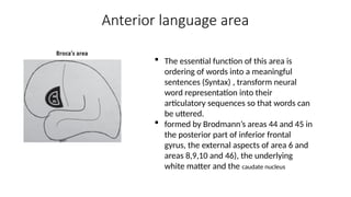
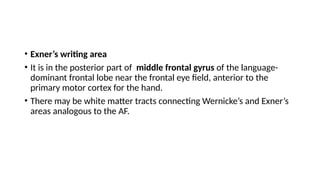
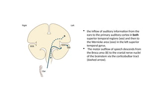
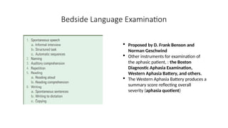
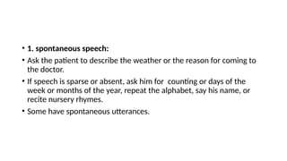
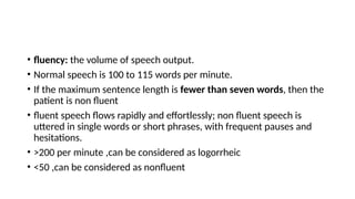
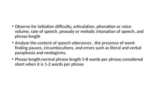
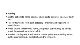
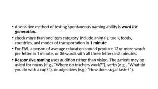
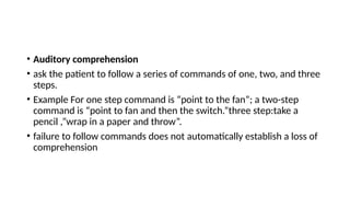
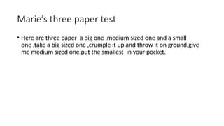
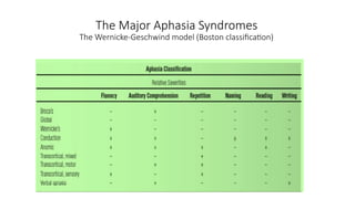
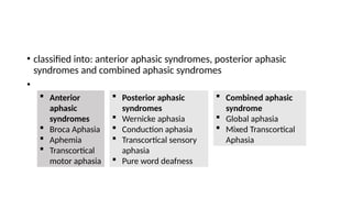
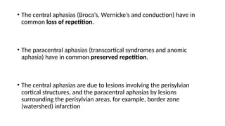
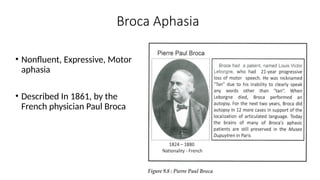
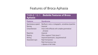
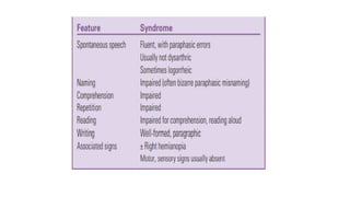
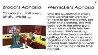
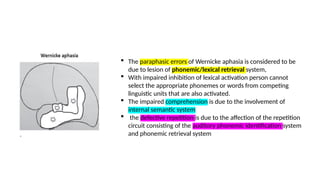
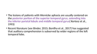
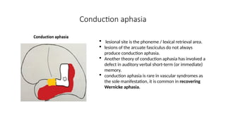
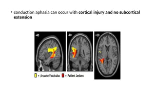
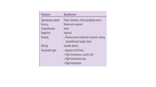
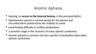
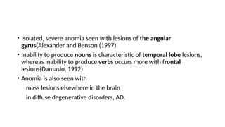
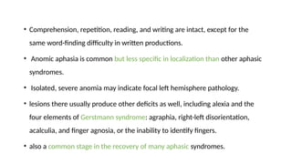
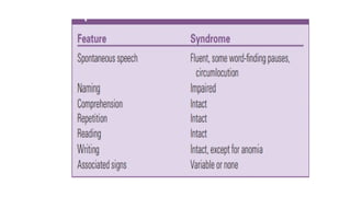
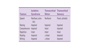
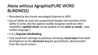
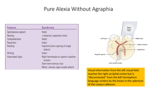
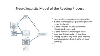
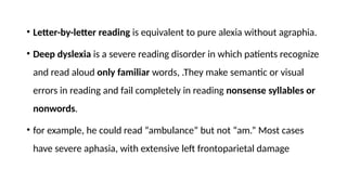
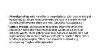
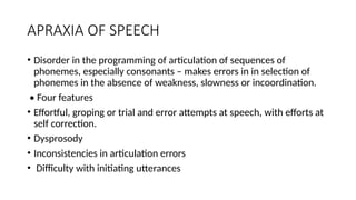
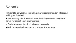
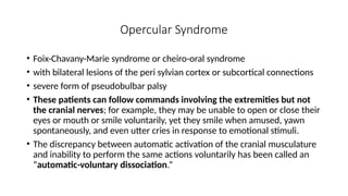
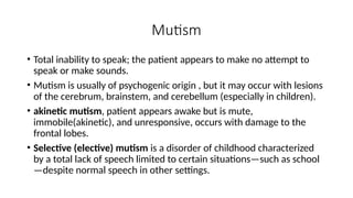
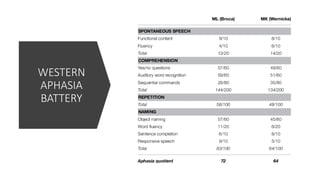
APHASIA
DR ATHIRA T K
SENIOR RESIDENT
GMCT
INTRODUCTION
• Language is a complex system of communication for expressing
thoughts and ideas by speech ,symbols, by writing or by gestures and
pantomime.
• It is a function of the cerebral cortex.
• The ability to prduce language has recently been linked to a gene –
FOXP2gene.
• Speech is the articulation and phonation of language sounds
LINGUISTIC ASPECTS OF LANGUAGE
• Linguistic communication requires:
• retention, recall, and visualization of the symbols
• reception and interpretation of these acts
• motor acts necessary for execution
• three cortical levels involved in language comprehension:
• 1. level of arrival, a function of the primary cortical reception areas
• 2. second level is that of knowing, or gnosis
• 3. third level is recognition of symbols in the form of words, and
association of learned symbols
• There are also three levels of motor speech function.
• 1. emotional level: Most primitive.
• 2. automatic level: concerned with casual, automatic speech
• 3.propositional, volitional, symbolic, or intellectualized language:
The highest level. most easily disrupted and most difficult to repair
• Aphasia is disorder of language that is acquired (previously intact)
secondary to brain damage, usually affects other language functions
such as reading and writing(ALEXANDER AND BENSON 1997)
• dysphasia is congenital or developmental language disorders
• Aphasia is distinguished from motor speech disorders, dysarthria,
dysphonia (voice disorders), stuttering, and apraxia of speech.
linguistic components
• Phonemes are the smallest distinctive sound units
• morphology :use of appropriate word endings and connector words
for grammatical categories such as tenses, possessives, and singular
versus plural
• semantics refers to word meanings
• lexicon is the internal dictionary (or vocabulary of a person. )
• syntax is the grammatical construction of phrases and sentences.
• Discourse :use of these elements to create organized and logical
expression of thoughts.
• Pragmatics : proper use of speech and language in a conversational
setting, including pausing while others are speaking, taking turns
properly, and responding to questions, as to express emotions, to
request, to give commands etc.
• Prosody is the intonation of speech and includes stress, pitch, melody,
rhythm, spacing between words (juncture) and the interval and
pauses in conversational language.
• Grammar includes both morphology and syntax.
Neuroanatomical basis
• 1.the reception and processing of spoken language take place in the
auditory system,
• 2.The decoding of sounds into linguistic information involves the left
superior temporal gyrus and sulcus.
• 3. auditory information is transmitted via direct and indirect dorsal
pathways to Broca area in the posterior inferior frontal gyrus, for
both repetition and spontaneous speech.
• 4. programming the neurons in the adjacent motor cortex, for the
mouth and larynx, for speech production
• 5.The inferior parietal lobule,(the supramarginal gyrus) may also be
involved in encoding of speech sounds for production.
• 6.Reading requires the perception of visual language stimuli by the
occipital cortex, followed by processing into auditory language
information.
• 7. Writing involves the activation of motor neurons projecting to the
arm and hand.
Centres important in language
A, Angular gyrus.
B, Broca’s area.
EC, Exner’s writing center;
SP, Superior parietal
lobule, which with the
PCG (postcentral gyrus) is
important in tactile
recognition;
T, Pars triangularis;
W, Wernicke’s area.
LANGUAGE AREAS AND CEREBRAL DOMINANCE:
• Broca’s area in the posterior part of inferior frontal gyrus
• Wernicke’s area in the posterior part of superior temporal gyrus
• The left cerebral hemisphere is dominant for language in 99% of right
handers, and 60% to 70% of left handers.
• Of the remaining left handers, about half are right hemisphere
dominant and about half have mixed dominance
• crossed aphasia: aphasia with right hemisphere lesion , in right
handed people with right hemispheric dominance
• Shifted sinistrals (anomalous dextrals) are naturally left-handed
individuals forced by parents or teachers early in life to function right
handed, primarily for writing.
• In bilinguals, the cerebral representation of some functions is similar
for both languages.
• Pitres’ law states that recovery from aphasia will be best for the
language most used.
• Ribot’s rule holds that recovery will be best for the native language.
• most patients show parallel recovery in both languages.
Posterior Language Areas
Wernicke’s area.
consists of at least three areas.
The blue striped area constitutes
the Heschl’s gyrus (primary
auditory cortex).
• 1. Auditory phoneme perception system
• This system analyses the auditory input for phonemic content
independent of word meaning.
• It is adjacent to superior temporal sulcus in both hemispheres.
• 2. Internal semantic system
• It is the word meaning system
• Includes mainly the anterolateral temporal lobe (including middle
temporal gyrus and ventral temporal lobe) and angular gyrus.
• medial parietal lobe, medial prefrontal cortex and inferior prefrontal
regions are also involved
• 3. Phonological/lexical retrieval system
• It processes for the sequence of phonemes to form the words to be
spoken.
• It is carried out by Wernicke’s area, (Brodmann area 22) and the
supramarginal gyrus of parietal lobe (area 40)
Comprehension
involves mapping
sequences of the
phonemes received in
the phonemic
perception system onto
the word meaning in the
internal semantic system
Speech repetition :
involves input from auditory
phoneme system to
phonological retrieval system
Reading aloud :
involves direct mapping
of visual word forms
from fusiform gyrus to
phonological/ lexical
retrieval system
Spontaneous speech
involves retrieval of
word meaning from
semantic system onto
phoneme
retrieval system.
• visual information is also processed in these Posterior Language
Areas akin to the spoken language.
• The fusiform gyrus function similar to the phoneme perception
system in visual language.
Anterior language area
 The essential function of this area is
ordering of words into a meaningful
sentences (Syntax) , transform neural
word representation into their
articulatory sequences so that words can
be uttered.
 formed by Brodmann’s areas 44 and 45 in
the posterior part of inferior frontal
gyrus, the external aspects of area 6 and
areas 8,9,10 and 46), the underlying
white matter and the caudate nucleus
• Exner’s writing area
• It is in the posterior part of middle frontal gyrus of the language-
dominant frontal lobe near the frontal eye field, anterior to the
primary motor cortex for the hand.
• There may be white matter tracts connecting Wernicke’s and Exner’s
areas analogous to the AF.
 the inflow of auditory information from the
ears to the primary auditory cortex in both
superior temporal regions (xxx) and then to
the Wernicke area (ooo) in the left superior
temporal gyrus.
 The motor outflow of speech descends from
the Broca area (B) to the cranial nerve nuclei
of the brainstem via the corticobulbar tract
(dashed arrow).
EXAMINATION OF THE PATIENT WITH
APHASIA
Bedside Language Examination
 Proposed by D. Frank Benson and
Norman Geschwind
 Other instruments for examination of
the aphasic patient, : the Boston
Diagnostic Aphasia Examination,
Western Aphasia Battery, and others.
 The Western Aphasia Battery produces a
summary score reflecting overall
severity (aphasia quotient)
• 1. spontaneous speech:
• Ask the patient to describe the weather or the reason for coming to
the doctor.
• If speech is sparse or absent, ask him for counting or days of the
week or months of the year, repeat the alphabet, say his name, or
recite nursery rhymes.
• Some have spontaneous utterances.
• fluency: the volume of speech output.
• Normal speech is 100 to 115 words per minute.
• If the maximum sentence length is fewer than seven words, then the
patient is non fluent
• fluent speech flows rapidly and effortlessly; non fluent speech is
uttered in single words or short phrases, with frequent pauses and
hesitations.
• >200 per minute ,can be considered as logorrheic
• <50 ,can be considered as nonfluent
• Observe for initiation difficulty, articulation, phonation or voice
volume, rate of speech, prosody or melodic intonation of speech, and
phrase length
• Analyze the content of speech utterances , the presence of word-
finding pauses, circumlocutions, and errors such as literal and verbal
paraphasia and neologisms.
• Phrase length:normal phrase length 5-8 words per phrase,considered
short when it is 1-2 words per phrase
• Naming
• ask the patient to name objects, object parts, pictures, colors, or body
parts.
• Test for few items from each category , anomia can be specific to
word classes.
• When unable to retrieve a name, an aphasic patient may be able to
select the correct name from a list.
• Another naming test is to have the patient point to something named
by the examiner (e.g., the telephone, the window).
• A sensitive method of testing spontaneous naming ability is word list
generation.
• check more than one item category; include animals, tools, foods,
countries, and modes of transportation in 1 minute
• For FAS, a person of average education should produce 12 or more words
per letter in 1 minute, or 36 words with all three letters in 3 minutes.
• Responsive naming uses audition rather than vision. The patient may be
asked for nouns (e.g., “Where do teachers work?”), verbs (e.g., “What do
you do with a cup?”), or adjectives (e.g., “How does sugar taste?”).
• Auditory comprehension
• ask the patient to follow a series of commands of one, two, and three
steps.
• Example For one step command is “point to the fan”; a two-step
command is “point to fan and then the switch.”three step:take a
pencil ,”wrap in a paper and throw”.
• failure to follow commands does not automatically establish a loss of
comprehension
Marie’s three paper test
• Here are three paper a big one ,medium sized one and a small
one ,take a big sized one ,crumple it up and throw it on ground,give
me medium sized one,put the smallest in your pocket.
• The patient must hear the command, understand the language the
examiner speaks, and possess the motor ability to execute it,
including the absence of apraxia
• Patients with a left hemisphere lesion may have apraxia for functions
of their nonparetic left hand. (sympathetic apraxia, because of
involvement of fibers that transmit information from the language
areas on the left to the motor areas on the right ).
• Both comprehension and retention are evaluated by telling a short
story and then asking questions about it.
• Patients with impaired comprehension have particular difficulty with
passive constructions (e.g., “The lion was killed by the tiger; which
animal is dead?” and possessives (e.g., “Is my wife’s brother a man or
a woman?”).
• Patients who are unable to comprehend spoken or written language
may understand pantomime, gestures, and symbols.
• Imitation, is a more lower-level function than comprehension.
• Repetition
• inability to repeat is proportional to the defect in comprehension or
fluency.
• A patient’s repetition span (i.e., the number of words he can repeat) is
usually two more than his digit span.
• A popular phrase for testing repetition in aphasia is “no ifs, ands, or
buts.”
• A better repetition test is “they heard him speak on the radio last
night’ (modified from the Boston diagnostic aphasia examination).
• Patients with impaired repetition may omit words, change the word
order, or commit paraphasic errors.
• Dysarthric patients have difficulty with rapid and variable sequences
of consonants, such as “Methodist Episcopal,” whereas people with
aphasia have difficulty with grammatically complex sentences, like
“no ifs, ands, or buts”
• Repetition is preserved in anomic, transcortical, and some cases of
subcortical aphasia.
• Reading :
• Written language is perceived by the visual system and the
information conveyed to the perisylvian language centers.
• Dysfunction of the language centers or interruption of the
connections with the visual system may cause an inability to read
(alexia).
• pure word blindness: alexia without any accompanying inability to
comprehend speech
• Dyslexia is developmental or congenital defect of reading.
• Reading should be Tested aloud and for comprehension.
• Consider the patient’s premorbid reading ability.
• Judge reading ability by having the patient follow a written command
such as close your eyes
• For patients unable to read aloud, use questions that can be answered
by “yes” or “no,” or by gestures.
• Oral reading (visual input-oral output) is comparable to copying (visual
input-manual output), repetition (auditory input-oral output), and
transcribing dictation (auditory input-manual output), and may be
preserved despite impaired reading comprehension.
• Writing:
• analysis of expressive language and of spelling,
• may be the most sensitive indicator of mild aphasia
• In all aphasias, reading and writing are typically worse than
understanding and speaking, probably because they are secondarily
acquired skills
• Patients who are aphasic in speech are also aphasic in writing, but writing
may be preserved in patients with dysarthria or verbal apraxia.
• Spontaneous writing, such as a sentence describing why the patient has
come for examination, is sensitive for the detection of language difficulty.
• When spontaneous writing fails, writing to dictation and copying
should be tested.
• The ability to write to dictation is analogous to the ability to repeat
verbal material.
• It also assesses the ability to transfer information from the visual
system to the language areas, test the connections between the
receptive language areas and Exner’s writing center.
• An inability to copy may be due to apraxia.
The Major Aphasia Syndromes
The Wernicke-Geschwind model (Boston classification)
• classified into: anterior aphasic syndromes, posterior aphasic
syndromes and combined aphasic syndromes
•
 Anterior
aphasic
syndromes
 Broca Aphasia
 Aphemia
 Transcortical
motor aphasia
 Posterior aphasic
syndromes
 Wernicke aphasia
 Conduction aphasia
 Transcortical sensory
aphasia
 Pure word deafness
 Combined aphasic
syndrome
 Global aphasia
 Mixed Transcortical
Aphasia
• The central aphasias (Broca’s, Wernicke’s and conduction) have in
common loss of repetition.
• The paracentral aphasias (transcortical syndromes and anomic
aphasia) have in common preserved repetition.
• The central aphasias are due to lesions involving the perisylvian
cortical structures, and the paracentral aphasias by lesions
surrounding the perisylvian areas, for example, border zone
(watershed) infarction
Broca Aphasia
• Nonfluent, Expressive, Motor
aphasia
• Described In 1861, by the
French physician Paul Broca
• The patient speaks hesitantly, meaning-containing nouns and verbs
but omitting small grammatical words and morphemes. This pattern
is called agrammatism or telegraphic speech. An example is “wife
come hospital.”
• They make many phonemic errors, with substitution of phonemes
usually differing only slightly from the correct target (e.g., p for b).
• Naming is deficient, but the patient often manifests a “tip of the
tongue” phenomenon, getting out the first letter or phoneme of the
correct name.
• Auditory comprehension intact, with some deficiency, in the
comprehension of complex syntax.
• Patients may be mute or may produce only single words, often with
dysarthria and apraxia of speech
• Repetition is hesitant in these patients, resembling their spontaneous
speech.
• Reading is often impaired, despite relatively preserved auditory
comprehension.
• Writing is virtually always deficient in Broca aphasia
Associated neurological deficits include
• Right hemiparesis, hemisensory loss,
• Apraxia of the oral apparatus and the nonparalyzed (left) limbs.
• Patients with Broca aphasia are typically aware of the deficit
• An important clinical feature of Broca aphasia is its frequent
association with depression (Robinson 1997).and frustrated by their
deficits.
• At times, they become withdrawn and refuse help or therapy.
Usually, the depression lifts as the deficit recovers, but it may be a
limiting factor in rehabilitation
Features of Broca Aphasia
The lesions 
Broca area in the posterior part of the inferior frontal gyrus, along with damage
to adjacent cortex and subcortical white matter.
• Most patients with lasting Broca aphasia, including Broca original cases, have
much larger left frontoparietal lesions, including most of the territory of the
upper division of the left middle cerebral artery.
• Such patients typically evolve from global to Broca aphasia over weeks to
months.
• Patients who manifest Broca aphasia immediately after their strokes, by
contrast, have smaller lesions of the inferior frontal region, and their deficits
generally resolve quickly.
• lesions restricted to the lower precentral gyrus produced only
dysarthria and mild expressive disturbance.(In computed tomography (CT) scan
analyses at the Boston Veterans Administration Medical Center)
• Lesions involving the traditional Broca area (Brodmann areas 44 and
45) resulted in difficulty initiating speech
• lesions combining Broca area, the lower precentral gyrus, and
subcortical white matter yielded the full syndrome of Broca aphasia
(Alexander et al., 1990).
• In studies by the same group, damage to two subcortical white matter sites—the rostral
subcallosal fasciculus deep to the Broca area and the periventricular white matter adjacent to the
body of the left lateral ventricle—was required to cause permanent nonfluency.
lesional site includes not only area 44 and
45 but also surrounding frontal fields (the
external aspects of area 6 and areas 8,9,10
and 46) and the underlying white matter
and caudate nucleus
When damage is restricted to Broca’s area
alone, true Broca aphasia does not develop.
They develop a mild and transient disturbance
of speech with impaired prosody and
articulatory agility called Broca’s area aphasia or
aphemia ..
Wernicke’s Aphasia(Fluent, Receptive, Sensory,
Posterior, Post Rolandic)
 speech pattern is effortless and sometimes even excessively fluent
(logorrhea)
 It is meaningless because of paragrammatisms and paraphasias which
include phonemic and verbal paraphasias, and neologisms.
 When the language is full of neologisms it is called jargon (devil)
speech
 Writing samples are especially useful in the detection of mild
Wernicke's aphasia.
• Naming in Wernicke aphasia is deficient, often with bizarre, paraphasic
substitutions for the correct name.
• Auditory comprehension is impaired, sometimes even for simple
nonsense questions.
• Repetition is impaired; whispering a phrase in the patient’s ear, as in a
hearing test, may help cue the patient to attempt repetition.
• Reading comprehension is usually affected similarly to auditory
comprehension, but occasional patients show greater deficits in one modality
versus the other.
• Writing is also impaired, but in a manner quite different from that of
Broca aphasia.
• The patient usually has no hemiparesis and can grasp the pen and write
easily.
• Written productions are even more abnormal than oral ones, however,
in that spelling errors are also evident.
• Associated signs are limited in Wernicke aphasia
• most patients have no elementary motor or sensory deficits, although a
partial or complete right homonymous hemianopia may be present.
• The psychiatric manifestations of Wernicke aphasia:
• Depression is less common; many Wernicke aphasics seem unaware of or
unconcerned about their communicative deficits.
• With time, some patients become angry or paranoid about the inability of family
members and medical staff to understand them.
• The agitated patient, speaking gibberish and with no gross neurologic deficit, is
frequently thought to be psychotic.
• This behavior, similarly to depression, may hinder rehabilitative efforts
• in its mildest form, there are mild paraphasias and minimal difficulty understanding grammatically
complex material (mini-Wernicke’s).
 The paraphasic errors of Wernicke aphasia is considered to be
due to lesion of phonemic/lexical retrieval system,
 With impaired inhibition of lexical activation person cannot
select the appropriate phonemes or words from competing
linguistic units that are also activated.
 The impaired comprehension is due to the involvement of
internal semantic system
 the defective repetition is due to the affection of the repetition
circuit consisting of the auditory phonemic identification system
and phonemic retrieval system
.
• The lesions of patients with Wernicke aphasia are usually centered on
the posterior portion of the superior temporal gyrus, extending into
the inferior parietal lobule and middle temporal gyrus( Kertesz et al.,
1993)
• Recent literature (see Binder, 2015; Bonilha et al., 2017) has suggested
that auditory comprehension is subserved by wider regions of the left
temporal lobe.
• Electrical stimulation of the Wernicke area produces consistent
interruption of auditory comprehension, supporting the importance
of this region for decoding auditory language (Boatman et al., 1995).
• A receptive speech area in the left inferior temporal gyrus has also
been suggested by electrical stimulation studies and by a few
descriptions of patients with seizures involving this area (Kirshner et
al., 1995).
• In terms of vascular anatomy, Wernicke aphasia is generally
associated with the inferior division of the left middle cerebral artery
Global Aphasia(Total, Expressive-Receptive, Complete)
 It is a summation of the deficits of Broca aphasia and Wernicke
aphasia.
 Spontaneous speech, naming and repetition may be limited to a
single preservative word (e.g. yes, yes) or non-word utterance.
 The lesions involve both the inferior frontal and the superior
temporal regions and often the parietal lobe.
• Speech is nonfluent or mute, but comprehension is also poor, as are
naming, repetition, reading, and writing.
• Most patients have dense right hemiparesis, hemisensory loss, and
often hemianopia, although occasional patients have little
hemiparesis.
• Milder aphasic syndromes in which all modalities of language are
affected are often called mixed aphasias.
• The lesions of patients with global aphasia are usually large,
involving both the inferior frontal and the superior temporal regions
and often much of the parietal lobe in between.
• This lesion represents most of the territory of the left middle cerebral
artery.
• Patients in whom the superior temporal gyrus is spared tend to
recover their auditory comprehension and to evolve toward the
syndrome of Broca aphasia.
• Recovery in global aphasia may be prolonged; global aphasics may
recover more during the second 6 months than during the first 6
months after a stroke (Sarno and Levita, 1979).
Pure Word Deafness(auditory verbal agnosia)
• Isolated loss of auditory comprehension and repetition, without any
abnormality of speech, naming, reading, or writing.
• mild aphasic deficits, (paraphasic speech).
• They lose the ability to understand spoken words, repeat words and
write down from dictation.
• But can recognize nonverbal sounds, such as animal cries
• Hearing for pure tones reserved, (different from cortical deafness).
 There is involvement of auditory phoneme
identification system in pure word deafness.
 the anatomical substrate is a bilateral lesion, isolating
Wernicke area from input from the primary auditory
cortex, in the bilateral Heschl gyri.
 Pure word deafness is thus an example of a
“disconnection syndrome,” (deficit results from loss of
white matter connections rather than of gray matter
language centers. )
 Some cases of pure word deafness, however, have unilateral,
left temporal lesions, if the lesion is placed such as to
disconnect Wernicke area from primary auditory cortex of
both hemispheres
Conduction(Associative, Commissural,
Central, Deep) Aphasia
• Striking deficit of repetition.
• Most patients have relatively fluent spontaneous speech but make literal
paraphasic errors and hesitate frequently for self-correction.
• Naming may be impaired, but auditory comprehension is preserved.
• Repetition may be disturbed to seemingly ridiculous extremes, such that a
patient who can express himself or herself at a sentence level and comprehend
conversation may be unable to repeat even single words.
• Reading and writing are somewhat variable, but reading aloud may
share some of the same difficulty as repeating.
• Associated deficits include hemianopia in some patients; right-sided
sensory loss may be present, but right hemiparesis is usually mild or
absent.
• Some patients have limb apraxia, creating a misimpression that
comprehension is impaired.
• The lesions of conduction aphasia usually involve either the superior
temporal or the inferior parietal regions.
• Benson and associates suggested that patients with limb apraxia have
parietal lesions, whereas those without apraxia have temporal lesions
(Benson et al., 1973).
• Conduction aphasia may represent a stage of recovery in patients with
Wernicke aphasia in whom the damage to the superior temporal
gyrus is not complete.
Conduction(Associative, Commissural, Central, Deep) Aphasia
 Described by Carl Wernicke.
 It is a classical disconnection syndrome, (Geschwind)
 the arcuate fasciculus, is the site of disconnection.
white matter tract traveling from the deep temporal lobe, around the sylvian
fissure to the frontal lobe, as the site of disconnection.
Anatomical involvement of the arcuate fasciculus is present in most, if not all, cases of conduction
aphasia, but some doubt has been raised about the importance of the arcuate fasciculus to
conduction aphasia or even to repetition (Bernal and Ardila, 2009).
• In cases of conduction aphasia, there is usually also cortical involvement of the
supramarginal gyrus or temporal lobe.
• The supramarginal gyrus appears to be involved in auditory immediate memory
and in phoneme perception related to word meaning, as well as phoneme
generation (Hickok and Poeppel, 2000).
• Lesions in this area are associated with conduction aphasia and phonemic
paraphasic errors.
• The etiology is usually an embolic occlusion of a terminal branch of the MCA,
usually involve either the superior temporal or the inferior parietal regions.
Conduction aphasia
 lesional site is the phoneme / lexical retrieval area.
 lesions of the arcuate fasciculus do not always
produce conduction aphasia.
 Another theory of conduction aphasia has involved a
defect in auditory verbal short-term (or immediate)
memory.
 conduction aphasia is rare in vascular syndromes as
the sole manifestation, it is common in recovering
Wernicke aphasia.
• conduction aphasia can occur with cortical injury and no subcortical
extension
Anomic Aphasia
 Naming, or access to the internal lexicon, is the principal deficit.
 Spontaneous speech is normal except for the pauses and
circumlocutions produced by the inability to name
 word-finding difficulty in written productions.
 a common stage in the recovery of many aphasic syndromes
 Anomic aphasia is common but less specific in localization than other
aphasic syndromes.
• Isolated, severe anomia seen with lesions of the angular
gyrus(Alexander and Benson (1997)
• Inability to produce nouns is characteristic of temporal lobe lesions,
whereas inability to produce verbs occurs more with frontal
lesions(Damasio, 1992)
• Anomia is also seen with
mass lesions elsewhere in the brain
in diffuse degenerative disorders, AD.
• Comprehension, repetition, reading, and writing are intact, except for the
same word-finding difficulty in written productions.
• Anomic aphasia is common but less specific in localization than other aphasic
syndromes.
• Isolated, severe anomia may indicate focal left hemisphere pathology.
• lesions there usually produce other deficits as well, including alexia and the
four elements of Gerstmann syndrome: agraphia, right-left disorientation,
acalculia, and finger agnosia, or the inability to identify fingers.
• also a common stage in the recovery of many aphasic syndromes.
Transcortical Aphasias
 Repetition is normal, presumably because the causative lesions do not
disrupt the perisylvian language circuit from the Wernicke area through the
arcuate fasciculus to the Broca area.
 These lesions disrupt connections from other cortical centers into the
language circuit (hence the name “transcortical”).
 Associated with watershed lesions, between the ACA & MCA and MCA &
PCA distributions
• Mixed transcortical aphasia, or the syndrome of the isolation of the
speech area, is a global aphasia with preserved repetition , often
echolalia, but has no propositional speech or comprehension
• This syndrome is rare, occurring predominantly in large, watershed
infarctions of the left hemisphere or both hemispheres that spare the
perisylvian cortex, or in advanced dementias.
• Transcortical motor aphasia  analogue of Broca aphasia in which speech
is hesitant or telegraphic, comprehension is relatively spared, but repetition
is fluent.
• lesions in the frontal lobe, anterior to the Broca area, in the deep frontal
white matter, or in the medial frontal region, in the vicinity of the
supplementary motor area.
• All of these lesion sites are within the territory of the anterior cerebral
artery, separating this syndrome from the aphasia syndromes of the middle
cerebral artery (Broca, Wernicke, global, and conduction).
• The third transcortical syndrome, transcortical sensory aphasia
• An analogue of Wernicke aphasia in which fluent, paraphasic speech,
paraphasic naming, impaired auditory and reading comprehension,
and abnormal writing coexist with normal repetition.
• This syndrome is relatively uncommon, occurring in strokes of the left
temporo-occipital area and in dementias.
Subcortical (extrasylvian) aphasia
• Language disorders that arise from lesions involving the thalamus, caudate,
putamen, periventricular white matter, or internal capsule of the language
dominant hemisphere.
• cortical hypoperfusion is common in subcortical aphasia.
• The presentation of a difficult-to-classify aphasic syndrome, in the presence
of dysarthria and right hemiparesis, should lead to suspicion of a subcortical
lesion
• two major groups of aphasic symptomatology
1 aphasia with thalamic lesions
2 aphasia with lesions of the subcortical white matter and basal ganglia.
• Left thalamic hemorrhages frequently produce a Wernicke-like fluent aphasia, with better
comprehension than cortical Wernicke aphasia.
• A fluctuating or “dichotomous” state has been described, alternating between an alert state
with nearly normal language and a drowsy state in which the patient mumbles paraphasically
and comprehends poorly.
• Luria has called this a quasi-aphasic abnormality of vigilance, in that the thalamus plays a role
in alerting the language cortex.
• The thalamus, a relay for the reticular activating system, appears to
alert the language cortex, and lesions of the dominant thalamus
frequently produce fluent aphasia.
• Nuclei of the basal ganglia involved in motor functions, especially the
caudate nucleus and putamen, lesions produce motor aphasia.
• In both forms, repetition is relatively preserved, and the patients
usually have an accompanying hemiplegia
• The anterior subcortical aphasia syndrome” (caudate or striato
capsular aphasia) : dysarthria, decreased fluency, mildly impaired
repetition, poor naming and mild disturbance or preserved
comprehension, but not telegraphic (Mega and Alexander, 1994)
• most closely resembles Broca aphasia, transcortical motor aphasia but
with greater dysarthria and less language dysfunction.
• The most common lesion is an infarct involving the anterior putamen,
caudate nucleus, and anterior limb of the internal capsule.
• In the posterior syndrome(putamen and deep temporal white matter
(temporal isthmus), there is fluent speech without dysarthria, poor
comprehension, and poor naming.
• resembles Wernicke’s or transcortical sensory aphasia
• associated with fluent, paraphasic speech and impaired comprehension
resembling Wernicke aphasia (Naeser et al., 1990).
• In both forms, repetition is relatively preserved, and the patients usually
have an accompanying hemiplegia
Alexia
• Alexia, or acquired inability to read.
• Abnormalities of reading with comprehension produce alexia,
whereas abnormalities of reading aloud produce paralexia.
• There are three types of alexia
• 1. Alexia without agraphia (posterior alexia or pure alexia)
• 2. Alexia with agraphia (central alexia)
• 3. Third alexia (anterior alexia)
Alexia without Agraphia(PURE WORD
BLINDNESS)
• Described by the French neurologist Dejerine in 1892.
• loss of ability to read and comprehend despite full retention of the
ability to write. But the patient readily recognize words by other
modalities.(spelled aloud, words spelled on the palm or words , copy
written language )
• It is a linguistic blindfolding
• may result from damage to pathway conveying visual input from both
hemispheres to the dominant angular gyrus(intact), disconnected
from the visual centers.
Pure Alexia Without Agraphia
Visual information from the left visual field
reaches the right occipital cortex but is
“disconnected” from the left hemisphere
language centers by the lesion in the splenium
of the corpus callosum.
• Patients can write but cannot read their own writing. They cannot read words at a glance, as
normal readers do.
• On bedside examination, speech, auditory comprehension, and repetition are normal.
• Naming may be deficient, especially for colors.
• Patients initially cannot read at all; as they recover, they learn to read letter by letter, spelling
out words laboriously.
• By contrast, they quickly understand words spelled orally to them, and they can spell normally.
• Associated deficits include a right hemianopia or right upper quadrant defect in nearly all
patients and, frequently, a deficit of shortterm memory.
• The causative lesion in pure alexia is nearly always a stroke in the
territory of the left posterior cerebral artery, with infarction of the
medial occipital lobe, often the splenium of the corpus callosum, and
often the medial temporal lobe.
• Dejerine postulated a disconnection between the intact right visual
cortex and left hemisphere language centers, particularly the angular
gyrus.
• Geschwind later rediscovered this disconnection hypothesis.
• Although Damasio and Damasio (1983) found splenial involvement in
only 2 of 16 cases, they postulated a disconnection within the deep
white matter of the left occipital lobe
Alexia with Agraphia(central alexia)
Lesions of angular gyrus producing alexia with
agraphia
• The second classic alexia syndrome
• described by Dejerine in 1891
• an acquired illiteracy, in which a previously educated patient is
rendered unable to read or write.
• The oral language modalities of speech, naming, auditory
comprehension, and repetition are largely intact, but many cases
manifest a fluent, paraphasic speech pattern with impaired naming.
• Associated deficits include right hemianopia and elements of
Gerstmann syndrome: agraphia, acalculia, right–left disorientation,
and finger agnosia.
• The lesions typically involve the inferior parietal lobule, especially the
angular gyrus.
• Etiologies include strokes in the territory of the angular branch of the
left middle cerebral artery or mass lesions in the same region.
Third Alexia or frontal alexia(APHASIC ALEXIA)
• Seen in Broca’s aphasia
• Impaired ability to comprehend syntactic structures. (in
understanding sentences when the meaning depends on syntax).
• For example, they have difficulty in understanding the sentence
“Ravana was killed by Rama”, whereas they have no difficulty in
understanding the sentence “Rama killed Ravana”.
site of lesion for the pure alexia
A lesion in left occipital lobe
along with lesion of the
adjacent splenium of corpus
callosum produce this
syndrome.
‘visual word form area’ lesion
producing pure alexia
The ‘visual word form area’ is
disconnected from the ‘higher’
language areas ( the angular
gyrus)
• Examination of Reading
• 1. Reading comprehension
Matching written words with objects
Carrying out written commands
paragraph comprehension
• 2. Reading aloud
Letters
Words
Sentences
• 3. Comprehension (reading) of symbolic system
• e.g. Traffic signals
• Neurolinguists and cognitive psychologists have divided alexias
according to breakdowns in specific stages of the reading process.
• The linguistic concepts of surface structure versus the deep meanings
of words have been instrumental in these new classifications.
• Four patterns of alexia (or dyslexia) have been recognized:
1 Letter-by-letter reading
2 deep
3 phonological
4 surface dyslexia
Neurolinguistic Model of the Reading Process
 there are three separate routes to reading:
 1 is the phonological (or grapheme-phoneme
conversion) route;
 2 is the semantic (or lexical-semantic-
phonological) route; and
 3 is the nonlexical phonological route.
 in surface dyslexia, only 1 is functional
 In deep dyslexia, only route 2 can operate;
 in phonological dyslexia, 3 is the principal
pathway;
 .
• Letter-by-letter reading is equivalent to pure alexia without agraphia.
• Deep dyslexia is a severe reading disorder in which patients recognize
and read aloud only familiar words, .They make semantic or visual
errors in reading and fail completely in reading nonsense syllables or
nonwords.
• for example, he could read “ambulance” but not “am.” Most cases
have severe aphasia, with extensive left frontoparietal damage
• Phonological dyslexia is similar to deep dyslexia, with poor reading of
nonwords, but single nouns and verbs are read in a nearly normal
fashion, and semantic errors are rare. (SEQUINS AS SEQUENCE )
• surface dyslexia, spared ability to read by grapheme-phoneme
conversion but inability to recognize long words, at a glance, or
irregular words. These patients can read nonsense syllables but not
words of irregular spelling, such as “colonel” or “yacht.” Their errors
tend to be phonological rather than semantic or visual (e.g.,
pronouncing rough and though alike)
Agraphia
• writing may be affected either in isolation (pure agraphia, left frontal
or parietal lesions) or in association with aphasia (aphasic agraphia).
• writing can be impaired by motor disorders, by apraxia, and by
visuospatial deficits.
• phonological agraphia involves the inability to convert phonemes
into graphemes or to write pronounceable nonsense syllables, in the
presence of ability to write familiar words.
• Deep dysgraphia is similar to phonological agraphia, but the patient
can write nouns and verbs better than articles, prepositions,
adjectives, and adverbs.
• In lexical or surface dysgraphia, patients can write regularly spelled
words and pronounceable nonsense words but not irregularly spelled
words. These patients have intact phoneme-grapheme conversion but
cannot write by a whole-word or “lexical” strategy.
Neurolinguistic Model of Writing and the Agraphia
 In deep agraphia, only the semantic
(phonological-semantic-lexical) route
(1) is operative;
 in phonological agraphia, route (2), the
nonlexical phonological route produces
written words directly from spoken
words;
 in surface agraphia, only route (3), the
phoneme-grapheme pathway, can be
used to generate writing
NONDOMINANT HEMISPHERE LANGUAGE
DISTURBANCES
• Non–right-handers, are thought to have some speech function in the
nondominant hemisphere.
• Some of the recovery from aphasia and the persistence of emotional
and automatic speech suggest some language function may be
present in the minor hemisphere.
• Lesions of the nondominant hemisphere cause speech disturbances
that affect the nonlinguistic elements of language.
NONDOMINANT HEMISPHERE LANGUAGE
DISTURBANCES
• Affective aspects of language are impaired, such that the speech
sounds flat and unemotional
• There is loss or impairment of the rhythm and emotional elements of
language. (aprosody)
• understand what is said, but not how it is said.
• Motor aprosodia involves loss of expressive emotion with
preservation of emotional comprehension;
• sensory aprosodia involves loss of comprehension of affective
language, also called affective agnosia
• Dysprosody, hypo prosody or aprosody, may occur with right
hemisphere lesions. Patients lose the ability to convey emotion in
speech or to detect the emotion expressed by others.
• have difficulty in
understanding figurative meanings and in distinguishing the implicit
and implied meaning of a phrase such as “Can you tell me the time?”
processing nonliteral, context bound, complex aspects of language,
such as understanding figurative language, stories, and jokes
vital aspects of human communication as metaphor, humor, sarcasm,
irony, and related constituents of language that transcend the literal
meaning of words are sensitive to right hemisphere dysfunction.
ETIOLOGY
1.Vascular lesions, ischemic strokes, most common causes
• the sudden onset of Wernicke aphasia , with an embolus to the inferior
division of the left MCA
• Global aphasia may be caused by an embolus to the MCA stem, thrombosis of
the internal carotid artery, or even a hemorrhage into the deep basal ganglia.
• transcortical motor aphasia is specific to the ACA teritory and pure alexia
without agraphia is specific to the posterior cerebral artery territory.
• hemorrhages compress cerebral tissue without necessarily destroying it, the
ultimate recovery from aphasia is often better in hemorrhages than in
ischemic strokes
• 2.Traumatic brain injury.
• 3.Tumors of the left hemisphere (Aphasia secondary to an enlarging
tumor may be difficult to distinguish from a diffuse encephalopathy
or early dementia)
• 4.Seizures can be associated with aphasia in children as part of the
Landau-Kleffner syndrome or in adults as either an ictal or postictal
Todd phenomenon.
• 5.Migraine cause transitory aphasia. Wernicke aphasia may be seen in
a migraine attack, usually with complete recovery over a few hours.
• 6. psychogenic, often stuttering or stammering..
• 7.Infections of the nervous system.
• Brain abscesses
• Chronic infections, such as tuberculosis or syphilis,
• Herpes simplex encephalitis has a predilection for the temporal lobe
and orbital frontal cortex, and aphasia can be an early symptom,
often a permanent sequela in survivors of herpes encephalitis.
• AIDS dementia complex
• 8.Degenrative conditions:
• Alzheimer’s disease: resembles transcortical sensory aphasia,
• Parkinson’s disease : motor speech abnormalities related to the
extrapyramidal dysfunction.
• When dementia complicates, aphasia may also develop.
• PPA : In contrast to the aphasia of Alzheimer’s disease, the non fluent
variant of PPA tends to affect the anterior speech areas initially,
resulting in impaired and anomia but relative preservation of
comprehension.
RECOVERY AND REHABILITATION
• Patients with aphasia from acute disorders(stroke), generally show
spontaneous improvement over days, weeks, and months. (greatest
recovery occurs during the first few weeks and months
• The aphasia type often changes during recovery: global aphasia
evolves into Broca aphasia, and Wernicke aphasia into conduction or
anomic aphasia
• aphasia recovers best when left hemisphere areas.
• Speech-language therapy: script training, response elaboration
training, constraint-induced aphasia therapy, speech entrainment, and
melodic intonation therapy
• pharmacological agents :
• dopaminergic drug bromocriptine promotes spontaneous speech
output in transcortical motor aphasia.
• memantine and constraint-induced aphasia therapy (CIAT).
• stimulation techniques such as transcranial magnetic stimulation and
direct cortical stimulation
•DIFFERENTIAL DIAGNOSIS
Dysarthria
• The abnormal articulation of sounds or phonemes, or abnormal
neuromuscular activation of the speech muscles, affecting the speed,
strength, timing, range, or accuracy of movements involving speech
• The most consistent finding in dysarthria is the distortion of
consonant sounds.
• Dysarthria can affect not only articulation but also phonation,
breathing, or prosody (emotional tone) of speech.
• Total loss of ability to articulate is called anarthria
classification of dysarthria
APRAXIA OF SPEECH
• Disorder in the programming of articulation of sequences of
phonemes, especially consonants – makes errors in in selection of
phonemes in the absence of weakness, slowness or incoordination.
• Four features
• Effortful, groping or trial and error attempts at speech, with efforts at
self correction.
• Dysprosody
• Inconsistencies in articulation errors
• Difficulty with initiating utterances
Aphemia
• Patient to be wordless (mute) but leaves comprehension intact and
writing undisturbed.
• Anatomically, this is believed to be a disconnection of the motor
cortex for speech from lower centers.
• Controversy whether its equivalent to apraxia.
• Lesions around primary motor cortex or Broca`s area
Opercular Syndrome
• Foix-Chavany-Marie syndrome or cheiro-oral syndrome
• with bilateral lesions of the peri sylvian cortex or subcortical connections
• severe form of pseudobulbar palsy
• These patients can follow commands involving the extremities but not
the cranial nerves; for example, they may be unable to open or close their
eyes or mouth or smile voluntarily, yet they smile when amused, yawn
spontaneously, and even utter cries in response to emotional stimuli.
• The discrepancy between automatic activation of the cranial musculature
and inability to perform the same actions voluntarily has been called an
“automatic-voluntary dissociation.”
Mutism
• Total inability to speak; the patient appears to make no attempt to
speak or make sounds.
• Mutism is usually of psychogenic origin , but it may occur with lesions
of the cerebrum, brainstem, and cerebellum (especially in children).
• akinetic mutism, patient appears awake but is mute,
immobile(akinetic), and unresponsive, occurs with damage to the
frontal lobes.
• Selective (elective) mutism is a disorder of childhood characterized
by a total lack of speech limited to certain situations—such as school
—despite normal speech in other settings.
Stuttering (spasmophemia)
• faulty, spasmodic, interrupted speech with involuntary hesitations in
which the speaker is unable to produce the next expected sound.
• The flow of speech is broken by pauses during which articulation is
entirely arrested.
• Difficulty in enunciating syllables and joining them together.
• There may be localized cramps, spasms, and tic-like contractions of
the muscles essential to articulation,
NONORGANIC SPEECH DISORDERS
• hysterical aphonia, there is profound speech difficulty but no
disturbance of coughing or respiration.
• Manic patients may have a rapid flow of words (pressured speech),
often with an abrupt change of subject.
• In depression speech may be slow, sometimes with mutism.
• in hysterical mutism there may be the appearance of great effort
without the production of so much as a tone; in simulated mutism the
patient does not even make an effort.
• In schizophrenia there may be hesitancy with blocking, or negativism
with resulting mutism (alalia).
• common nonorganic dysphonia seen in children and adolescents are
the whispering syndrome, seen primarily in girls, and mutational
falsetto (hysterical high-pitched voice), seen primarily in boys.
• Palilalia, echolalia, and perseveration are often manifestations of
psychosis, but they can occur with organic lesions(frontal lobes).
• Palilalia is the repetition of one’s own speech.
• Echolalia is the meaningless repetition of heard words.
• Perseveration is the persistence of one reply or one idea in response
to various questions.
• Idioglossia is imperfect articulation with utterance of meaningless
sounds; the individual may speak with a vocabulary all his own.
(partial deafness, aphasia, and congenital word deafness.)
WESTERN
APHASIA
BATTERY
Thank you

Different types of aphasia with examples.pptx

  • 1.
    APHASIA DR ATHIRA TK SENIOR RESIDENT GMCT
  • 2.
    INTRODUCTION • Language isa complex system of communication for expressing thoughts and ideas by speech ,symbols, by writing or by gestures and pantomime. • It is a function of the cerebral cortex. • The ability to prduce language has recently been linked to a gene – FOXP2gene. • Speech is the articulation and phonation of language sounds
  • 3.
    LINGUISTIC ASPECTS OFLANGUAGE • Linguistic communication requires: • retention, recall, and visualization of the symbols • reception and interpretation of these acts • motor acts necessary for execution
  • 4.
    • three corticallevels involved in language comprehension: • 1. level of arrival, a function of the primary cortical reception areas • 2. second level is that of knowing, or gnosis • 3. third level is recognition of symbols in the form of words, and association of learned symbols
  • 5.
    • There arealso three levels of motor speech function. • 1. emotional level: Most primitive. • 2. automatic level: concerned with casual, automatic speech • 3.propositional, volitional, symbolic, or intellectualized language: The highest level. most easily disrupted and most difficult to repair
  • 6.
    • Aphasia isdisorder of language that is acquired (previously intact) secondary to brain damage, usually affects other language functions such as reading and writing(ALEXANDER AND BENSON 1997) • dysphasia is congenital or developmental language disorders • Aphasia is distinguished from motor speech disorders, dysarthria, dysphonia (voice disorders), stuttering, and apraxia of speech.
  • 7.
    linguistic components • Phonemesare the smallest distinctive sound units • morphology :use of appropriate word endings and connector words for grammatical categories such as tenses, possessives, and singular versus plural • semantics refers to word meanings • lexicon is the internal dictionary (or vocabulary of a person. ) • syntax is the grammatical construction of phrases and sentences. • Discourse :use of these elements to create organized and logical expression of thoughts.
  • 8.
    • Pragmatics :proper use of speech and language in a conversational setting, including pausing while others are speaking, taking turns properly, and responding to questions, as to express emotions, to request, to give commands etc. • Prosody is the intonation of speech and includes stress, pitch, melody, rhythm, spacing between words (juncture) and the interval and pauses in conversational language. • Grammar includes both morphology and syntax.
  • 10.
    Neuroanatomical basis • 1.thereception and processing of spoken language take place in the auditory system, • 2.The decoding of sounds into linguistic information involves the left superior temporal gyrus and sulcus. • 3. auditory information is transmitted via direct and indirect dorsal pathways to Broca area in the posterior inferior frontal gyrus, for both repetition and spontaneous speech. • 4. programming the neurons in the adjacent motor cortex, for the mouth and larynx, for speech production
  • 11.
    • 5.The inferiorparietal lobule,(the supramarginal gyrus) may also be involved in encoding of speech sounds for production. • 6.Reading requires the perception of visual language stimuli by the occipital cortex, followed by processing into auditory language information. • 7. Writing involves the activation of motor neurons projecting to the arm and hand.
  • 12.
    Centres important inlanguage A, Angular gyrus. B, Broca’s area. EC, Exner’s writing center; SP, Superior parietal lobule, which with the PCG (postcentral gyrus) is important in tactile recognition; T, Pars triangularis; W, Wernicke’s area.
  • 13.
    LANGUAGE AREAS ANDCEREBRAL DOMINANCE: • Broca’s area in the posterior part of inferior frontal gyrus • Wernicke’s area in the posterior part of superior temporal gyrus • The left cerebral hemisphere is dominant for language in 99% of right handers, and 60% to 70% of left handers. • Of the remaining left handers, about half are right hemisphere dominant and about half have mixed dominance
  • 14.
    • crossed aphasia:aphasia with right hemisphere lesion , in right handed people with right hemispheric dominance • Shifted sinistrals (anomalous dextrals) are naturally left-handed individuals forced by parents or teachers early in life to function right handed, primarily for writing.
  • 15.
    • In bilinguals,the cerebral representation of some functions is similar for both languages. • Pitres’ law states that recovery from aphasia will be best for the language most used. • Ribot’s rule holds that recovery will be best for the native language. • most patients show parallel recovery in both languages.
  • 16.
    Posterior Language Areas Wernicke’sarea. consists of at least three areas. The blue striped area constitutes the Heschl’s gyrus (primary auditory cortex).
  • 17.
    • 1. Auditoryphoneme perception system • This system analyses the auditory input for phonemic content independent of word meaning. • It is adjacent to superior temporal sulcus in both hemispheres.
  • 18.
    • 2. Internalsemantic system • It is the word meaning system • Includes mainly the anterolateral temporal lobe (including middle temporal gyrus and ventral temporal lobe) and angular gyrus. • medial parietal lobe, medial prefrontal cortex and inferior prefrontal regions are also involved
  • 19.
    • 3. Phonological/lexicalretrieval system • It processes for the sequence of phonemes to form the words to be spoken. • It is carried out by Wernicke’s area, (Brodmann area 22) and the supramarginal gyrus of parietal lobe (area 40)
  • 20.
    Comprehension involves mapping sequences ofthe phonemes received in the phonemic perception system onto the word meaning in the internal semantic system Speech repetition : involves input from auditory phoneme system to phonological retrieval system Reading aloud : involves direct mapping of visual word forms from fusiform gyrus to phonological/ lexical retrieval system Spontaneous speech involves retrieval of word meaning from semantic system onto phoneme retrieval system.
  • 21.
    • visual informationis also processed in these Posterior Language Areas akin to the spoken language. • The fusiform gyrus function similar to the phoneme perception system in visual language.
  • 22.
    Anterior language area The essential function of this area is ordering of words into a meaningful sentences (Syntax) , transform neural word representation into their articulatory sequences so that words can be uttered.  formed by Brodmann’s areas 44 and 45 in the posterior part of inferior frontal gyrus, the external aspects of area 6 and areas 8,9,10 and 46), the underlying white matter and the caudate nucleus
  • 23.
    • Exner’s writingarea • It is in the posterior part of middle frontal gyrus of the language- dominant frontal lobe near the frontal eye field, anterior to the primary motor cortex for the hand. • There may be white matter tracts connecting Wernicke’s and Exner’s areas analogous to the AF.
  • 24.
     the inflowof auditory information from the ears to the primary auditory cortex in both superior temporal regions (xxx) and then to the Wernicke area (ooo) in the left superior temporal gyrus.  The motor outflow of speech descends from the Broca area (B) to the cranial nerve nuclei of the brainstem via the corticobulbar tract (dashed arrow).
  • 25.
    EXAMINATION OF THEPATIENT WITH APHASIA
  • 26.
    Bedside Language Examination Proposed by D. Frank Benson and Norman Geschwind  Other instruments for examination of the aphasic patient, : the Boston Diagnostic Aphasia Examination, Western Aphasia Battery, and others.  The Western Aphasia Battery produces a summary score reflecting overall severity (aphasia quotient)
  • 27.
    • 1. spontaneousspeech: • Ask the patient to describe the weather or the reason for coming to the doctor. • If speech is sparse or absent, ask him for counting or days of the week or months of the year, repeat the alphabet, say his name, or recite nursery rhymes. • Some have spontaneous utterances.
  • 28.
    • fluency: thevolume of speech output. • Normal speech is 100 to 115 words per minute. • If the maximum sentence length is fewer than seven words, then the patient is non fluent • fluent speech flows rapidly and effortlessly; non fluent speech is uttered in single words or short phrases, with frequent pauses and hesitations. • >200 per minute ,can be considered as logorrheic • <50 ,can be considered as nonfluent
  • 29.
    • Observe forinitiation difficulty, articulation, phonation or voice volume, rate of speech, prosody or melodic intonation of speech, and phrase length • Analyze the content of speech utterances , the presence of word- finding pauses, circumlocutions, and errors such as literal and verbal paraphasia and neologisms. • Phrase length:normal phrase length 5-8 words per phrase,considered short when it is 1-2 words per phrase
  • 30.
    • Naming • askthe patient to name objects, object parts, pictures, colors, or body parts. • Test for few items from each category , anomia can be specific to word classes. • When unable to retrieve a name, an aphasic patient may be able to select the correct name from a list. • Another naming test is to have the patient point to something named by the examiner (e.g., the telephone, the window).
  • 31.
    • A sensitivemethod of testing spontaneous naming ability is word list generation. • check more than one item category; include animals, tools, foods, countries, and modes of transportation in 1 minute • For FAS, a person of average education should produce 12 or more words per letter in 1 minute, or 36 words with all three letters in 3 minutes. • Responsive naming uses audition rather than vision. The patient may be asked for nouns (e.g., “Where do teachers work?”), verbs (e.g., “What do you do with a cup?”), or adjectives (e.g., “How does sugar taste?”).
  • 32.
    • Auditory comprehension •ask the patient to follow a series of commands of one, two, and three steps. • Example For one step command is “point to the fan”; a two-step command is “point to fan and then the switch.”three step:take a pencil ,”wrap in a paper and throw”. • failure to follow commands does not automatically establish a loss of comprehension
  • 33.
    Marie’s three papertest • Here are three paper a big one ,medium sized one and a small one ,take a big sized one ,crumple it up and throw it on ground,give me medium sized one,put the smallest in your pocket.
  • 34.
    • The patientmust hear the command, understand the language the examiner speaks, and possess the motor ability to execute it, including the absence of apraxia • Patients with a left hemisphere lesion may have apraxia for functions of their nonparetic left hand. (sympathetic apraxia, because of involvement of fibers that transmit information from the language areas on the left to the motor areas on the right ).
  • 35.
    • Both comprehensionand retention are evaluated by telling a short story and then asking questions about it. • Patients with impaired comprehension have particular difficulty with passive constructions (e.g., “The lion was killed by the tiger; which animal is dead?” and possessives (e.g., “Is my wife’s brother a man or a woman?”). • Patients who are unable to comprehend spoken or written language may understand pantomime, gestures, and symbols. • Imitation, is a more lower-level function than comprehension.
  • 36.
    • Repetition • inabilityto repeat is proportional to the defect in comprehension or fluency. • A patient’s repetition span (i.e., the number of words he can repeat) is usually two more than his digit span. • A popular phrase for testing repetition in aphasia is “no ifs, ands, or buts.” • A better repetition test is “they heard him speak on the radio last night’ (modified from the Boston diagnostic aphasia examination).
  • 37.
    • Patients withimpaired repetition may omit words, change the word order, or commit paraphasic errors. • Dysarthric patients have difficulty with rapid and variable sequences of consonants, such as “Methodist Episcopal,” whereas people with aphasia have difficulty with grammatically complex sentences, like “no ifs, ands, or buts” • Repetition is preserved in anomic, transcortical, and some cases of subcortical aphasia.
  • 38.
    • Reading : •Written language is perceived by the visual system and the information conveyed to the perisylvian language centers. • Dysfunction of the language centers or interruption of the connections with the visual system may cause an inability to read (alexia). • pure word blindness: alexia without any accompanying inability to comprehend speech • Dyslexia is developmental or congenital defect of reading.
  • 39.
    • Reading shouldbe Tested aloud and for comprehension. • Consider the patient’s premorbid reading ability. • Judge reading ability by having the patient follow a written command such as close your eyes • For patients unable to read aloud, use questions that can be answered by “yes” or “no,” or by gestures. • Oral reading (visual input-oral output) is comparable to copying (visual input-manual output), repetition (auditory input-oral output), and transcribing dictation (auditory input-manual output), and may be preserved despite impaired reading comprehension.
  • 40.
    • Writing: • analysisof expressive language and of spelling, • may be the most sensitive indicator of mild aphasia • In all aphasias, reading and writing are typically worse than understanding and speaking, probably because they are secondarily acquired skills • Patients who are aphasic in speech are also aphasic in writing, but writing may be preserved in patients with dysarthria or verbal apraxia. • Spontaneous writing, such as a sentence describing why the patient has come for examination, is sensitive for the detection of language difficulty.
  • 41.
    • When spontaneouswriting fails, writing to dictation and copying should be tested. • The ability to write to dictation is analogous to the ability to repeat verbal material. • It also assesses the ability to transfer information from the visual system to the language areas, test the connections between the receptive language areas and Exner’s writing center. • An inability to copy may be due to apraxia.
  • 42.
    The Major AphasiaSyndromes The Wernicke-Geschwind model (Boston classification)
  • 43.
    • classified into:anterior aphasic syndromes, posterior aphasic syndromes and combined aphasic syndromes •  Anterior aphasic syndromes  Broca Aphasia  Aphemia  Transcortical motor aphasia  Posterior aphasic syndromes  Wernicke aphasia  Conduction aphasia  Transcortical sensory aphasia  Pure word deafness  Combined aphasic syndrome  Global aphasia  Mixed Transcortical Aphasia
  • 44.
    • The centralaphasias (Broca’s, Wernicke’s and conduction) have in common loss of repetition. • The paracentral aphasias (transcortical syndromes and anomic aphasia) have in common preserved repetition. • The central aphasias are due to lesions involving the perisylvian cortical structures, and the paracentral aphasias by lesions surrounding the perisylvian areas, for example, border zone (watershed) infarction
  • 45.
    Broca Aphasia • Nonfluent,Expressive, Motor aphasia • Described In 1861, by the French physician Paul Broca
  • 46.
    • The patientspeaks hesitantly, meaning-containing nouns and verbs but omitting small grammatical words and morphemes. This pattern is called agrammatism or telegraphic speech. An example is “wife come hospital.” • They make many phonemic errors, with substitution of phonemes usually differing only slightly from the correct target (e.g., p for b). • Naming is deficient, but the patient often manifests a “tip of the tongue” phenomenon, getting out the first letter or phoneme of the correct name.
  • 47.
    • Auditory comprehensionintact, with some deficiency, in the comprehension of complex syntax. • Patients may be mute or may produce only single words, often with dysarthria and apraxia of speech • Repetition is hesitant in these patients, resembling their spontaneous speech. • Reading is often impaired, despite relatively preserved auditory comprehension. • Writing is virtually always deficient in Broca aphasia
  • 48.
    Associated neurological deficitsinclude • Right hemiparesis, hemisensory loss, • Apraxia of the oral apparatus and the nonparalyzed (left) limbs. • Patients with Broca aphasia are typically aware of the deficit • An important clinical feature of Broca aphasia is its frequent association with depression (Robinson 1997).and frustrated by their deficits. • At times, they become withdrawn and refuse help or therapy. Usually, the depression lifts as the deficit recovers, but it may be a limiting factor in rehabilitation
  • 49.
  • 50.
    The lesions  Brocaarea in the posterior part of the inferior frontal gyrus, along with damage to adjacent cortex and subcortical white matter. • Most patients with lasting Broca aphasia, including Broca original cases, have much larger left frontoparietal lesions, including most of the territory of the upper division of the left middle cerebral artery. • Such patients typically evolve from global to Broca aphasia over weeks to months. • Patients who manifest Broca aphasia immediately after their strokes, by contrast, have smaller lesions of the inferior frontal region, and their deficits generally resolve quickly.
  • 51.
    • lesions restrictedto the lower precentral gyrus produced only dysarthria and mild expressive disturbance.(In computed tomography (CT) scan analyses at the Boston Veterans Administration Medical Center) • Lesions involving the traditional Broca area (Brodmann areas 44 and 45) resulted in difficulty initiating speech • lesions combining Broca area, the lower precentral gyrus, and subcortical white matter yielded the full syndrome of Broca aphasia (Alexander et al., 1990). • In studies by the same group, damage to two subcortical white matter sites—the rostral subcallosal fasciculus deep to the Broca area and the periventricular white matter adjacent to the body of the left lateral ventricle—was required to cause permanent nonfluency.
  • 53.
    lesional site includesnot only area 44 and 45 but also surrounding frontal fields (the external aspects of area 6 and areas 8,9,10 and 46) and the underlying white matter and caudate nucleus When damage is restricted to Broca’s area alone, true Broca aphasia does not develop. They develop a mild and transient disturbance of speech with impaired prosody and articulatory agility called Broca’s area aphasia or aphemia ..
  • 54.
    Wernicke’s Aphasia(Fluent, Receptive,Sensory, Posterior, Post Rolandic)  speech pattern is effortless and sometimes even excessively fluent (logorrhea)  It is meaningless because of paragrammatisms and paraphasias which include phonemic and verbal paraphasias, and neologisms.  When the language is full of neologisms it is called jargon (devil) speech  Writing samples are especially useful in the detection of mild Wernicke's aphasia.
  • 56.
    • Naming inWernicke aphasia is deficient, often with bizarre, paraphasic substitutions for the correct name. • Auditory comprehension is impaired, sometimes even for simple nonsense questions. • Repetition is impaired; whispering a phrase in the patient’s ear, as in a hearing test, may help cue the patient to attempt repetition. • Reading comprehension is usually affected similarly to auditory comprehension, but occasional patients show greater deficits in one modality versus the other.
  • 57.
    • Writing isalso impaired, but in a manner quite different from that of Broca aphasia. • The patient usually has no hemiparesis and can grasp the pen and write easily. • Written productions are even more abnormal than oral ones, however, in that spelling errors are also evident. • Associated signs are limited in Wernicke aphasia • most patients have no elementary motor or sensory deficits, although a partial or complete right homonymous hemianopia may be present.
  • 58.
    • The psychiatricmanifestations of Wernicke aphasia: • Depression is less common; many Wernicke aphasics seem unaware of or unconcerned about their communicative deficits. • With time, some patients become angry or paranoid about the inability of family members and medical staff to understand them. • The agitated patient, speaking gibberish and with no gross neurologic deficit, is frequently thought to be psychotic. • This behavior, similarly to depression, may hinder rehabilitative efforts • in its mildest form, there are mild paraphasias and minimal difficulty understanding grammatically complex material (mini-Wernicke’s).
  • 61.
     The paraphasicerrors of Wernicke aphasia is considered to be due to lesion of phonemic/lexical retrieval system,  With impaired inhibition of lexical activation person cannot select the appropriate phonemes or words from competing linguistic units that are also activated.  The impaired comprehension is due to the involvement of internal semantic system  the defective repetition is due to the affection of the repetition circuit consisting of the auditory phonemic identification system and phonemic retrieval system .
  • 62.
    • The lesionsof patients with Wernicke aphasia are usually centered on the posterior portion of the superior temporal gyrus, extending into the inferior parietal lobule and middle temporal gyrus( Kertesz et al., 1993) • Recent literature (see Binder, 2015; Bonilha et al., 2017) has suggested that auditory comprehension is subserved by wider regions of the left temporal lobe.
  • 63.
    • Electrical stimulationof the Wernicke area produces consistent interruption of auditory comprehension, supporting the importance of this region for decoding auditory language (Boatman et al., 1995). • A receptive speech area in the left inferior temporal gyrus has also been suggested by electrical stimulation studies and by a few descriptions of patients with seizures involving this area (Kirshner et al., 1995). • In terms of vascular anatomy, Wernicke aphasia is generally associated with the inferior division of the left middle cerebral artery
  • 65.
    Global Aphasia(Total, Expressive-Receptive,Complete)  It is a summation of the deficits of Broca aphasia and Wernicke aphasia.  Spontaneous speech, naming and repetition may be limited to a single preservative word (e.g. yes, yes) or non-word utterance.  The lesions involve both the inferior frontal and the superior temporal regions and often the parietal lobe.
  • 66.
    • Speech isnonfluent or mute, but comprehension is also poor, as are naming, repetition, reading, and writing. • Most patients have dense right hemiparesis, hemisensory loss, and often hemianopia, although occasional patients have little hemiparesis. • Milder aphasic syndromes in which all modalities of language are affected are often called mixed aphasias. • The lesions of patients with global aphasia are usually large, involving both the inferior frontal and the superior temporal regions and often much of the parietal lobe in between.
  • 67.
    • This lesionrepresents most of the territory of the left middle cerebral artery. • Patients in whom the superior temporal gyrus is spared tend to recover their auditory comprehension and to evolve toward the syndrome of Broca aphasia. • Recovery in global aphasia may be prolonged; global aphasics may recover more during the second 6 months than during the first 6 months after a stroke (Sarno and Levita, 1979).
  • 69.
    Pure Word Deafness(auditoryverbal agnosia) • Isolated loss of auditory comprehension and repetition, without any abnormality of speech, naming, reading, or writing. • mild aphasic deficits, (paraphasic speech). • They lose the ability to understand spoken words, repeat words and write down from dictation. • But can recognize nonverbal sounds, such as animal cries • Hearing for pure tones reserved, (different from cortical deafness).
  • 70.
     There isinvolvement of auditory phoneme identification system in pure word deafness.  the anatomical substrate is a bilateral lesion, isolating Wernicke area from input from the primary auditory cortex, in the bilateral Heschl gyri.  Pure word deafness is thus an example of a “disconnection syndrome,” (deficit results from loss of white matter connections rather than of gray matter language centers. )  Some cases of pure word deafness, however, have unilateral, left temporal lesions, if the lesion is placed such as to disconnect Wernicke area from primary auditory cortex of both hemispheres
  • 71.
    Conduction(Associative, Commissural, Central, Deep)Aphasia • Striking deficit of repetition. • Most patients have relatively fluent spontaneous speech but make literal paraphasic errors and hesitate frequently for self-correction. • Naming may be impaired, but auditory comprehension is preserved. • Repetition may be disturbed to seemingly ridiculous extremes, such that a patient who can express himself or herself at a sentence level and comprehend conversation may be unable to repeat even single words.
  • 72.
    • Reading andwriting are somewhat variable, but reading aloud may share some of the same difficulty as repeating. • Associated deficits include hemianopia in some patients; right-sided sensory loss may be present, but right hemiparesis is usually mild or absent. • Some patients have limb apraxia, creating a misimpression that comprehension is impaired.
  • 73.
    • The lesionsof conduction aphasia usually involve either the superior temporal or the inferior parietal regions. • Benson and associates suggested that patients with limb apraxia have parietal lesions, whereas those without apraxia have temporal lesions (Benson et al., 1973). • Conduction aphasia may represent a stage of recovery in patients with Wernicke aphasia in whom the damage to the superior temporal gyrus is not complete.
  • 74.
    Conduction(Associative, Commissural, Central,Deep) Aphasia  Described by Carl Wernicke.  It is a classical disconnection syndrome, (Geschwind)  the arcuate fasciculus, is the site of disconnection. white matter tract traveling from the deep temporal lobe, around the sylvian fissure to the frontal lobe, as the site of disconnection. Anatomical involvement of the arcuate fasciculus is present in most, if not all, cases of conduction aphasia, but some doubt has been raised about the importance of the arcuate fasciculus to conduction aphasia or even to repetition (Bernal and Ardila, 2009).
  • 75.
    • In casesof conduction aphasia, there is usually also cortical involvement of the supramarginal gyrus or temporal lobe. • The supramarginal gyrus appears to be involved in auditory immediate memory and in phoneme perception related to word meaning, as well as phoneme generation (Hickok and Poeppel, 2000). • Lesions in this area are associated with conduction aphasia and phonemic paraphasic errors. • The etiology is usually an embolic occlusion of a terminal branch of the MCA, usually involve either the superior temporal or the inferior parietal regions.
  • 76.
    Conduction aphasia  lesionalsite is the phoneme / lexical retrieval area.  lesions of the arcuate fasciculus do not always produce conduction aphasia.  Another theory of conduction aphasia has involved a defect in auditory verbal short-term (or immediate) memory.  conduction aphasia is rare in vascular syndromes as the sole manifestation, it is common in recovering Wernicke aphasia.
  • 77.
    • conduction aphasiacan occur with cortical injury and no subcortical extension
  • 79.
    Anomic Aphasia  Naming,or access to the internal lexicon, is the principal deficit.  Spontaneous speech is normal except for the pauses and circumlocutions produced by the inability to name  word-finding difficulty in written productions.  a common stage in the recovery of many aphasic syndromes  Anomic aphasia is common but less specific in localization than other aphasic syndromes.
  • 80.
    • Isolated, severeanomia seen with lesions of the angular gyrus(Alexander and Benson (1997) • Inability to produce nouns is characteristic of temporal lobe lesions, whereas inability to produce verbs occurs more with frontal lesions(Damasio, 1992) • Anomia is also seen with mass lesions elsewhere in the brain in diffuse degenerative disorders, AD.
  • 81.
    • Comprehension, repetition,reading, and writing are intact, except for the same word-finding difficulty in written productions. • Anomic aphasia is common but less specific in localization than other aphasic syndromes. • Isolated, severe anomia may indicate focal left hemisphere pathology. • lesions there usually produce other deficits as well, including alexia and the four elements of Gerstmann syndrome: agraphia, right-left disorientation, acalculia, and finger agnosia, or the inability to identify fingers. • also a common stage in the recovery of many aphasic syndromes.
  • 83.
    Transcortical Aphasias  Repetitionis normal, presumably because the causative lesions do not disrupt the perisylvian language circuit from the Wernicke area through the arcuate fasciculus to the Broca area.  These lesions disrupt connections from other cortical centers into the language circuit (hence the name “transcortical”).  Associated with watershed lesions, between the ACA & MCA and MCA & PCA distributions
  • 84.
    • Mixed transcorticalaphasia, or the syndrome of the isolation of the speech area, is a global aphasia with preserved repetition , often echolalia, but has no propositional speech or comprehension • This syndrome is rare, occurring predominantly in large, watershed infarctions of the left hemisphere or both hemispheres that spare the perisylvian cortex, or in advanced dementias.
  • 85.
    • Transcortical motoraphasia  analogue of Broca aphasia in which speech is hesitant or telegraphic, comprehension is relatively spared, but repetition is fluent. • lesions in the frontal lobe, anterior to the Broca area, in the deep frontal white matter, or in the medial frontal region, in the vicinity of the supplementary motor area. • All of these lesion sites are within the territory of the anterior cerebral artery, separating this syndrome from the aphasia syndromes of the middle cerebral artery (Broca, Wernicke, global, and conduction).
  • 86.
    • The thirdtranscortical syndrome, transcortical sensory aphasia • An analogue of Wernicke aphasia in which fluent, paraphasic speech, paraphasic naming, impaired auditory and reading comprehension, and abnormal writing coexist with normal repetition. • This syndrome is relatively uncommon, occurring in strokes of the left temporo-occipital area and in dementias.
  • 88.
    Subcortical (extrasylvian) aphasia •Language disorders that arise from lesions involving the thalamus, caudate, putamen, periventricular white matter, or internal capsule of the language dominant hemisphere. • cortical hypoperfusion is common in subcortical aphasia. • The presentation of a difficult-to-classify aphasic syndrome, in the presence of dysarthria and right hemiparesis, should lead to suspicion of a subcortical lesion
  • 89.
    • two majorgroups of aphasic symptomatology 1 aphasia with thalamic lesions 2 aphasia with lesions of the subcortical white matter and basal ganglia. • Left thalamic hemorrhages frequently produce a Wernicke-like fluent aphasia, with better comprehension than cortical Wernicke aphasia. • A fluctuating or “dichotomous” state has been described, alternating between an alert state with nearly normal language and a drowsy state in which the patient mumbles paraphasically and comprehends poorly. • Luria has called this a quasi-aphasic abnormality of vigilance, in that the thalamus plays a role in alerting the language cortex.
  • 90.
    • The thalamus,a relay for the reticular activating system, appears to alert the language cortex, and lesions of the dominant thalamus frequently produce fluent aphasia. • Nuclei of the basal ganglia involved in motor functions, especially the caudate nucleus and putamen, lesions produce motor aphasia. • In both forms, repetition is relatively preserved, and the patients usually have an accompanying hemiplegia
  • 91.
    • The anteriorsubcortical aphasia syndrome” (caudate or striato capsular aphasia) : dysarthria, decreased fluency, mildly impaired repetition, poor naming and mild disturbance or preserved comprehension, but not telegraphic (Mega and Alexander, 1994) • most closely resembles Broca aphasia, transcortical motor aphasia but with greater dysarthria and less language dysfunction. • The most common lesion is an infarct involving the anterior putamen, caudate nucleus, and anterior limb of the internal capsule.
  • 92.
    • In theposterior syndrome(putamen and deep temporal white matter (temporal isthmus), there is fluent speech without dysarthria, poor comprehension, and poor naming. • resembles Wernicke’s or transcortical sensory aphasia • associated with fluent, paraphasic speech and impaired comprehension resembling Wernicke aphasia (Naeser et al., 1990). • In both forms, repetition is relatively preserved, and the patients usually have an accompanying hemiplegia
  • 93.
    Alexia • Alexia, oracquired inability to read. • Abnormalities of reading with comprehension produce alexia, whereas abnormalities of reading aloud produce paralexia. • There are three types of alexia • 1. Alexia without agraphia (posterior alexia or pure alexia) • 2. Alexia with agraphia (central alexia) • 3. Third alexia (anterior alexia)
  • 95.
    Alexia without Agraphia(PUREWORD BLINDNESS) • Described by the French neurologist Dejerine in 1892. • loss of ability to read and comprehend despite full retention of the ability to write. But the patient readily recognize words by other modalities.(spelled aloud, words spelled on the palm or words , copy written language ) • It is a linguistic blindfolding • may result from damage to pathway conveying visual input from both hemispheres to the dominant angular gyrus(intact), disconnected from the visual centers.
  • 96.
    Pure Alexia WithoutAgraphia Visual information from the left visual field reaches the right occipital cortex but is “disconnected” from the left hemisphere language centers by the lesion in the splenium of the corpus callosum.
  • 97.
    • Patients canwrite but cannot read their own writing. They cannot read words at a glance, as normal readers do. • On bedside examination, speech, auditory comprehension, and repetition are normal. • Naming may be deficient, especially for colors. • Patients initially cannot read at all; as they recover, they learn to read letter by letter, spelling out words laboriously. • By contrast, they quickly understand words spelled orally to them, and they can spell normally. • Associated deficits include a right hemianopia or right upper quadrant defect in nearly all patients and, frequently, a deficit of shortterm memory.
  • 98.
    • The causativelesion in pure alexia is nearly always a stroke in the territory of the left posterior cerebral artery, with infarction of the medial occipital lobe, often the splenium of the corpus callosum, and often the medial temporal lobe. • Dejerine postulated a disconnection between the intact right visual cortex and left hemisphere language centers, particularly the angular gyrus. • Geschwind later rediscovered this disconnection hypothesis.
  • 99.
    • Although Damasioand Damasio (1983) found splenial involvement in only 2 of 16 cases, they postulated a disconnection within the deep white matter of the left occipital lobe
  • 101.
    Alexia with Agraphia(centralalexia) Lesions of angular gyrus producing alexia with agraphia
  • 102.
    • The secondclassic alexia syndrome • described by Dejerine in 1891 • an acquired illiteracy, in which a previously educated patient is rendered unable to read or write. • The oral language modalities of speech, naming, auditory comprehension, and repetition are largely intact, but many cases manifest a fluent, paraphasic speech pattern with impaired naming.
  • 103.
    • Associated deficitsinclude right hemianopia and elements of Gerstmann syndrome: agraphia, acalculia, right–left disorientation, and finger agnosia. • The lesions typically involve the inferior parietal lobule, especially the angular gyrus. • Etiologies include strokes in the territory of the angular branch of the left middle cerebral artery or mass lesions in the same region.
  • 104.
    Third Alexia orfrontal alexia(APHASIC ALEXIA) • Seen in Broca’s aphasia • Impaired ability to comprehend syntactic structures. (in understanding sentences when the meaning depends on syntax). • For example, they have difficulty in understanding the sentence “Ravana was killed by Rama”, whereas they have no difficulty in understanding the sentence “Rama killed Ravana”.
  • 105.
    site of lesionfor the pure alexia A lesion in left occipital lobe along with lesion of the adjacent splenium of corpus callosum produce this syndrome. ‘visual word form area’ lesion producing pure alexia The ‘visual word form area’ is disconnected from the ‘higher’ language areas ( the angular gyrus)
  • 106.
    • Examination ofReading • 1. Reading comprehension Matching written words with objects Carrying out written commands paragraph comprehension • 2. Reading aloud Letters Words Sentences • 3. Comprehension (reading) of symbolic system • e.g. Traffic signals
  • 107.
    • Neurolinguists andcognitive psychologists have divided alexias according to breakdowns in specific stages of the reading process. • The linguistic concepts of surface structure versus the deep meanings of words have been instrumental in these new classifications. • Four patterns of alexia (or dyslexia) have been recognized: 1 Letter-by-letter reading 2 deep 3 phonological 4 surface dyslexia
  • 108.
    Neurolinguistic Model ofthe Reading Process  there are three separate routes to reading:  1 is the phonological (or grapheme-phoneme conversion) route;  2 is the semantic (or lexical-semantic- phonological) route; and  3 is the nonlexical phonological route.  in surface dyslexia, only 1 is functional  In deep dyslexia, only route 2 can operate;  in phonological dyslexia, 3 is the principal pathway;  .
  • 109.
    • Letter-by-letter readingis equivalent to pure alexia without agraphia. • Deep dyslexia is a severe reading disorder in which patients recognize and read aloud only familiar words, .They make semantic or visual errors in reading and fail completely in reading nonsense syllables or nonwords. • for example, he could read “ambulance” but not “am.” Most cases have severe aphasia, with extensive left frontoparietal damage
  • 110.
    • Phonological dyslexiais similar to deep dyslexia, with poor reading of nonwords, but single nouns and verbs are read in a nearly normal fashion, and semantic errors are rare. (SEQUINS AS SEQUENCE ) • surface dyslexia, spared ability to read by grapheme-phoneme conversion but inability to recognize long words, at a glance, or irregular words. These patients can read nonsense syllables but not words of irregular spelling, such as “colonel” or “yacht.” Their errors tend to be phonological rather than semantic or visual (e.g., pronouncing rough and though alike)
  • 111.
    Agraphia • writing maybe affected either in isolation (pure agraphia, left frontal or parietal lesions) or in association with aphasia (aphasic agraphia). • writing can be impaired by motor disorders, by apraxia, and by visuospatial deficits.
  • 112.
    • phonological agraphiainvolves the inability to convert phonemes into graphemes or to write pronounceable nonsense syllables, in the presence of ability to write familiar words. • Deep dysgraphia is similar to phonological agraphia, but the patient can write nouns and verbs better than articles, prepositions, adjectives, and adverbs. • In lexical or surface dysgraphia, patients can write regularly spelled words and pronounceable nonsense words but not irregularly spelled words. These patients have intact phoneme-grapheme conversion but cannot write by a whole-word or “lexical” strategy.
  • 113.
    Neurolinguistic Model ofWriting and the Agraphia  In deep agraphia, only the semantic (phonological-semantic-lexical) route (1) is operative;  in phonological agraphia, route (2), the nonlexical phonological route produces written words directly from spoken words;  in surface agraphia, only route (3), the phoneme-grapheme pathway, can be used to generate writing
  • 114.
    NONDOMINANT HEMISPHERE LANGUAGE DISTURBANCES •Non–right-handers, are thought to have some speech function in the nondominant hemisphere. • Some of the recovery from aphasia and the persistence of emotional and automatic speech suggest some language function may be present in the minor hemisphere. • Lesions of the nondominant hemisphere cause speech disturbances that affect the nonlinguistic elements of language.
  • 115.
    NONDOMINANT HEMISPHERE LANGUAGE DISTURBANCES •Affective aspects of language are impaired, such that the speech sounds flat and unemotional • There is loss or impairment of the rhythm and emotional elements of language. (aprosody) • understand what is said, but not how it is said.
  • 116.
    • Motor aprosodiainvolves loss of expressive emotion with preservation of emotional comprehension; • sensory aprosodia involves loss of comprehension of affective language, also called affective agnosia • Dysprosody, hypo prosody or aprosody, may occur with right hemisphere lesions. Patients lose the ability to convey emotion in speech or to detect the emotion expressed by others.
  • 117.
    • have difficultyin understanding figurative meanings and in distinguishing the implicit and implied meaning of a phrase such as “Can you tell me the time?” processing nonliteral, context bound, complex aspects of language, such as understanding figurative language, stories, and jokes vital aspects of human communication as metaphor, humor, sarcasm, irony, and related constituents of language that transcend the literal meaning of words are sensitive to right hemisphere dysfunction.
  • 118.
    ETIOLOGY 1.Vascular lesions, ischemicstrokes, most common causes • the sudden onset of Wernicke aphasia , with an embolus to the inferior division of the left MCA • Global aphasia may be caused by an embolus to the MCA stem, thrombosis of the internal carotid artery, or even a hemorrhage into the deep basal ganglia. • transcortical motor aphasia is specific to the ACA teritory and pure alexia without agraphia is specific to the posterior cerebral artery territory. • hemorrhages compress cerebral tissue without necessarily destroying it, the ultimate recovery from aphasia is often better in hemorrhages than in ischemic strokes
  • 120.
    • 2.Traumatic braininjury. • 3.Tumors of the left hemisphere (Aphasia secondary to an enlarging tumor may be difficult to distinguish from a diffuse encephalopathy or early dementia) • 4.Seizures can be associated with aphasia in children as part of the Landau-Kleffner syndrome or in adults as either an ictal or postictal Todd phenomenon. • 5.Migraine cause transitory aphasia. Wernicke aphasia may be seen in a migraine attack, usually with complete recovery over a few hours.
  • 121.
    • 6. psychogenic,often stuttering or stammering.. • 7.Infections of the nervous system. • Brain abscesses • Chronic infections, such as tuberculosis or syphilis, • Herpes simplex encephalitis has a predilection for the temporal lobe and orbital frontal cortex, and aphasia can be an early symptom, often a permanent sequela in survivors of herpes encephalitis. • AIDS dementia complex
  • 122.
    • 8.Degenrative conditions: •Alzheimer’s disease: resembles transcortical sensory aphasia, • Parkinson’s disease : motor speech abnormalities related to the extrapyramidal dysfunction. • When dementia complicates, aphasia may also develop. • PPA : In contrast to the aphasia of Alzheimer’s disease, the non fluent variant of PPA tends to affect the anterior speech areas initially, resulting in impaired and anomia but relative preservation of comprehension.
  • 123.
    RECOVERY AND REHABILITATION •Patients with aphasia from acute disorders(stroke), generally show spontaneous improvement over days, weeks, and months. (greatest recovery occurs during the first few weeks and months • The aphasia type often changes during recovery: global aphasia evolves into Broca aphasia, and Wernicke aphasia into conduction or anomic aphasia • aphasia recovers best when left hemisphere areas.
  • 124.
    • Speech-language therapy:script training, response elaboration training, constraint-induced aphasia therapy, speech entrainment, and melodic intonation therapy • pharmacological agents : • dopaminergic drug bromocriptine promotes spontaneous speech output in transcortical motor aphasia. • memantine and constraint-induced aphasia therapy (CIAT). • stimulation techniques such as transcranial magnetic stimulation and direct cortical stimulation
  • 125.
  • 126.
    Dysarthria • The abnormalarticulation of sounds or phonemes, or abnormal neuromuscular activation of the speech muscles, affecting the speed, strength, timing, range, or accuracy of movements involving speech • The most consistent finding in dysarthria is the distortion of consonant sounds. • Dysarthria can affect not only articulation but also phonation, breathing, or prosody (emotional tone) of speech. • Total loss of ability to articulate is called anarthria
  • 127.
  • 128.
    APRAXIA OF SPEECH •Disorder in the programming of articulation of sequences of phonemes, especially consonants – makes errors in in selection of phonemes in the absence of weakness, slowness or incoordination. • Four features • Effortful, groping or trial and error attempts at speech, with efforts at self correction. • Dysprosody • Inconsistencies in articulation errors • Difficulty with initiating utterances
  • 129.
    Aphemia • Patient tobe wordless (mute) but leaves comprehension intact and writing undisturbed. • Anatomically, this is believed to be a disconnection of the motor cortex for speech from lower centers. • Controversy whether its equivalent to apraxia. • Lesions around primary motor cortex or Broca`s area
  • 130.
    Opercular Syndrome • Foix-Chavany-Mariesyndrome or cheiro-oral syndrome • with bilateral lesions of the peri sylvian cortex or subcortical connections • severe form of pseudobulbar palsy • These patients can follow commands involving the extremities but not the cranial nerves; for example, they may be unable to open or close their eyes or mouth or smile voluntarily, yet they smile when amused, yawn spontaneously, and even utter cries in response to emotional stimuli. • The discrepancy between automatic activation of the cranial musculature and inability to perform the same actions voluntarily has been called an “automatic-voluntary dissociation.”
  • 131.
    Mutism • Total inabilityto speak; the patient appears to make no attempt to speak or make sounds. • Mutism is usually of psychogenic origin , but it may occur with lesions of the cerebrum, brainstem, and cerebellum (especially in children). • akinetic mutism, patient appears awake but is mute, immobile(akinetic), and unresponsive, occurs with damage to the frontal lobes. • Selective (elective) mutism is a disorder of childhood characterized by a total lack of speech limited to certain situations—such as school —despite normal speech in other settings.
  • 132.
    Stuttering (spasmophemia) • faulty,spasmodic, interrupted speech with involuntary hesitations in which the speaker is unable to produce the next expected sound. • The flow of speech is broken by pauses during which articulation is entirely arrested. • Difficulty in enunciating syllables and joining them together. • There may be localized cramps, spasms, and tic-like contractions of the muscles essential to articulation,
  • 133.
    NONORGANIC SPEECH DISORDERS •hysterical aphonia, there is profound speech difficulty but no disturbance of coughing or respiration. • Manic patients may have a rapid flow of words (pressured speech), often with an abrupt change of subject. • In depression speech may be slow, sometimes with mutism. • in hysterical mutism there may be the appearance of great effort without the production of so much as a tone; in simulated mutism the patient does not even make an effort.
  • 134.
    • In schizophreniathere may be hesitancy with blocking, or negativism with resulting mutism (alalia). • common nonorganic dysphonia seen in children and adolescents are the whispering syndrome, seen primarily in girls, and mutational falsetto (hysterical high-pitched voice), seen primarily in boys. • Palilalia, echolalia, and perseveration are often manifestations of psychosis, but they can occur with organic lesions(frontal lobes). • Palilalia is the repetition of one’s own speech. • Echolalia is the meaningless repetition of heard words.
  • 135.
    • Perseveration isthe persistence of one reply or one idea in response to various questions. • Idioglossia is imperfect articulation with utterance of meaningless sounds; the individual may speak with a vocabulary all his own. (partial deafness, aphasia, and congenital word deafness.)
  • 136.
  • 137.

Editor's Notes

  • #2 . The development of language is universal to all healthy humans who are exposed to language during childhood and is integral to all human interaction The commanding position of the human being in animal kingdom can be attributed to the language and praxis development Both of which are locallised in left hemisphere and determine the cerebral do,inance There are evidence that higher animals can communicate with one another by vocalization and gestures This can be considered as an emotional language The propositional language in humans differs from the emotional language as the former consists of series of sounds and symbols to transfer ideas rather than emotions from one person to another.
  • #3 Aphasia can be considered not only as aneurological disorder but also as alinguistic disorder Different aspects of language function –phonological,morphological,syntactic or semantic may be abnormal in aphasia An understanding of language disorders requires an elementary review of linguistic components
  • #19 Logopenic variant of ppa classical disease which affects this sysytem
  • #20 Fusiform gyrus functions similar to the phoneme perception yytem in visual language
  • #23 In modern aphasiology this concept is not acceptedand writing is considered as a function of brocas area itself.
  • #27 10 sentences about a common object
  • #30 Unfamiliar bjects
  • #31 FAS
  • #33 Axial movements may be comprehended in otherwise profound comprehension defects
  • #45 In 1861, the French physician Paul Broca described a nonfluent speech disorder in two patients, one of whom could say only “tan.. tan”. He proposed the term aphemia, but aphasia was adopted In Broca aphasia, the speech pattern is nonfluent; on bedside examination, the patient speaks hesitantly, often producing the principal, meaning-containing nouns and verbs but omitting small grammatical words and morphemes
  • #46 the main functions of brocas area are: 1. Motor planning and programming of speech. (if Abnormal, apraxia of speech.) 2. Syntactical arrangements of morphemes to form meaningful sentences. (if Abnormal, agrammatism ,hallmark of Broca’s aphasia
  • #48 apraxia of speech can account for impaired fluency, distorted production of phonemes and disruption of prosody
  • #54 Wernicke aphasia may be considered a syndrome opposite to Broca aphasia, in that expressive speech is fluent, but comprehension is impaired. The). A speaker of a foreign language might notice nothing amiss, but a listener who shares the patient’s language detects speech empty of meaning, containing verbal paraphasias, neologisms, and jargon productions. In milder cases, the intended meaning of an utterance may be discerned, but the sentence goes awry with paraphasic substitutions Phrase and sentence length and prosody are normal.
  • #57 Writing samples are especially useful in the detection of mild Wernicke aphasia.
  • #62 in the acute phase, the ability to match a spoken word to a picture is quantitatively related to decreased perfusion of the Wernicke area on perfusion-weighted MRI, indicating less variability during the acute phase than after recovery has taken place (Hillis et al., 2001).
  • #71 One such patient could not repeat the word “boy” but said “I like girls better.”
  • #76 Another condition which produce syndrome mimicking conduction aphasia is logopenic variant of ppa which affects posterior part of superior temporal lobe and adjacent posterior parietal lobe.
  • #89 Whereas some skeptics have attributed thalamic aphasia to pressure on adjacent structures and secondary effects on the cortex, cases of thalamic aphasia have been described with small ischemic lesions, especially those involving the paramedian or anterior nuclei of the thalamus, in the territory of the tuberothalamic artery. Because these lesions produce little or no mass effect, such cases indicate that the thalamus and its connections play a definite role in language function (Carrerra and Bogousslavsky, 2006). A case report found fluent aphasia in a left-handed patient with a right thalamic hemorrhage, raising the possibility that language dominance extends to the level of the thalamus (Kirshner & Kistler, 1982)
  • #94 Left fusiform gyrus considered as most important neural parts for visual word recognition Visual word form area Here letters are stored as letter strings than whole word. Visual information reaches the left fusiform gyrus from both the occipital lobes.visual information from left occipital lobe reaches the fusiform gyrus directly whereas visual information from rt occipital lobe has to cross the corpus callsum to reach the lrft fusiform gyrus. High order processing of the words in the language area is needed for comprehension,the angular gyrus is considered as the area important for interpretation of visual verbal material. From the left fusiform gyrus the visual information is projected to the phoneme retrieval system directly.