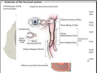
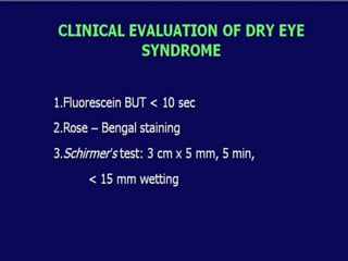
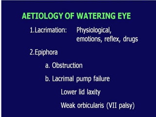
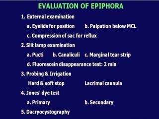
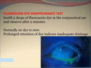
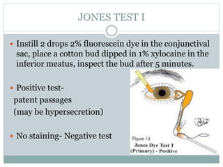
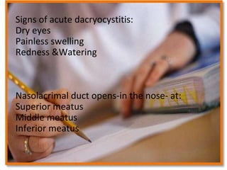
By/Mohamed Ahmed El –Shafie
Assistant Lecturer in ophthalmology department KafrELShiekh
University
The lacrimal system consists of two parts:
• Structures that secrete tear fluid.
• Structures that facilitate tear drainage.
LACRIMAL SYSTEM
It lies beneath the superior temporal margin of the
orbital bone in the lacrimal fossa of the frontal bone
It is neither visible nor palpable
A palpable lacrimal gland is usually a sign of a
pathologic change such as dacryoadenitis or tumors.
LACRIMAL GLAND ANATOMY
The tendon of the levator palpebrae muscle
divides the lacrimal gland into a larger
orbital part (two-thirds) and a smaller
palpebral part (one-third).
LACRIMAL GLAND PARTS
Several tiny accessory lacrimal glands
(glands of
Krause and Wolfring) located in the
superior fornix secrete additional serous
tear fluid.
ACCESSORY LACRIMAL GLAND
Tear Flow Physiology
may be due to one of two causes:
• Reduced tear production in certain systemic disorders
(such as Sjögren’s syndrome and rheumatoid arthritis) or as a
result of atrophy or destruction of the lacrimal gland.
• Altered composition of the tear film due to vitamin A
deficiency, medications (such as oral contraceptives), or
certain environmental influences (such as nicotine smoking, or
air conditioning).
The tear film breaks up too quickly and causes corneal
DRY EYE
Patients complain of
• Burning, reddened eyes, and excessive lacrimation
(reflex lacrimation) from only slight environmental
causes such as wind, cold, low humidity, or reading
for an extended period of time.
• A foreign body sensation is also present.
• May be accompanied by intense pain.
• Vision is usually minimally compromised .
DRY EYE SYMPTOMS
Bitot’s spots:
interpalpebral conjunctival foamy
patches associated with severe
vitamin A & xerophthalmia
FLUORESCEIN DYE DISAPPEARANCE TEST
Instill 2 drops of fluorescein dye in the conjunctival sac
and observe after 2 minutes
Normally no dye is seen
Prolonged retention of dye indicate inadequate drainage
JONES TEST I
 Instill 2 drops 2% fluorescein dye in the conjunctival
sac, place a cotton bud dipped in 1% xylocaine in the
inferior meatus, inspect the bud after 5 minutes.
 Positive test-
patent passages
(may be hypersecretion)
 No staining- Negative test
JONES TEST II
 If Jones I is negative, do Jones II
 Place a bud similarly and perform lacrimal syringing
 Positive test-
lacrimal pump failure
 Negative test-
mechanical obstruction
DACRYOCYSTOGRAPHY
 To visualize the passage and know the exact site,
nature and extent of block.
 Inject radioopaque material in the sac.
 Take X-rays
after 5 and 30 minutes
Congenital nasolacrimal duct obstruction
Delayed canalization
Can be bilateral
Reflux of purulent material from punctum
Acute dacryocystitis
 Acute inflammation of sac or pericystic area leading to
acute pain, swelling, redness and watering from the eye.
 No interference like syringing,probing etc.
Dacryocystorhinostomy
DCR
QuizSigns of acute dacryocystitis:
Dry eyes
Painless swelling
Redness &Watering
Nasolacrimal duct opens-in the nose- at:
Superior meatus
Middle meatus
Inferior meatus

Lacrimal system for undergraduates

  • 2.
    By/Mohamed Ahmed El–Shafie Assistant Lecturer in ophthalmology department KafrELShiekh University
  • 4.
    The lacrimal systemconsists of two parts: • Structures that secrete tear fluid. • Structures that facilitate tear drainage. LACRIMAL SYSTEM
  • 6.
    It lies beneaththe superior temporal margin of the orbital bone in the lacrimal fossa of the frontal bone It is neither visible nor palpable A palpable lacrimal gland is usually a sign of a pathologic change such as dacryoadenitis or tumors. LACRIMAL GLAND ANATOMY
  • 7.
    The tendon ofthe levator palpebrae muscle divides the lacrimal gland into a larger orbital part (two-thirds) and a smaller palpebral part (one-third). LACRIMAL GLAND PARTS
  • 8.
    Several tiny accessorylacrimal glands (glands of Krause and Wolfring) located in the superior fornix secrete additional serous tear fluid. ACCESSORY LACRIMAL GLAND
  • 11.
  • 12.
    may be dueto one of two causes: • Reduced tear production in certain systemic disorders (such as Sjögren’s syndrome and rheumatoid arthritis) or as a result of atrophy or destruction of the lacrimal gland. • Altered composition of the tear film due to vitamin A deficiency, medications (such as oral contraceptives), or certain environmental influences (such as nicotine smoking, or air conditioning). The tear film breaks up too quickly and causes corneal DRY EYE
  • 13.
    Patients complain of •Burning, reddened eyes, and excessive lacrimation (reflex lacrimation) from only slight environmental causes such as wind, cold, low humidity, or reading for an extended period of time. • A foreign body sensation is also present. • May be accompanied by intense pain. • Vision is usually minimally compromised . DRY EYE SYMPTOMS
  • 16.
    Bitot’s spots: interpalpebral conjunctivalfoamy patches associated with severe vitamin A & xerophthalmia
  • 22.
    FLUORESCEIN DYE DISAPPEARANCETEST Instill 2 drops of fluorescein dye in the conjunctival sac and observe after 2 minutes Normally no dye is seen Prolonged retention of dye indicate inadequate drainage
  • 25.
    JONES TEST I Instill 2 drops 2% fluorescein dye in the conjunctival sac, place a cotton bud dipped in 1% xylocaine in the inferior meatus, inspect the bud after 5 minutes.  Positive test- patent passages (may be hypersecretion)  No staining- Negative test
  • 26.
    JONES TEST II If Jones I is negative, do Jones II  Place a bud similarly and perform lacrimal syringing  Positive test- lacrimal pump failure  Negative test- mechanical obstruction
  • 27.
    DACRYOCYSTOGRAPHY  To visualizethe passage and know the exact site, nature and extent of block.  Inject radioopaque material in the sac.  Take X-rays after 5 and 30 minutes
  • 28.
    Congenital nasolacrimal ductobstruction Delayed canalization Can be bilateral Reflux of purulent material from punctum
  • 29.
    Acute dacryocystitis  Acuteinflammation of sac or pericystic area leading to acute pain, swelling, redness and watering from the eye.  No interference like syringing,probing etc.
  • 31.
  • 32.
    QuizSigns of acutedacryocystitis: Dry eyes Painless swelling Redness &Watering Nasolacrimal duct opens-in the nose- at: Superior meatus Middle meatus Inferior meatus