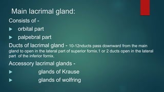
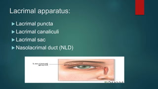
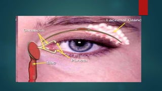
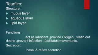
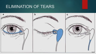
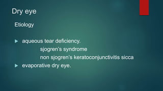
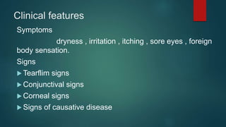
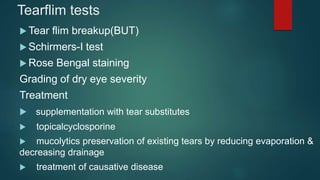
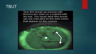
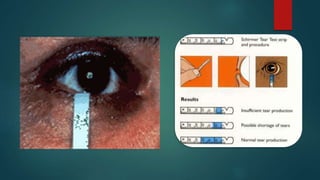
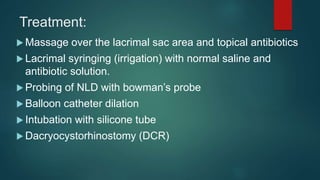
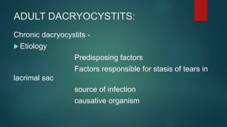
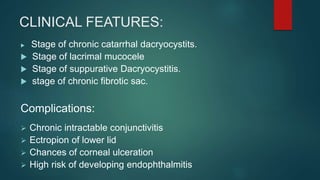
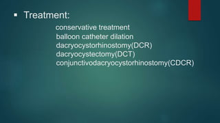
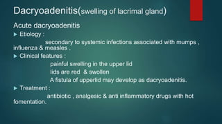
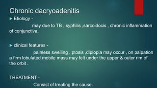
Lacrimalsystem
Applied anatomy
Lacrimal apparatus comprises
 main lacrimal gland,
 accessory lacrimal glands,
 lacrimal passages.
Main lacrimal gland:
Consists of -
 orbital part
 palpebral part
Ducts of lacrimal gland - 10-12nducts pass downward from the main
gland to open in the lateral part of superior fornix.1 or 2 ducts open in the lateral
part of the inferior fornix.
Accessory lacrimal glands -
 glands of Krause
 glands of wolfring
Structure, blood supply and nerve supply:
Nerve supply -
 sensory supply
 sympathetic supply
 secretomotor fibers
Lacrimal apparatus:
 Lacrimal puncta
 Lacrimal canaliculi
 Lacrimal sac
 Nasolacrimal duct (NLD)
Tearflim:
Structure :
 mucus layer
 aqueous layer
 lipid layer
Functions :
act as lubricant ,provide Oxygen , wash out
debris ,prevent infection , facilitates movements.
Secretion:
basal & reflex secretion.
ELIMINATION OF TEARS
Dry eye
Etiology
 aqueous tear deficiency.
sjogren’s syndrome
non sjogren’s keratoconjunctivitis sicca
 evaporative dry eye.
Clinical features
Symptoms
dryness , irritation , itching , sore eyes , foreign
body sensation.
Signs
 Tearflim signs
 Conjunctival signs
 Corneal signs
 Signs of causative disease
Tearflim tests
 Tear flim breakup(BUT)
 Schirmers-I test
 Rose Bengal staining
Grading of dry eye severity
Treatment
 supplementation with tear substitutes
 topicalcyclosporine
 mucolytics preservation of existing tears by reducing evaporation &
decreasing drainage
 treatment of causative disease
TBUT
SJOGREN’S SYNDROME
ETIOLOGY
CHARACTERISTIC FEATURES -
 Primary sjogren’s syndrome
 secondary sjogren’s syndrome
PATHOLOGICAL FEATURES -
focal accumulation , infiltration of lymphocytes , plasma
cells with destruction of lacrimal & salivary glandular tissues.
WATERING EYE
ETIOLOGY
 causes of hyperlacrimation.
 primary hyperlacrimation
 reflex hyperlacrimation
 central lacrimation
Causes of epiphora:
 Physiological cause -
lacrimal pump failure
 Mechanical obstruction -
obstruction in lacrimal passage at the level of
punctum , canaliculus , lacrimal sac or naso lacrimal duct.
Clinical evaluation:
 Ocular examination with diffuse illumination.
 Regurgitation test.
 Fluorescein dye disappearance test.
 Lacrimal syringing test.
 Jones dye test.
 Dacryosystogaaphy.
 Radio nucleotide dacryocystography.
Dacryocystitis:
inflammation of lacrimal sac.
Congenital Dacryocystitis: (Dacryocystitis neonatorum)
 inflammation of lacrimal sac occurring in newborn infants.
Etiology:
 membranous occlusion.
 other causes of congenital NLD.
 common bacteria.
Congenital dacryocystitis -
Clinical features:
 epiphora,
 regurgitation test,
 swelling.
Differential diagnosis:
 ophthalmia neonaturum,
 congenital glaucoma.
Treatment:
 Massage over the lacrimal sac area and topical antibiotics
 Lacrimal syringing (irrigation) with normal saline and
antibiotic solution.
 Probing of NLD with bowman’s probe
 Balloon catheter dilation
 Intubation with silicone tube
 Dacryocystorhinostomy (DCR)
ADULT DACRYOCYSTITS:
Chronic dacryocystits -
 Etiology
Predisposing factors
Factors responsible for stasis of tears in
lacrimal sac
source of infection
causative organism
CLINICAL FEATURES:
 Stage of chronic catarrhal dacryocystits.
 Stage of lacrimal mucocele
 Stage of suppurative Dacryocystitis.
 stage of chronic fibrotic sac.
Complications:
 Chronic intractable conjunctivitis
 Ectropion of lower lid
 Chances of corneal ulceration
 High risk of developing endophthalmitis
 Treatment:
conservative treatment
balloon catheter dilation
dacryocystorhinostomy(DCR)
dacryocystectomy(DCT)
conjunctivodacryocystorhinostomy(CDCR)
Acute Dacryocystitis:
 Etiology-
as an acute exacerbation of chronic of chronic Dacryocystitis.
as an acute peridacryocystitis due to direct involvement from the
neighboring infected structures.
 Causative organisms-
streptococcus , haemolyticus , pneumococcus & staphylococcus .
 Clinical features-
stage of cellulitis
stage of lacrimal abscess
stage of fistula formation
Acute dacryocystitis: stage of lacrimal abscess and stage of
external lacrimal fistula .
treatment
cellulitis stage
systemic & topical antibiotics ,anti inflammatory drugs &
hot fomentation.
stage of lacrimal abscess.
small incision to squeeze out the pus formation
external lacrimal fistula.
systemic antibiotics , fistulectomy along with DCT or DCR
operation.
Surgical steps of external dacryocystorhinostomy: A, skin incision; B,
exposure of bony lacrimal fossa; C, preparation of bony osteum and exposure of nasal mucosa; D,
preparation of flaps of the nasal mucosa and lacrimal sac; E, suturing of posterior flaps; F, suturing of
anterior flaps.
Dacryoadenitis(swelling of lacrimal gland)
Acute dacryoadenitis
 Etiology :
secondary to systemic infections associated with mumps ,
influenza & measles .
 Clinical features :
painful swelling in the upper lid
lids are red & swollen
A fistula of upperlid may develop as dacryoadenitis.
 Treatment :
antibiotic , analgesic & anti inflammatory drugs with hot
fomentation.
Chronic dacryoadenitis
 Etiology -
may due to TB , syphilis ,sarcoidocis , chronic inflammation
of conjunctiva.
 clinical features -
painless swelling , ptosis ,diplopia may occur , on palpation
a firm lobulated mobile mass may felt under the upper & outer rim of
the orbit .
TREATMENT -
Consist of treating the cause.
A patient with bilateral dacryoadenitis: s-shaped curve of upper
eyelid.
Thank you

lacrimal apparatus - diseases.

  • 1.
  • 3.
    Applied anatomy Lacrimal apparatuscomprises  main lacrimal gland,  accessory lacrimal glands,  lacrimal passages.
  • 4.
    Main lacrimal gland: Consistsof -  orbital part  palpebral part Ducts of lacrimal gland - 10-12nducts pass downward from the main gland to open in the lateral part of superior fornix.1 or 2 ducts open in the lateral part of the inferior fornix. Accessory lacrimal glands -  glands of Krause  glands of wolfring
  • 5.
    Structure, blood supplyand nerve supply: Nerve supply -  sensory supply  sympathetic supply  secretomotor fibers
  • 6.
    Lacrimal apparatus:  Lacrimalpuncta  Lacrimal canaliculi  Lacrimal sac  Nasolacrimal duct (NLD)
  • 8.
    Tearflim: Structure :  mucuslayer  aqueous layer  lipid layer Functions : act as lubricant ,provide Oxygen , wash out debris ,prevent infection , facilitates movements. Secretion: basal & reflex secretion.
  • 9.
  • 10.
    Dry eye Etiology  aqueoustear deficiency. sjogren’s syndrome non sjogren’s keratoconjunctivitis sicca  evaporative dry eye.
  • 11.
    Clinical features Symptoms dryness ,irritation , itching , sore eyes , foreign body sensation. Signs  Tearflim signs  Conjunctival signs  Corneal signs  Signs of causative disease
  • 12.
    Tearflim tests  Tearflim breakup(BUT)  Schirmers-I test  Rose Bengal staining Grading of dry eye severity Treatment  supplementation with tear substitutes  topicalcyclosporine  mucolytics preservation of existing tears by reducing evaporation & decreasing drainage  treatment of causative disease
  • 13.
  • 15.
    SJOGREN’S SYNDROME ETIOLOGY CHARACTERISTIC FEATURES-  Primary sjogren’s syndrome  secondary sjogren’s syndrome PATHOLOGICAL FEATURES - focal accumulation , infiltration of lymphocytes , plasma cells with destruction of lacrimal & salivary glandular tissues.
  • 16.
    WATERING EYE ETIOLOGY  causesof hyperlacrimation.  primary hyperlacrimation  reflex hyperlacrimation  central lacrimation
  • 17.
    Causes of epiphora: Physiological cause - lacrimal pump failure  Mechanical obstruction - obstruction in lacrimal passage at the level of punctum , canaliculus , lacrimal sac or naso lacrimal duct.
  • 18.
    Clinical evaluation:  Ocularexamination with diffuse illumination.  Regurgitation test.  Fluorescein dye disappearance test.  Lacrimal syringing test.  Jones dye test.  Dacryosystogaaphy.  Radio nucleotide dacryocystography.
  • 19.
    Dacryocystitis: inflammation of lacrimalsac. Congenital Dacryocystitis: (Dacryocystitis neonatorum)  inflammation of lacrimal sac occurring in newborn infants. Etiology:  membranous occlusion.  other causes of congenital NLD.  common bacteria.
  • 20.
  • 21.
    Clinical features:  epiphora, regurgitation test,  swelling. Differential diagnosis:  ophthalmia neonaturum,  congenital glaucoma.
  • 22.
    Treatment:  Massage overthe lacrimal sac area and topical antibiotics  Lacrimal syringing (irrigation) with normal saline and antibiotic solution.  Probing of NLD with bowman’s probe  Balloon catheter dilation  Intubation with silicone tube  Dacryocystorhinostomy (DCR)
  • 23.
    ADULT DACRYOCYSTITS: Chronic dacryocystits-  Etiology Predisposing factors Factors responsible for stasis of tears in lacrimal sac source of infection causative organism
  • 24.
    CLINICAL FEATURES:  Stageof chronic catarrhal dacryocystits.  Stage of lacrimal mucocele  Stage of suppurative Dacryocystitis.  stage of chronic fibrotic sac. Complications:  Chronic intractable conjunctivitis  Ectropion of lower lid  Chances of corneal ulceration  High risk of developing endophthalmitis
  • 25.
     Treatment: conservative treatment ballooncatheter dilation dacryocystorhinostomy(DCR) dacryocystectomy(DCT) conjunctivodacryocystorhinostomy(CDCR)
  • 26.
    Acute Dacryocystitis:  Etiology- asan acute exacerbation of chronic of chronic Dacryocystitis. as an acute peridacryocystitis due to direct involvement from the neighboring infected structures.  Causative organisms- streptococcus , haemolyticus , pneumococcus & staphylococcus .  Clinical features- stage of cellulitis stage of lacrimal abscess stage of fistula formation
  • 27.
    Acute dacryocystitis: stageof lacrimal abscess and stage of external lacrimal fistula .
  • 28.
    treatment cellulitis stage systemic &topical antibiotics ,anti inflammatory drugs & hot fomentation. stage of lacrimal abscess. small incision to squeeze out the pus formation external lacrimal fistula. systemic antibiotics , fistulectomy along with DCT or DCR operation.
  • 29.
    Surgical steps ofexternal dacryocystorhinostomy: A, skin incision; B, exposure of bony lacrimal fossa; C, preparation of bony osteum and exposure of nasal mucosa; D, preparation of flaps of the nasal mucosa and lacrimal sac; E, suturing of posterior flaps; F, suturing of anterior flaps.
  • 30.
    Dacryoadenitis(swelling of lacrimalgland) Acute dacryoadenitis  Etiology : secondary to systemic infections associated with mumps , influenza & measles .  Clinical features : painful swelling in the upper lid lids are red & swollen A fistula of upperlid may develop as dacryoadenitis.  Treatment : antibiotic , analgesic & anti inflammatory drugs with hot fomentation.
  • 31.
    Chronic dacryoadenitis  Etiology- may due to TB , syphilis ,sarcoidocis , chronic inflammation of conjunctiva.  clinical features - painless swelling , ptosis ,diplopia may occur , on palpation a firm lobulated mobile mass may felt under the upper & outer rim of the orbit . TREATMENT - Consist of treating the cause.
  • 32.
    A patient withbilateral dacryoadenitis: s-shaped curve of upper eyelid.
  • 33.