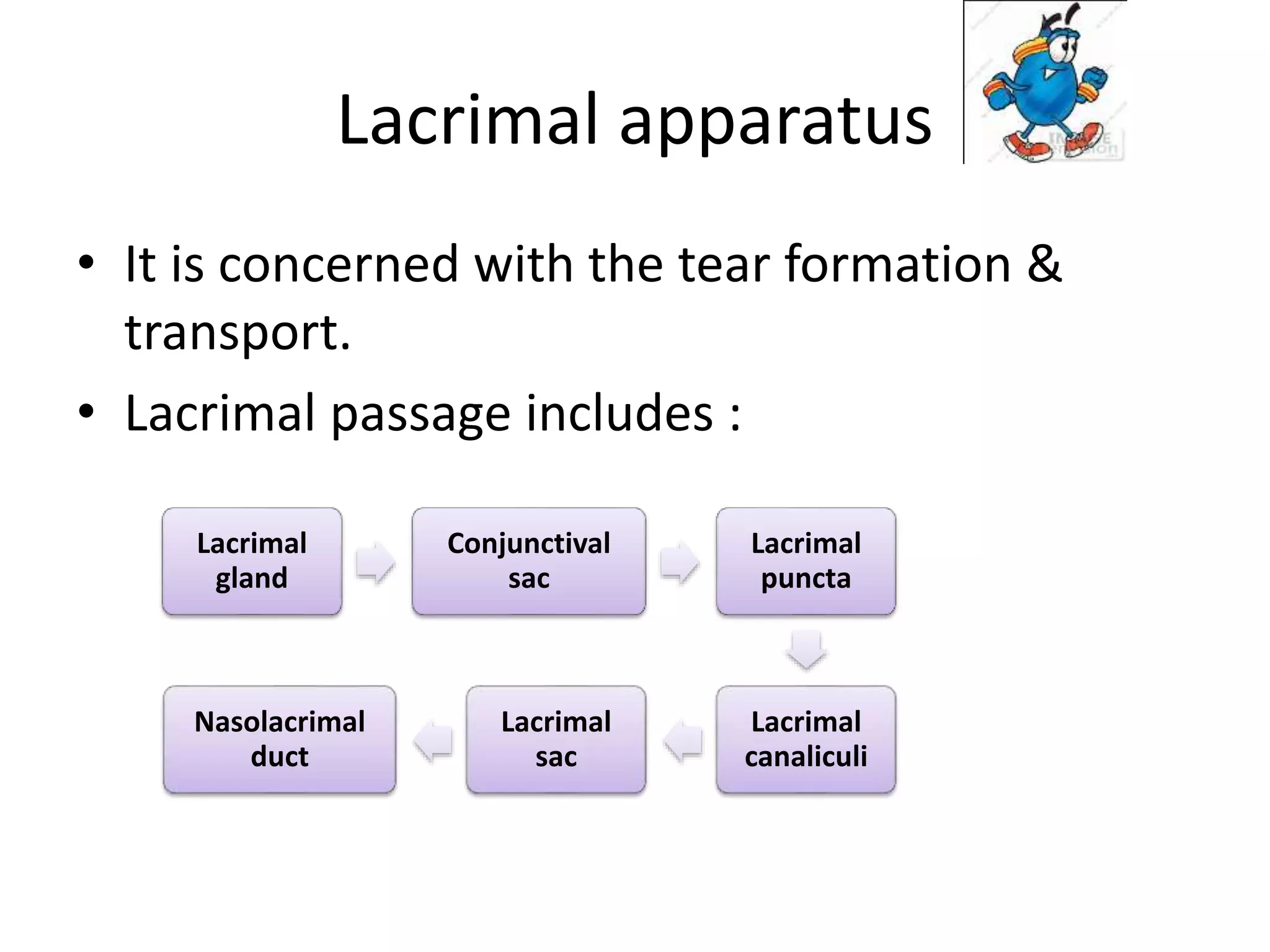
Lacrimal apparatus
• It is concerned with the tear formation &
transport.
• Lacrimal passage includes :
Lacrimal
gland
Conjunctival
sac
Lacrimal
puncta
Lacrimal
canaliculi
Lacrimal
sac
Nasolacrimal
duct
ANATOMY & PHYSIOLOGY
BRIEF REVIEW
 Secretory and drainage parts.
 Lacrimal gland [orbital and palpebral parts] and accessory lac
glands of Krause and Wolfring Seven to twelve ducts open in
the supero temporal part pf bulbar conjunctiva.
 Streams running across the eye ball lead to a smaller meniscus
along upper and another along the lower lid margin.
 Eye closure is from temporal to nasal side thus pushing the
tears medially in lacus lacrimalis.
 Here starts drainage system.
Secretory system
• It includes lacrimal gland, accessory glands
• Lacrimal gland is above & anterolateral to globe.
• Secretes tears into superior fornix.
• Tears moisten & lubricates the : cornea , conjunctiva.
• It contributes 43D of 50D of refractive power of eye .
• It consists of
• Large Orbital Part
• Smaller Palpebral
part
• Lateral expansion of levator separates
the parts
The orbital part
• Paired almond-shaped glands.
• It is present in a fossa on the anterolateral
area of orbit
• It has 2 surfaces, 2 borders, 2 extremities
– Superior surface
• Frontal bone
– Inferior surface
• Levator palpebrae superioris & lateral rectus
– Anterior border
• Septum orbitale
– Posterior border
• Contact with orbital fat , level with posterior
pole.
– Lateral extremity
• Rest on lateral rectus
– Medial extremity
• On levator
The palpebral part
• 1/3rd size of orbital part
• Superior fornix , seen on lid eversion.
• It is situated upon the course of ducts
• Related to levator superiorly, inferiorly to
superior fornix
• Posteriorly it continues with orbital part.
Accessory glands
• Are small, compound, branched, tubular
glands
• Located in the middle of lid (Wolfring glands)
or superior & inferior fornices (Krause
glands).
Blood supply
• Artery supply : Lacrimal artery , branch of
ophthalmic artery.
• Venous drainages : Ophthalmic Vein.
• Lymphatic drainage : Joins that of conjunctiva
& drain into the preauricular lymph nodes.
Nerve supply
• Sensory nerve supply : lacrimal nerve , branch
of ophthalmic division of Vth nerve
• Sympathetic nerve supply : carotid plexus
• Secretomotor fibers : superior salivary nucleus
Excretory system
the PUNCTA
• A small, round or oval orifice on the elevation, the
papilla lacrimalis.
• At medial end of lid margin at the junction of its
ciliated and non-ciliated parts.
• Upper punctum medial to lower, from the medial
canthus being 6 and 6.5 mm.
• The upper punctum opens inferoposteriorly, the lower
superoposteriorly.
THE LACRIMAL CANALICULI
• First vertical and then horizontal
• Vertical part is 2 mm & turns medially at right-
angle to become horizontal 8 mm
• At angle - dilatation or ampulla.
• The canaliculi pierce the fascia (i.e. the
periorbita covering the lacrimal sac) separately,
• Uniting to enter lacrimal sac.
• Stratified squamous epithelium supported by
elastic tissue.
THE LACRIMAL SAC
• A fold of mucosa at the junction of common canaliculus,
forms the valve of Rosenmuller, preventing reflex tears.
• It lies in the lacrimal fossa formed by the lacrimal bone,
when distended it is about 15mm long vertically and 5-
6mm wide.
• The upper portion or the fundus extends slightly above the
level of the medial palpebral ligament and the sac itself is
surrounded by fibers of orbicularis muscle.
• The sac, closed above and open below, is continuous with
the nasolacrimal duct.
THE NASOLACRIMAL DUCT
• The nasolacrimal duct, continuation of
lacrimal sac to the inferior meatus.
• 12 mm.
• It descends posteriolaterally, a surface
indication a line from medial canthus to first
upper molar.
• The most constant is the 'valve' of Hasner at
the lower end.
• It prevents sudden blast of air (when blowing
the nose) from entering the lacrimal sac.
physiology
• The tear film overlays corneal and conjunctival
epithelia.
• Tears produced by the ocular surface epithelia
and adnexa.
• Thickness of up to 40 µm,
• Volume of tears covering the ocular surface
range from 2.74 ± 2.0µL to 7 µL
• For mucous and aqueous layers, secretion is
regulated by neural reflexes.
• For the lipid layer, the blink itself regulates release
of pre-secreted meibomian gland .
• Tear secretion is balanced by drainage and
evaporation.
• Drainage is regulated by neural reflexes ,causing
vasodilation and vasoconstriction of blood sinus.
• Evaporation depends blink rate and temperature,
humidity, and wind speed.
The roles of the precorneal tear film
• To protect the cornea from drying;
• To maintain the refractive power of the cornea;
• To defend against eye infection;
• To allow gas to move between the air and the avascular
cornea;
• To support corneal dehydration (assisted by the tear
film hyperosmolality).
• Consists of four layers
– Glycocalyx
– Mucous layer
– Aqueous layer.
– Lipid layers
Glycocalyx
Function
• The membrane-spanning mucins function to
hydrate the ocular surface and serve as a
barrier to pathogens.
• Membrane-spanning mucins appear to be
altered in dry eye
Mucous layer
Structure
• The mucous layer backbone is the gel-forming mucin ,
synthesized and secreted by conjunctival goblet cells.
Function
• To resistance of the eye to infection by providing protection
against microorganisms.
• Mucins serve as wetting agents that keep the apical
epithelia hydrated.
Aqueous layer
• Lacrimal gland produce aqueous layer.
• Other ocular surface epithelia also contribute to the
aqueous layer, eg. conjunctiva, accessory lacrimal glands
• 7µm thick.
• Without the lubrication , the shearing forces produced on
blinking will cause accumulative ocular surface damage.
• Composed of water, with many solutes,
including dissolved mucins, electrolytes and
proteins.
• The osmotic pressure : concentrations of
sodium, potassium and chloride ions.
• The tear film’s osmotic pressure is important
in the control of cornea–tear film water flux.
• Bicarbonate and carbonate : pH buffering,
maintaining the pH at 7.3–7.6 when the eyes
open & 6.8 eyes closed.
Aqueous layer function
• Aqueous deficiency dry eye.
• Protection from bacterial infection
• Reflex secretion washes away noxious
substances.
• Protects against changes in pH.
Lipid layer
• Meibomian glands, modified sebaceous glands, that
line the upper and lower eyelids.
• Meibomian gland lipids are stored in vesicles.
• The secretory product contains a complex mixture of
lipids and proteins and is termed meibum.
• Meibum is released on to the ocular surface in small
amounts with each blink.
• 0.1m in thickness
Function
• Hydrophobic barrier to prevent tear
overflow.
• The meibum forms a water-tight seal of the
apposed lid margins during sleep.
• Reduce tear evaporation .
• Lipids enhance the stability of the tear
film and provide a smooth optical.
DISTRIBUTION OF THE TEARS
• Conjunctival fornices, preocular tear film, and
marginal tear strips.
• Marginal tear strips are wedge shaped tear
menisci, borders of upper and lower lids.
• Apposed lacrimal puncta dip into marginal
strip of tears
• Anterior limit of the marginal strip is the
mucocutaneous junction of the lid,
CONDUCTION OF THE TEARS
LACRIMAL PUMP MECHANISM:
• Tears are lost from the conjunctiva sac by
absorption, evaporation, and nasolacrimal
system.
• This is related to the size of the palpebral
aperture, the blink rate, ambient temperature
and humidity.
• Tears flow
– the upper and lower marginal strips → upper
and lower canaliculi (capillarity + suction)
• Eyes close
– Pretarsal orbicularis oculi compresses the
ampullae + shortens and compresses canaliculi
+ puncta medially.
– Lacrimal part of the orbicularis oculi, contracts
→ compresses the sac,(positive pressure)
tears → nasolacrimal duct → nose.
• Eyes open
– Muscles relax → canaliculi and sac
expand(negative pressure) + capillarity= tears
into sac.
LACRIMAL SYSTEM.pptx

LACRIMAL SYSTEM.pptx

  • 1.
    Lacrimal apparatus • Itis concerned with the tear formation & transport. • Lacrimal passage includes : Lacrimal gland Conjunctival sac Lacrimal puncta Lacrimal canaliculi Lacrimal sac Nasolacrimal duct
  • 3.
    ANATOMY & PHYSIOLOGY BRIEFREVIEW  Secretory and drainage parts.  Lacrimal gland [orbital and palpebral parts] and accessory lac glands of Krause and Wolfring Seven to twelve ducts open in the supero temporal part pf bulbar conjunctiva.  Streams running across the eye ball lead to a smaller meniscus along upper and another along the lower lid margin.  Eye closure is from temporal to nasal side thus pushing the tears medially in lacus lacrimalis.  Here starts drainage system.
  • 5.
    Secretory system • Itincludes lacrimal gland, accessory glands • Lacrimal gland is above & anterolateral to globe. • Secretes tears into superior fornix. • Tears moisten & lubricates the : cornea , conjunctiva. • It contributes 43D of 50D of refractive power of eye .
  • 6.
    • It consistsof • Large Orbital Part • Smaller Palpebral part • Lateral expansion of levator separates the parts
  • 7.
    The orbital part •Paired almond-shaped glands. • It is present in a fossa on the anterolateral area of orbit • It has 2 surfaces, 2 borders, 2 extremities – Superior surface • Frontal bone – Inferior surface • Levator palpebrae superioris & lateral rectus
  • 8.
    – Anterior border •Septum orbitale – Posterior border • Contact with orbital fat , level with posterior pole. – Lateral extremity • Rest on lateral rectus – Medial extremity • On levator
  • 9.
    The palpebral part •1/3rd size of orbital part • Superior fornix , seen on lid eversion. • It is situated upon the course of ducts • Related to levator superiorly, inferiorly to superior fornix • Posteriorly it continues with orbital part.
  • 10.
    Accessory glands • Aresmall, compound, branched, tubular glands • Located in the middle of lid (Wolfring glands) or superior & inferior fornices (Krause glands).
  • 11.
    Blood supply • Arterysupply : Lacrimal artery , branch of ophthalmic artery. • Venous drainages : Ophthalmic Vein. • Lymphatic drainage : Joins that of conjunctiva & drain into the preauricular lymph nodes.
  • 12.
    Nerve supply • Sensorynerve supply : lacrimal nerve , branch of ophthalmic division of Vth nerve • Sympathetic nerve supply : carotid plexus • Secretomotor fibers : superior salivary nucleus
  • 13.
    Excretory system the PUNCTA •A small, round or oval orifice on the elevation, the papilla lacrimalis. • At medial end of lid margin at the junction of its ciliated and non-ciliated parts. • Upper punctum medial to lower, from the medial canthus being 6 and 6.5 mm. • The upper punctum opens inferoposteriorly, the lower superoposteriorly.
  • 15.
    THE LACRIMAL CANALICULI •First vertical and then horizontal • Vertical part is 2 mm & turns medially at right- angle to become horizontal 8 mm • At angle - dilatation or ampulla. • The canaliculi pierce the fascia (i.e. the periorbita covering the lacrimal sac) separately, • Uniting to enter lacrimal sac. • Stratified squamous epithelium supported by elastic tissue.
  • 16.
    THE LACRIMAL SAC •A fold of mucosa at the junction of common canaliculus, forms the valve of Rosenmuller, preventing reflex tears. • It lies in the lacrimal fossa formed by the lacrimal bone, when distended it is about 15mm long vertically and 5- 6mm wide. • The upper portion or the fundus extends slightly above the level of the medial palpebral ligament and the sac itself is surrounded by fibers of orbicularis muscle. • The sac, closed above and open below, is continuous with the nasolacrimal duct.
  • 17.
    THE NASOLACRIMAL DUCT •The nasolacrimal duct, continuation of lacrimal sac to the inferior meatus. • 12 mm. • It descends posteriolaterally, a surface indication a line from medial canthus to first upper molar.
  • 18.
    • The mostconstant is the 'valve' of Hasner at the lower end. • It prevents sudden blast of air (when blowing the nose) from entering the lacrimal sac.
  • 19.
    physiology • The tearfilm overlays corneal and conjunctival epithelia. • Tears produced by the ocular surface epithelia and adnexa. • Thickness of up to 40 µm, • Volume of tears covering the ocular surface range from 2.74 ± 2.0µL to 7 µL
  • 20.
    • For mucousand aqueous layers, secretion is regulated by neural reflexes. • For the lipid layer, the blink itself regulates release of pre-secreted meibomian gland . • Tear secretion is balanced by drainage and evaporation. • Drainage is regulated by neural reflexes ,causing vasodilation and vasoconstriction of blood sinus. • Evaporation depends blink rate and temperature, humidity, and wind speed.
  • 21.
    The roles ofthe precorneal tear film • To protect the cornea from drying; • To maintain the refractive power of the cornea; • To defend against eye infection; • To allow gas to move between the air and the avascular cornea; • To support corneal dehydration (assisted by the tear film hyperosmolality).
  • 22.
    • Consists offour layers – Glycocalyx – Mucous layer – Aqueous layer. – Lipid layers
  • 23.
    Glycocalyx Function • The membrane-spanningmucins function to hydrate the ocular surface and serve as a barrier to pathogens. • Membrane-spanning mucins appear to be altered in dry eye
  • 24.
    Mucous layer Structure • Themucous layer backbone is the gel-forming mucin , synthesized and secreted by conjunctival goblet cells. Function • To resistance of the eye to infection by providing protection against microorganisms. • Mucins serve as wetting agents that keep the apical epithelia hydrated.
  • 25.
    Aqueous layer • Lacrimalgland produce aqueous layer. • Other ocular surface epithelia also contribute to the aqueous layer, eg. conjunctiva, accessory lacrimal glands • 7µm thick. • Without the lubrication , the shearing forces produced on blinking will cause accumulative ocular surface damage.
  • 26.
    • Composed ofwater, with many solutes, including dissolved mucins, electrolytes and proteins. • The osmotic pressure : concentrations of sodium, potassium and chloride ions. • The tear film’s osmotic pressure is important in the control of cornea–tear film water flux. • Bicarbonate and carbonate : pH buffering, maintaining the pH at 7.3–7.6 when the eyes open & 6.8 eyes closed.
  • 27.
    Aqueous layer function •Aqueous deficiency dry eye. • Protection from bacterial infection • Reflex secretion washes away noxious substances. • Protects against changes in pH.
  • 28.
    Lipid layer • Meibomianglands, modified sebaceous glands, that line the upper and lower eyelids. • Meibomian gland lipids are stored in vesicles. • The secretory product contains a complex mixture of lipids and proteins and is termed meibum. • Meibum is released on to the ocular surface in small amounts with each blink. • 0.1m in thickness
  • 29.
    Function • Hydrophobic barrierto prevent tear overflow. • The meibum forms a water-tight seal of the apposed lid margins during sleep. • Reduce tear evaporation . • Lipids enhance the stability of the tear film and provide a smooth optical.
  • 31.
    DISTRIBUTION OF THETEARS • Conjunctival fornices, preocular tear film, and marginal tear strips. • Marginal tear strips are wedge shaped tear menisci, borders of upper and lower lids. • Apposed lacrimal puncta dip into marginal strip of tears • Anterior limit of the marginal strip is the mucocutaneous junction of the lid,
  • 33.
    CONDUCTION OF THETEARS LACRIMAL PUMP MECHANISM: • Tears are lost from the conjunctiva sac by absorption, evaporation, and nasolacrimal system. • This is related to the size of the palpebral aperture, the blink rate, ambient temperature and humidity.
  • 34.
    • Tears flow –the upper and lower marginal strips → upper and lower canaliculi (capillarity + suction) • Eyes close – Pretarsal orbicularis oculi compresses the ampullae + shortens and compresses canaliculi + puncta medially. – Lacrimal part of the orbicularis oculi, contracts → compresses the sac,(positive pressure) tears → nasolacrimal duct → nose. • Eyes open – Muscles relax → canaliculi and sac expand(negative pressure) + capillarity= tears into sac.