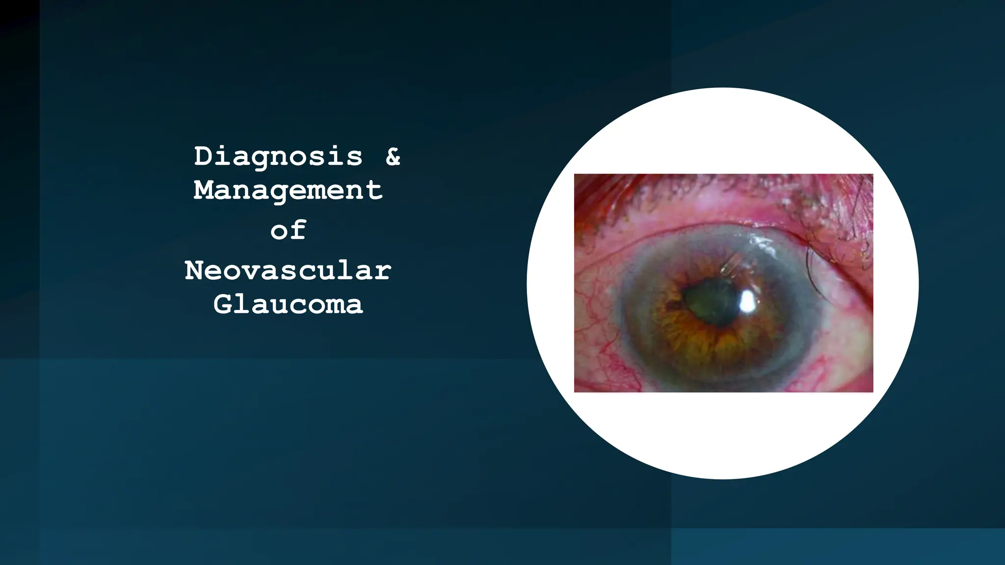
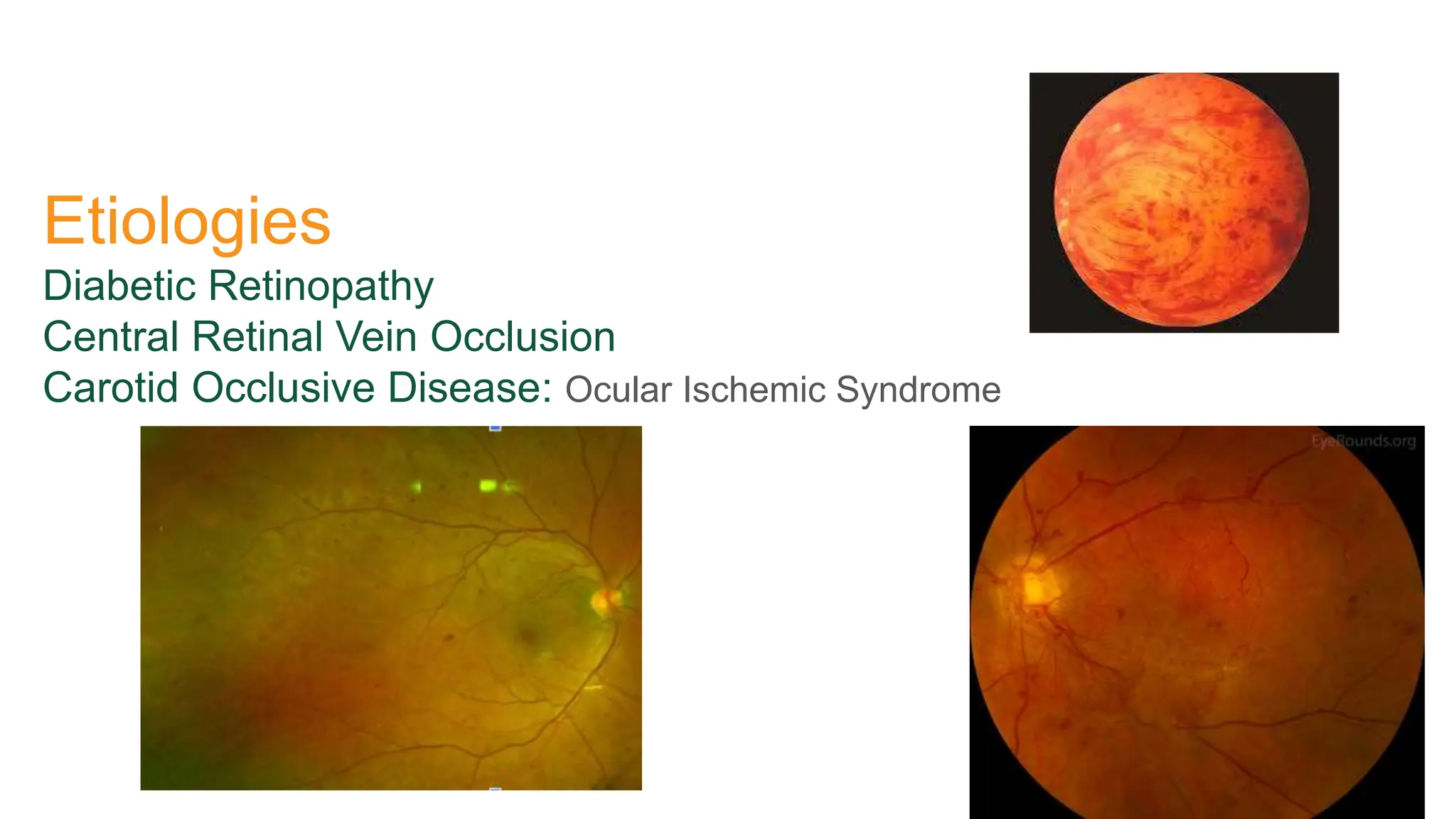
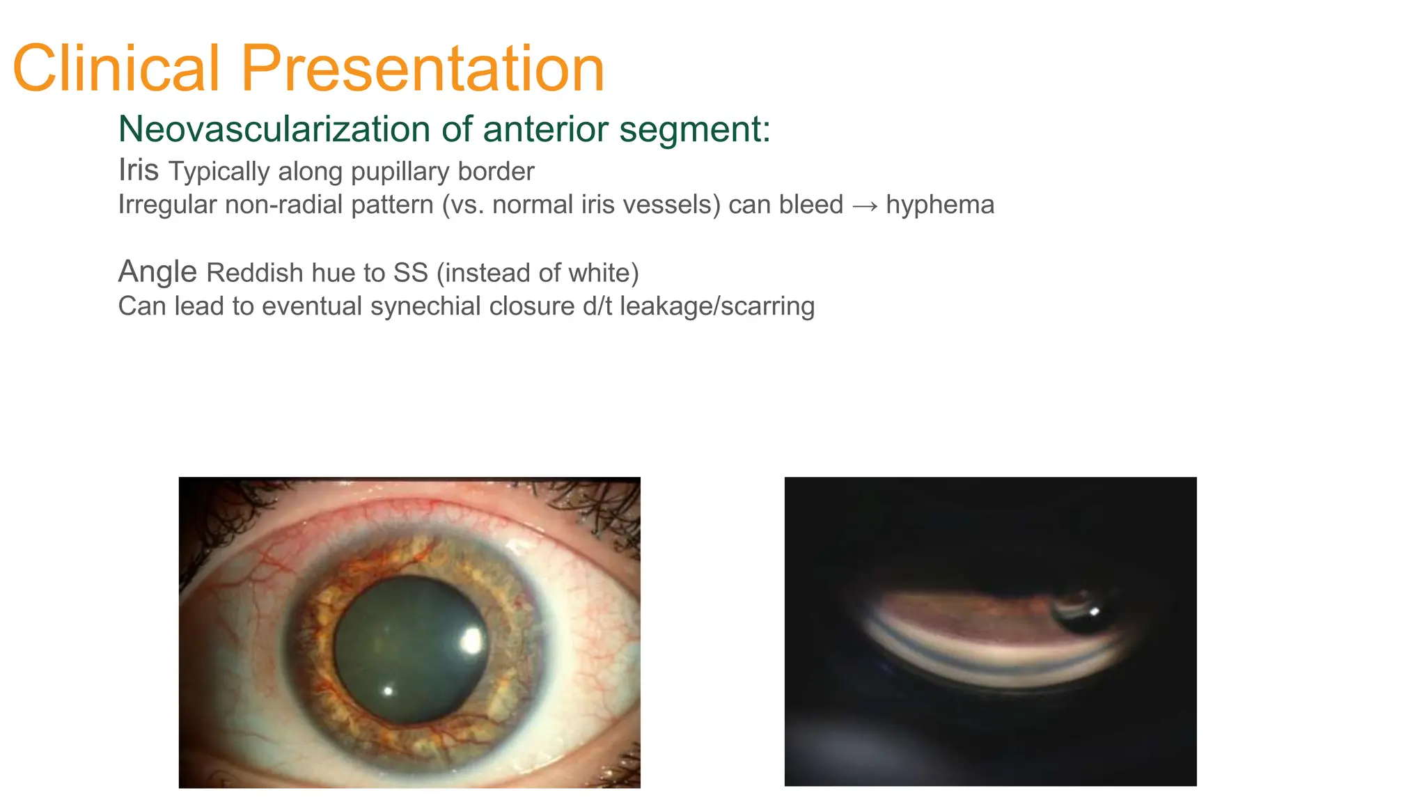
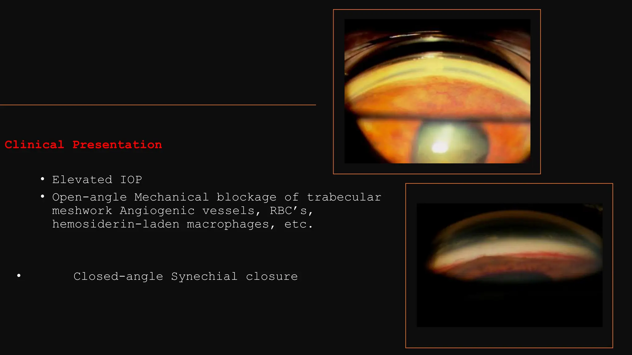
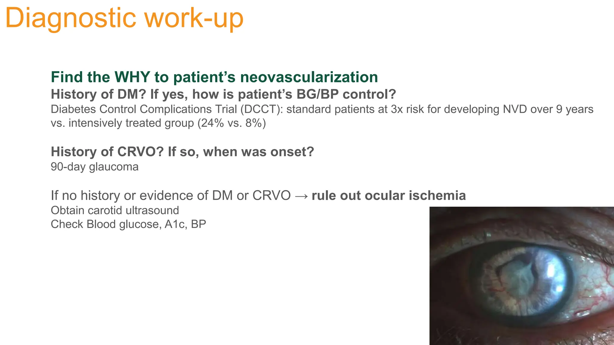
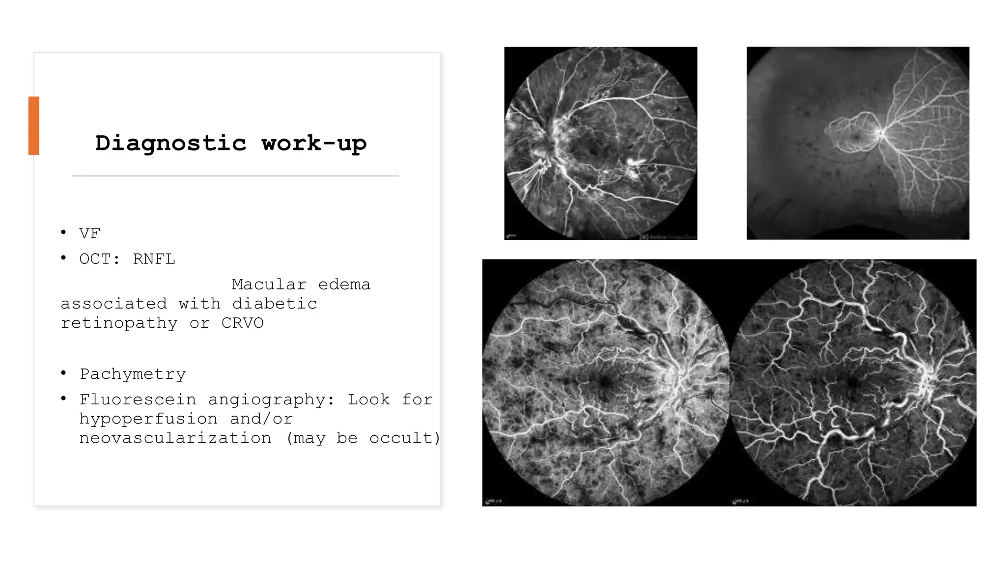
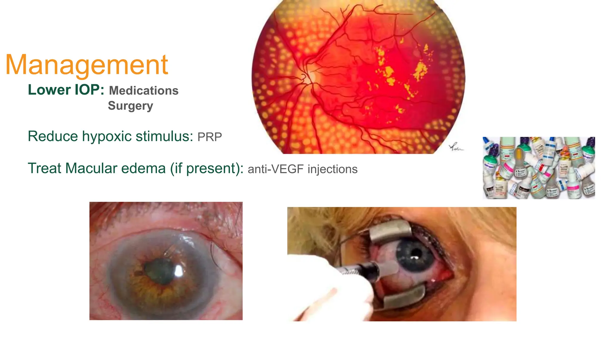
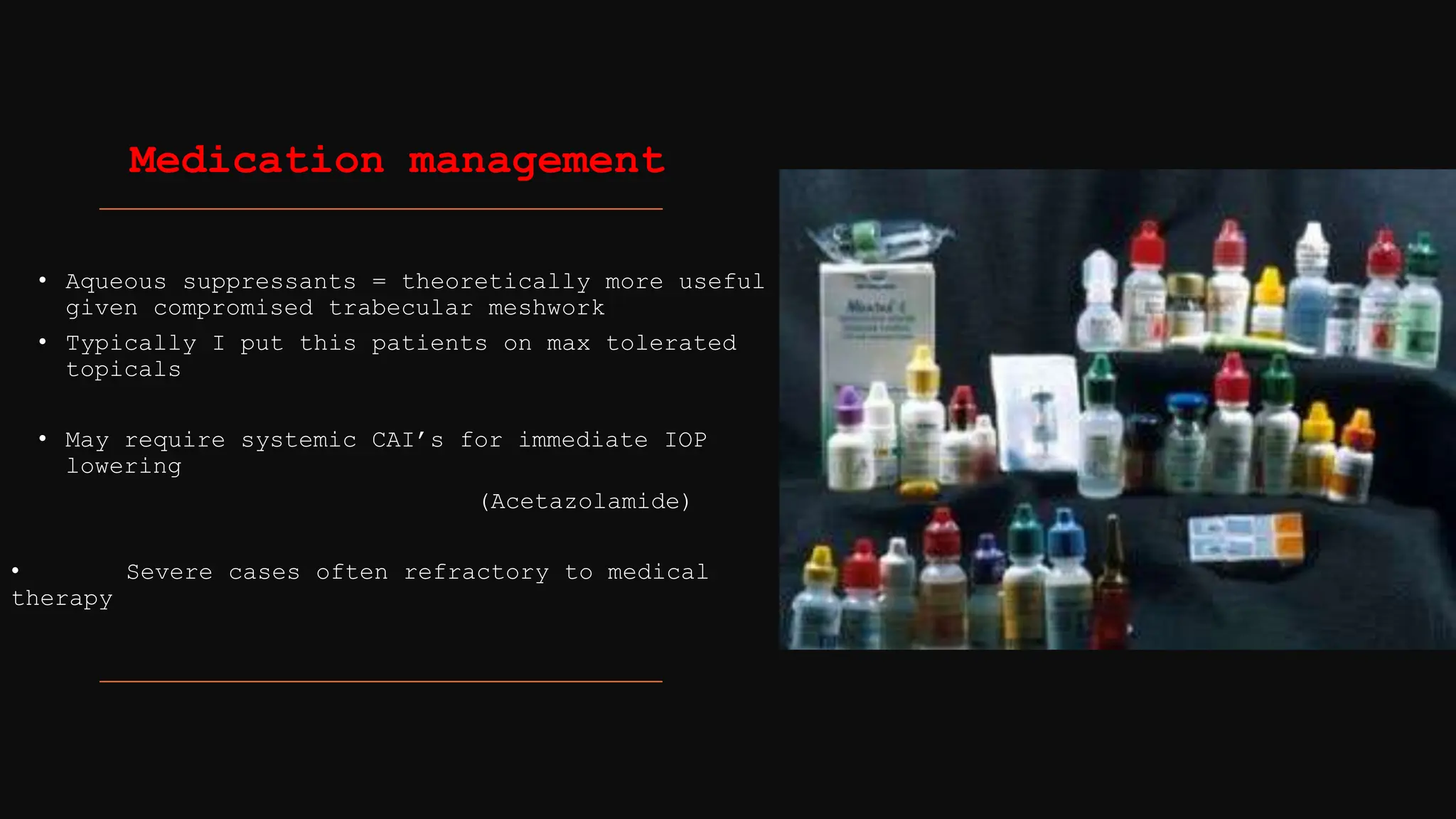
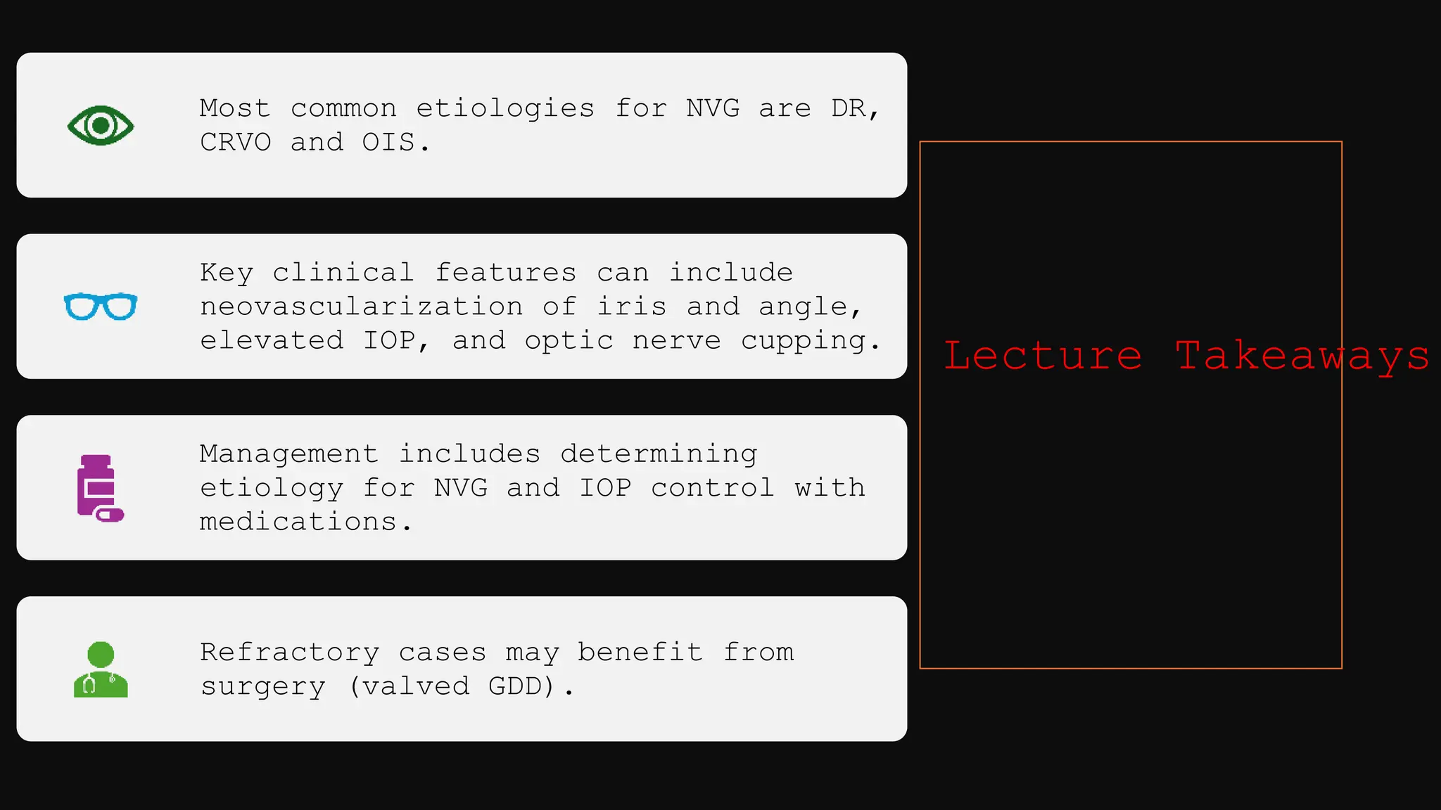
Diagnosis &
Management
of
Neovascular
Glaucoma
History
First described in association with iris rubeosis following CRVO (1906)
“NVG” defined as elevated IOP in context of new blood vessel and connective tissue growth (1963)
Also known as: Hemorrhagic glaucoma
Congestive glaucoma
Thrombotic glaucoma
Rubeotic glaucoma
● Normal eye:
• balance between pro- and anti-angiogenic growth factors
• Pro-angiogenic growth factors: Vascular endothelial growth factor
(VEGF) & Angiopoietin-2 (Ang-2)
• Anti-angiogenic growth factors: Pigment epithelial derived growth
factor (PDEF)
●Normal Blood vessel anatomy:
• non-fenestrated endothelial cells with tight intercellular junctions
Pathophysiology
Ischemic eye:
• pro-angiogenic >> anti-angiogenic
• Retina ischemia/hypoxia → release of VEGF +
Angiopoietin-2 → activation + proliferation +
migration of endothelial cells → angiogenesis with
fibrovascular membrane formation
Neovascular vessels:
• Little/absent muscle layer and little adventitial
structures → prone to leakage/breakage
Etiologies
Diabetic Retinopathy
Central Retinal Vein Occlusion
Carotid Occlusive Disease: Ocular Ischemic Syndrome
Clinical Presentation
Neovascularization of anterior segment:
Iris Typically along pupillary border
Irregular non-radial pattern (vs. normal iris vessels) can bleed → hyphema
Angle Reddish hue to SS (instead of white)
Can lead to eventual synechial closure d/t leakage/scarring
Clinical Presentation
• Elevated IOP
• Open-angle Mechanical blockage of trabecular
meshwork Angiogenic vessels, RBC’s,
hemosiderin-laden macrophages, etc.
• Closed-angle Synechial closure
Diagnostic work-up
Find the WHY to patient’s neovascularization
History of DM? If yes, how is patient’s BG/BP control?
Diabetes Control Complications Trial (DCCT): standard patients at 3x risk for developing NVD over 9 years
vs. intensively treated group (24% vs. 8%)
History of CRVO? If so, when was onset?
90-day glaucoma
If no history or evidence of DM or CRVO → rule out ocular ischemia
Obtain carotid ultrasound
Check Blood glucose, A1c, BP
• VF
• OCT: RNFL
Macular edema
associated with diabetic
retinopathy or CRVO
• Pachymetry
• Fluorescein angiography: Look for
hypoperfusion and/or
neovascularization (may be occult)
Diagnostic work-up
Management
Lower IOP: Medications
Surgery
Reduce hypoxic stimulus: PRP
Treat Macular edema (if present): anti-VEGF injections
• Aqueous suppressants = theoretically more useful
given compromised trabecular meshwork
• Typically I put this patients on max tolerated
topicals
• May require systemic CAI’s for immediate IOP
lowering
(Acetazolamide)
• Severe cases often refractory to medical
therapy
Medication management
Surgical Management
• Glaucoma drainage device (GDD) > Trabeculectomy
Risk of scarring of trabeculectomy flap from NV
• Valved GDD (Ahmed) > non-valved GDD (Baerveldt)
Immediate IOP lowering
Less risk of hypotony
• Cyclophotocoagulation (CPC) in refractory cases:
I typically reserve this for patient’s have AGV
already.
Micropulse also option - less chance of hypotony than
CPC
Which of the following is NOT considered a COMMON cause of
Neovascular glaucoma?
• A.Ocular ischemic syndrome
• B.Diabetic retinopathy
• C.Central retinal artery occlusion
• D.Central retinal vein occlusion
Which of the following is NOT a clinical sign of
neovascular glaucoma?
• A.Iris rubeosis
• B.Iris atrophy
• C.Angle vessels
• D.Optic Disc cupping
Which of the following is the optimal surgical intervention for
neovascular glaucoma?
• A.Cataract surgery with goniotomy
• B.Trabeculectomy with Mitomycin C
• C.Non-valved glaucoma drainage device (i.e. Baerveldt)
• D.Valved glaucoma drainage device (i.e. Ahmed)
Most common etiologies for NVG are DR,
CRVO and OIS.
Key clinical features can include
neovascularization of iris and angle,
elevated IOP, and optic nerve cupping.
Management includes determining
etiology for NVG and IOP control with
medications.
Refractory cases may benefit from
surgery (valved GDD).
Lecture Takeaways
How to deal with Neovascular glaucoma cases

How to deal with Neovascular glaucoma cases

  • 1.
  • 2.
    History First described inassociation with iris rubeosis following CRVO (1906) “NVG” defined as elevated IOP in context of new blood vessel and connective tissue growth (1963) Also known as: Hemorrhagic glaucoma Congestive glaucoma Thrombotic glaucoma Rubeotic glaucoma
  • 3.
    ● Normal eye: •balance between pro- and anti-angiogenic growth factors • Pro-angiogenic growth factors: Vascular endothelial growth factor (VEGF) & Angiopoietin-2 (Ang-2) • Anti-angiogenic growth factors: Pigment epithelial derived growth factor (PDEF) ●Normal Blood vessel anatomy: • non-fenestrated endothelial cells with tight intercellular junctions
  • 4.
    Pathophysiology Ischemic eye: • pro-angiogenic>> anti-angiogenic • Retina ischemia/hypoxia → release of VEGF + Angiopoietin-2 → activation + proliferation + migration of endothelial cells → angiogenesis with fibrovascular membrane formation Neovascular vessels: • Little/absent muscle layer and little adventitial structures → prone to leakage/breakage
  • 5.
    Etiologies Diabetic Retinopathy Central RetinalVein Occlusion Carotid Occlusive Disease: Ocular Ischemic Syndrome
  • 6.
    Clinical Presentation Neovascularization ofanterior segment: Iris Typically along pupillary border Irregular non-radial pattern (vs. normal iris vessels) can bleed → hyphema Angle Reddish hue to SS (instead of white) Can lead to eventual synechial closure d/t leakage/scarring
  • 8.
    Clinical Presentation • ElevatedIOP • Open-angle Mechanical blockage of trabecular meshwork Angiogenic vessels, RBC’s, hemosiderin-laden macrophages, etc. • Closed-angle Synechial closure
  • 9.
    Diagnostic work-up Find theWHY to patient’s neovascularization History of DM? If yes, how is patient’s BG/BP control? Diabetes Control Complications Trial (DCCT): standard patients at 3x risk for developing NVD over 9 years vs. intensively treated group (24% vs. 8%) History of CRVO? If so, when was onset? 90-day glaucoma If no history or evidence of DM or CRVO → rule out ocular ischemia Obtain carotid ultrasound Check Blood glucose, A1c, BP
  • 10.
    • VF • OCT:RNFL Macular edema associated with diabetic retinopathy or CRVO • Pachymetry • Fluorescein angiography: Look for hypoperfusion and/or neovascularization (may be occult) Diagnostic work-up
  • 11.
    Management Lower IOP: Medications Surgery Reducehypoxic stimulus: PRP Treat Macular edema (if present): anti-VEGF injections
  • 12.
    • Aqueous suppressants= theoretically more useful given compromised trabecular meshwork • Typically I put this patients on max tolerated topicals • May require systemic CAI’s for immediate IOP lowering (Acetazolamide) • Severe cases often refractory to medical therapy Medication management
  • 13.
    Surgical Management • Glaucomadrainage device (GDD) > Trabeculectomy Risk of scarring of trabeculectomy flap from NV • Valved GDD (Ahmed) > non-valved GDD (Baerveldt) Immediate IOP lowering Less risk of hypotony • Cyclophotocoagulation (CPC) in refractory cases: I typically reserve this for patient’s have AGV already. Micropulse also option - less chance of hypotony than CPC
  • 14.
    Which of thefollowing is NOT considered a COMMON cause of Neovascular glaucoma? • A.Ocular ischemic syndrome • B.Diabetic retinopathy • C.Central retinal artery occlusion • D.Central retinal vein occlusion
  • 15.
    Which of thefollowing is NOT a clinical sign of neovascular glaucoma? • A.Iris rubeosis • B.Iris atrophy • C.Angle vessels • D.Optic Disc cupping
  • 16.
    Which of thefollowing is the optimal surgical intervention for neovascular glaucoma? • A.Cataract surgery with goniotomy • B.Trabeculectomy with Mitomycin C • C.Non-valved glaucoma drainage device (i.e. Baerveldt) • D.Valved glaucoma drainage device (i.e. Ahmed)
  • 17.
    Most common etiologiesfor NVG are DR, CRVO and OIS. Key clinical features can include neovascularization of iris and angle, elevated IOP, and optic nerve cupping. Management includes determining etiology for NVG and IOP control with medications. Refractory cases may benefit from surgery (valved GDD). Lecture Takeaways

Editor's Notes