
Recommended
More Related Content
Similar to LACRIMAL glands APPARTUS lecture 3 lec.pptx
Similar to LACRIMAL glands APPARTUS lecture 3 lec.pptx (20)
Recently uploaded
Recently uploaded (20)
LACRIMAL glands APPARTUS lecture 3 lec.pptx
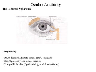
- 1. Ocular Anatomy The Lacrimal Apparatus Prepared by: Dr.Abdikarim Mustafa Ismail (Dr Goodman) Bsc. Optometry and visual science Msc public health (Epidemiology and Bio statistics)
- 2. The lacrimal apparatus (Tear System) It's a group of glands, sacs and ducts that makes new tears and drains old ones away. consists of two divisions. The first is the secretory part, which is responsible for the production of tears, and the second is the drainage part. The secretory part contains the main lacrimal gland, which is lodged in a fossa in the lateral part of the roof of the orbit, and two accessory lacrimal glands of Krause and Wolfring. The latter are present near the fornix. They are often called basal secretors because normally their secretions are sufficient to serve the purpose for which tears are meant without any need of drainage to the nose..
- 3. The main lacrimal gland is brought into action when there is severe emotional stress or physical irritation caused by inflammation in the anterior segment of the eye, causing an increase in the amount of secretion and a possible need for drainage to the nose The main lacrimal gland is divided into a larger orbital and a smaller palpebral part by the lateral horn of the aponeurosis of the levator palpebrae superioris. The secretory ducts reach the lateral part of the superior fornix. One or two ducts may open directly into the inferior fornix.
- 6. The tear film The tear film has three layers. The outermost is the lipid layer from the meibomian glands which regulates the evaporation from the surface of the cornea and conjunctiva and prevents the spilling of tears on the face. The middle layer is aqueous, having soluble minerals and proteins that originate in the main and accessory lacrimal glands. The third innermost layer is mucoid and comes from the goblet cells of the conjunctiva. It contains glycoprotein mucin
- 8. The surface of the eye is protected by a blinking reflex helped by the tears. The aqueous component dilutes the infectious material, mucus traps the debris, and the pumping action of the lids constantly flushes the tears to the tear duct. Tears contain antimicrobial substances, including lysozyme and antibodies.
- 9. The tear film has three layers. The unicellular goblet cells of the conjunctiva secrete glycoprotein in the form of mucin, forming the innermost layer. The intermediate watery layer comes from the main and accessory lacrimal glands. The outermost lipid layer is produced by the meibomian glands of the tarsus. 1. The lipid layer is a monomolecular film thought to regulate evaporation and forms a watertight seal when the lids are closed. 2. The aqueous layer from main and accessory lacrimal glands contains water-soluble salts and proteins. 3. The deep mucinous layer overlying the corneal and conjunctival epithelial cells is composed of glycoprotein.
- 12. Composition of Tears and Volume The normal tear volume is about 7 ± 2 µL in each eye. Albumin constitutes about 60 % of the total protein. Immunoglobulins IgA, IgG, and IgE as well as lysozymes make up the remaining 40 % Potassium, sodium, and chloride also occur in higher concentrations in tears than in plasma. Tears have a small amount of glucose (5 mg/dL) and urea (0.04 mg/dL), and changes in blood concentration produces changes in tear glucose and urea levels. The average pH of tears is 7.35, although a wide normal variation exists (5.20– 8.35). Under normal conditions, tear fluid is isotonic.
- 13. Dryness of the eye may result from any disease associated with deficiency of the tear film components (aqueous, mucin, or lipid), lid surface abnormalities, or epithelial abnormalities. Many of the causes of dry eye syndrome affect more than one component of the tear film or lead to ocular surface alterations that secondarily cause tear film instability. Dry spots may appear on the corneal and conjunctival epithelia.
- 14. Tear functions: (1)They make the corneal surface smooth by covering the microscopic irregularities of the corneal epithelium, which is essential for adequate refraction at the cornea. (2)They keep the corneal and conjunctival surfaces moist for normal function. (3)They provide nutrition to the corneal and conjunctival epithelia. (4)They catch flying dust articles and send them along the tear streak at the posterior sharp border of the lid margin.
- 15. (5) They inhibit the growth of microorganisms by the antimicrobial action of lysozyme. (6) The constant evaporation of the tear film from the surface of cornea and conjunctiva takes heat from the conjunctival sac, allowing its temperature to be lower than that of other parts of body
- 16. The drainage system The drainage system is located in the upper and lower puncta at the junction of the ciliary and lacrimal parts of the lid margin, which are continuous with the vertical and horizontal parts of the canaliculus. The canaliculi open separately into the medial wall of the lacrimal sac housed in the lacrimal fossa. The lacrimal sac is continuous below with the nasolacrimal duct. The lower end of the nasolacrimal duct opens into the inferior meatus of the nose.
- 18. Secretary ducts from the main lacrimal gland open into the lateral part of the superior fornix and one or two open into the inferior fornix. The involuntary act of blinking spreads the tears like a swab in front of the cornea and conjunctiva; they also reach the posterior sharp border of the lid margins The capillarity and suction caused by the contraction of the orbicularis oculi in blinking make tears enter the puncta, canaliculi, and lacrimal sac. Once the tears reach the lacrimal sac, the elasticity of the lacrimal sac and gravity help them to reach the nasolacrimal duct from where they enter the inferior meatus of the nose.
- 19. Drainage to the nose normally does not occur because of evaporation but only is seen during severe emotional stress or physical irritation caused by inflammation of the anterior segment of the eye when the main lacrimal gland comes into action.
- 20. Main and Accessory Lacrimal Glands The structures responsible for the production and drainage of tears are collectively called the lacrimal apparatus. It has two divisions: the production of tears by the main lacrimal and accessory lacrimal glands and the drainage of tears. The main gland is located in the lacrimal fossa, in the superior temporal quadrant of the orbit.
- 21. The accessory lacrimal glands of Krause and Wolfring are located in the conjunctiva of the superior fornix and the superior tarsal border. They have the same structure as the main gland. They are known as basal secretors because their secretions are normally sufficient to maintain the health of the cornea. The main gland comes into action in severe emotional situations, whereas routinely the secretions of the accessory glands are enough for normal functioning. Emotional or physical irritation caused by inflammation of the anterior segment may trigger an increase in secretion called lacrimation and may cause tears to flow copiously over the lid margin.
- 23. Drainage Channels for Tears The punctum is the hole present in a papilla at the junction of the ciliary and nonciliary (lacrimal) parts of the lid margins. The superior one is located in the upper lid and the inferior one is in the lower lid. The lower canaliculus is 6.5 mm and is longer than the upper one, which is 6.0 mm. Both open separately through the lacrimal fascia into the medial wall of the lacrimal sac ,The lacrimal sac is lodged in the lacrimal fossa in the medial wall of the orbit between the anterior and posterior lacrimal crests. The vertical and horizontal parts of the canaliculus are present in the lacrimal part of the lid margin, which encloses the inner canthus.
- 24. The lacrimal sac makes an angle with the upper end of the nasolacrimal duct. That explains why that angle is the common site of blockage of the lacrimal passages. If you put a small finger just below the inner canthus, it goes into the lacrimal fossa. Digital pressure is used as a regurgitation test when mucoid discharge can be seen through the upper or lower punctum in cases of chronic dacryocystitis. Acute dacryocystitis presents clinically as a painful red swelling just below the inner canthus or lacrimal fistula also can occur in complicated cases
- 26. Conduction of the Lacrimal Fluid Secreted by the lacrimal gland, tears pass through ducts mostly to the lateral part of the upper fornix and then descend to the strip of fluid at the posterior sharp border of the upper lid margin. They reach the upper punctum along this strip or directly under the upper lid. Closure of the lid may help them to reach the lower lid margin. Some ducts also open into the lower fornix. Tear fluid is prevented from spilling by oily secretions from the meibomian glands. Blinking spreads the tear film over the cornea and conjunctiva like a swab.
- 27. Tears enter the puncta is caused by negative pressure into the sac by contraction of the orbicularis. Once it reaches the lacrimal sac, elasticity of the sac and gravity help them to reach the nasolacrimal duct, which opens into the inferior meatus of the nose. Normally, the tears do not reach the nose because of constant evaporation. Drainage of the nose is needed during severe emotional stress or irritation caused by inflammation of the anterior segment of the eye
- 28. Tear Tests The Schirmer Test The Schirmer test is done with strip. The Schirmer strip is inserted into the lower conjunctival sac at the junction of the middle and temporal thirds of the lower lid. The moistened exposed portion is measured without anesthesia after 5 min. Less than 15 mm wetting is considered abnormal. This test is a screening test of tear production. False-positive or false-negative results do occur.
- 30. Tear Film Breakup Time The normal time is 15 s. Measurement of the tear film breakup time may be useful to estimate the mucin content of tears. For the method, apply a slightly moistened fluorescein strip to the bulbar conjunctiva and ask the patient to blink. The tear film is then scanned with a cobalt filter on the slit lamp while the patient refrains from blinking. The time that elapses before the first dry spot that appears in the corneal fluorescein layer is the “tear film breakup time.” It is definitely reduced in eyes with mucin deficiency.