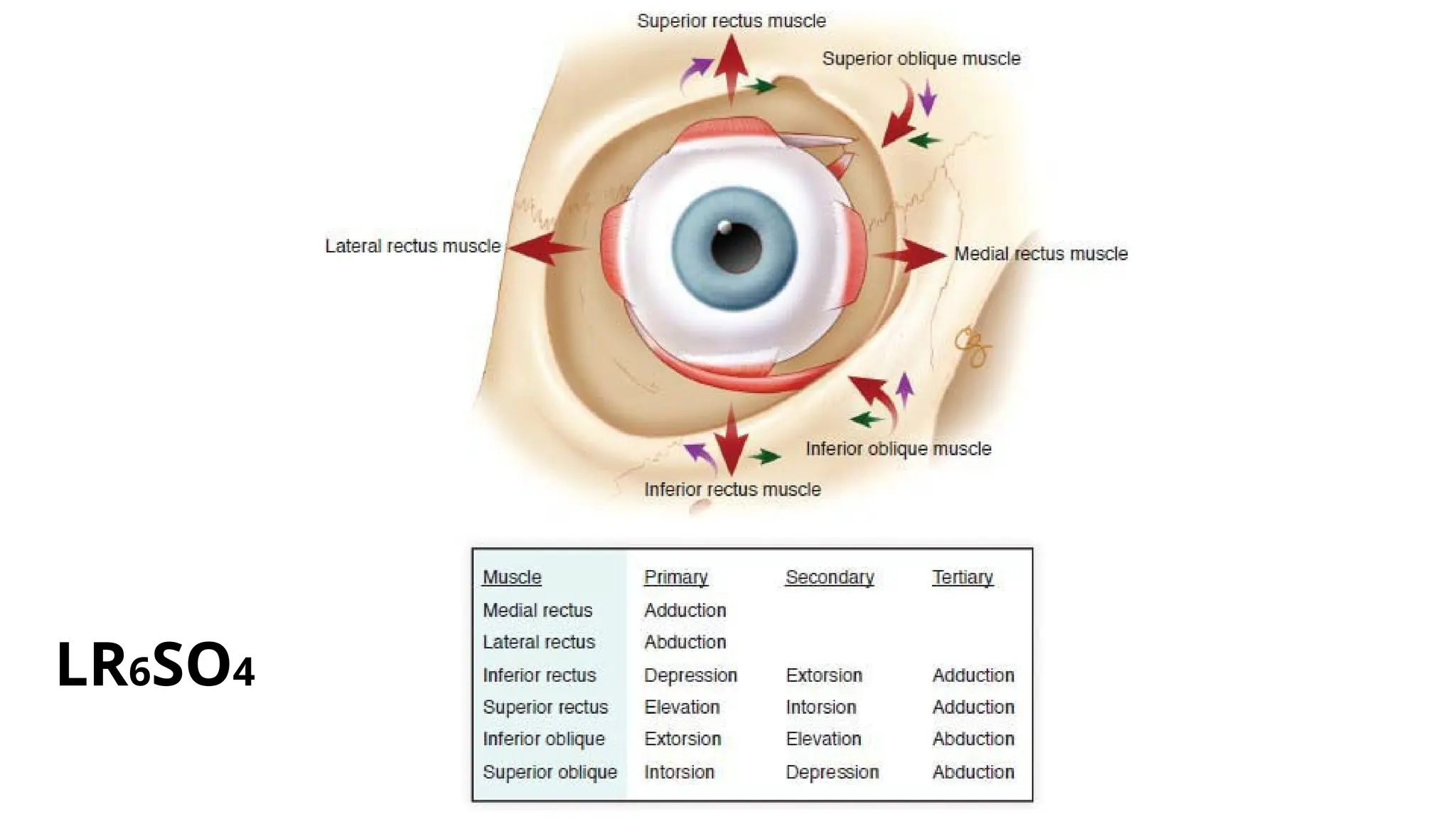
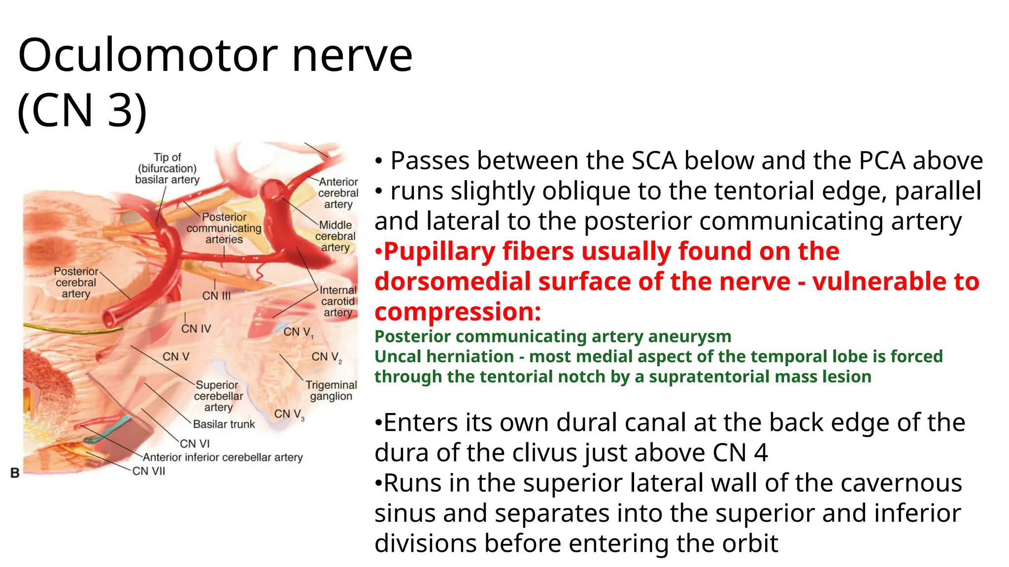
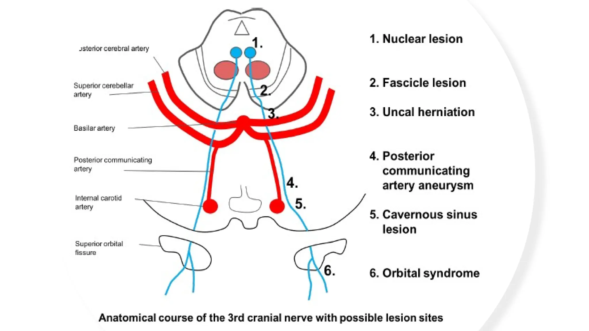
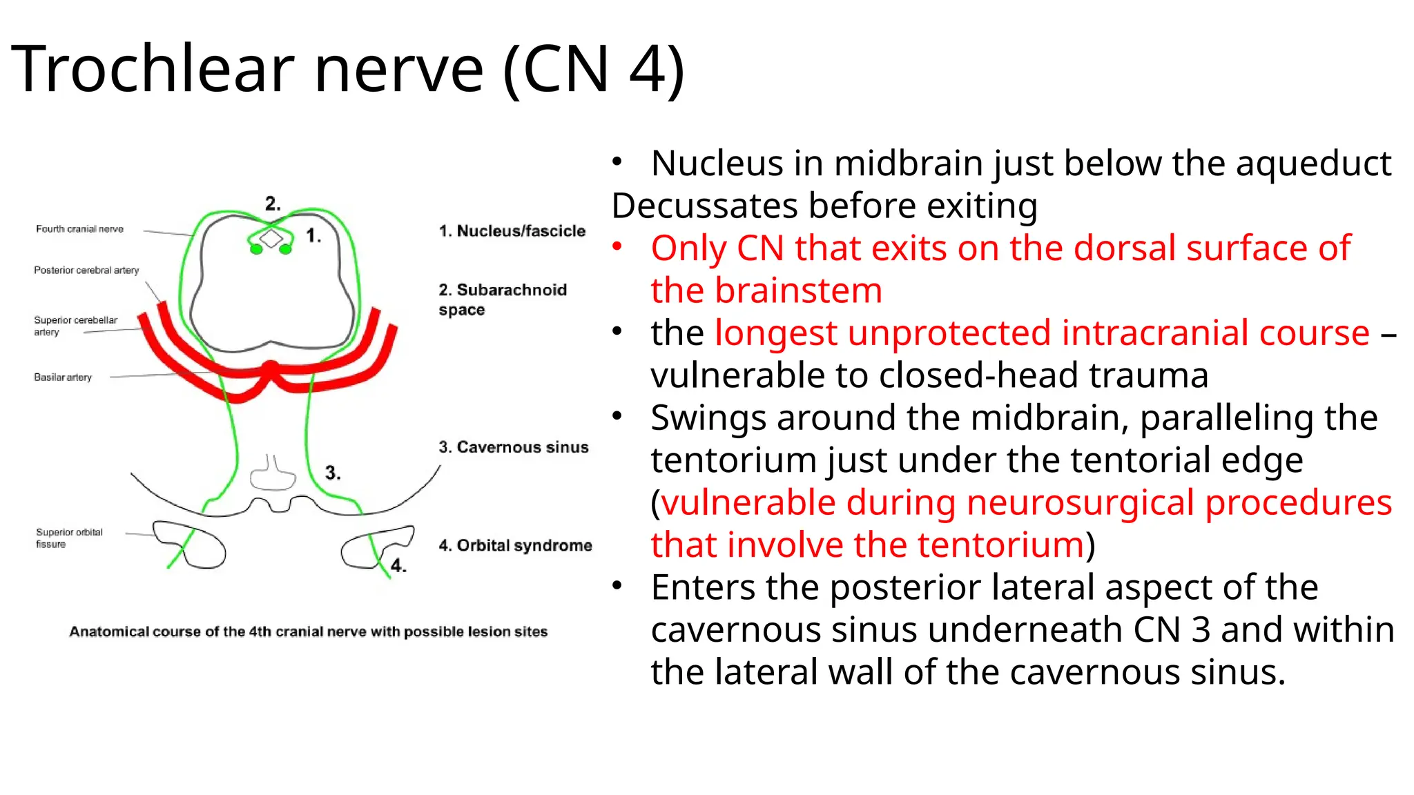
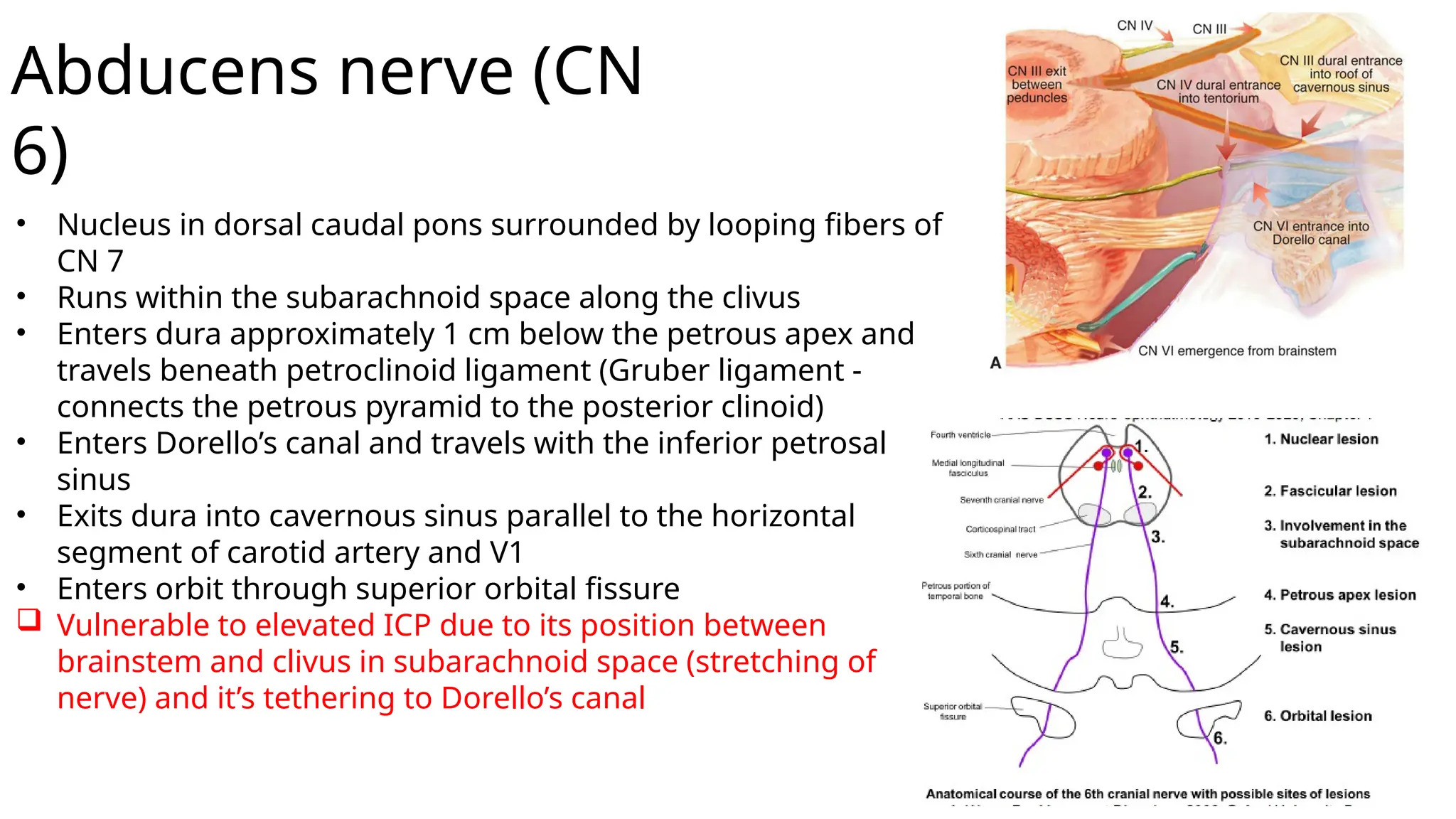
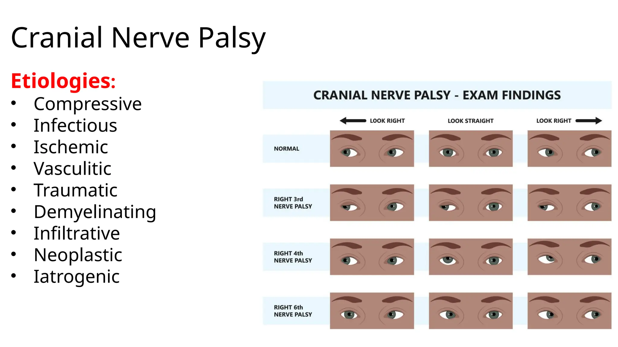
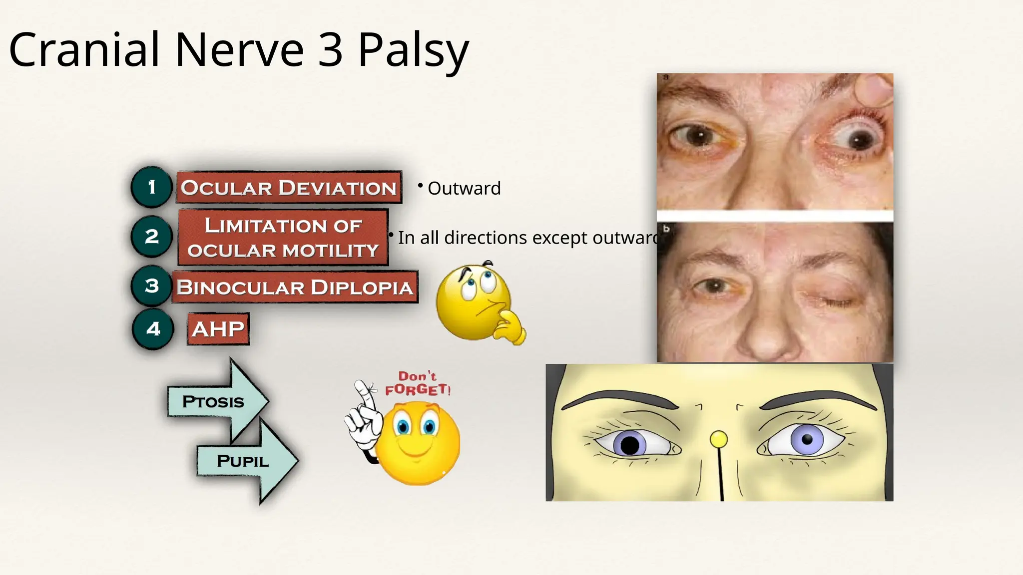
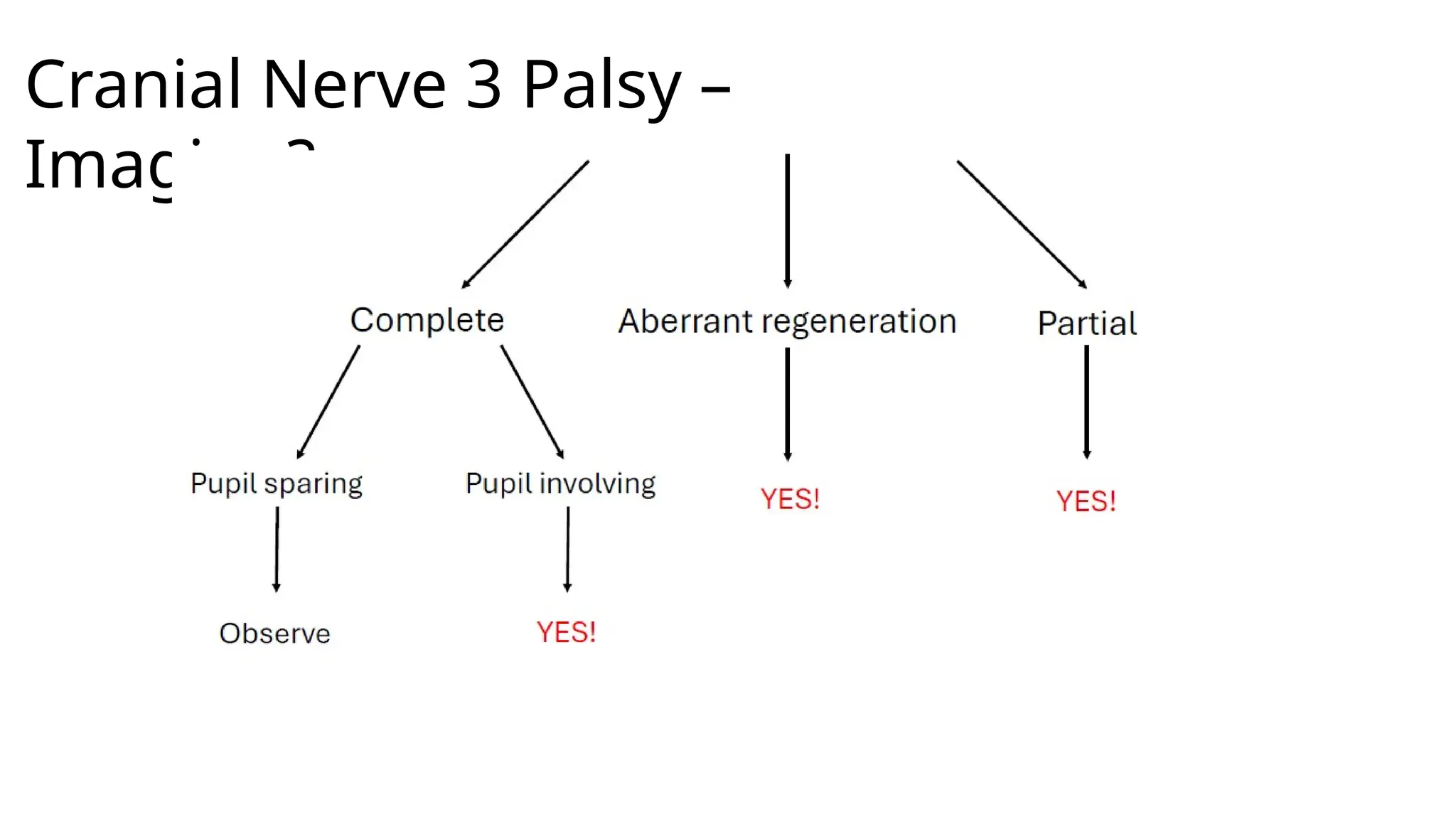
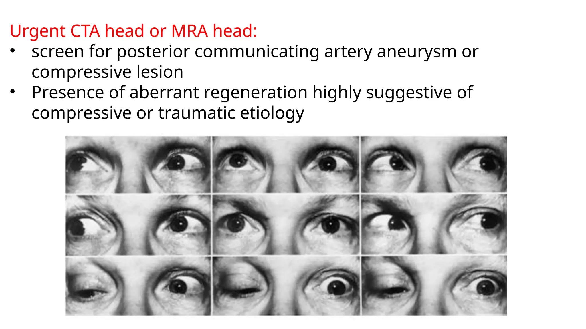
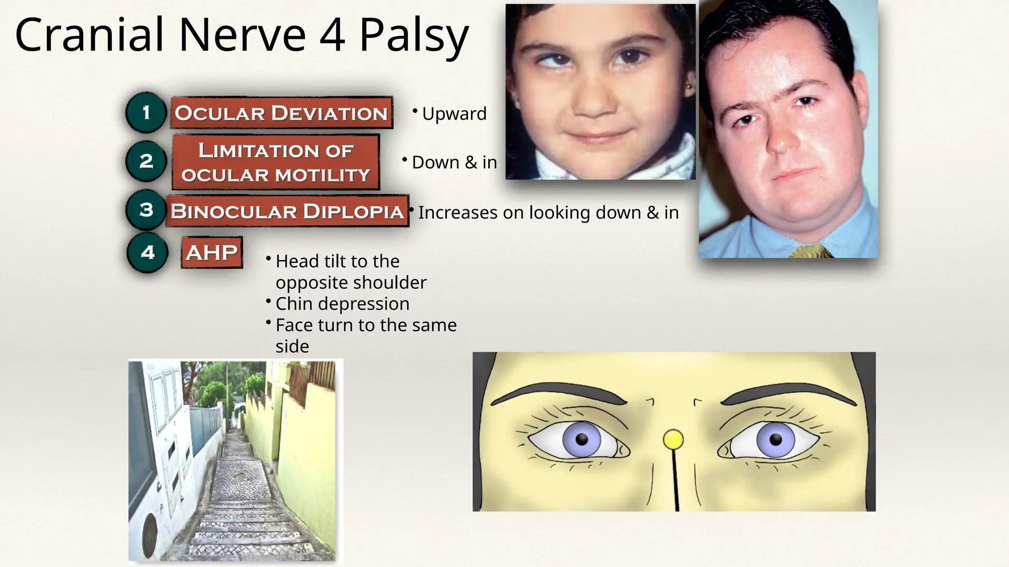
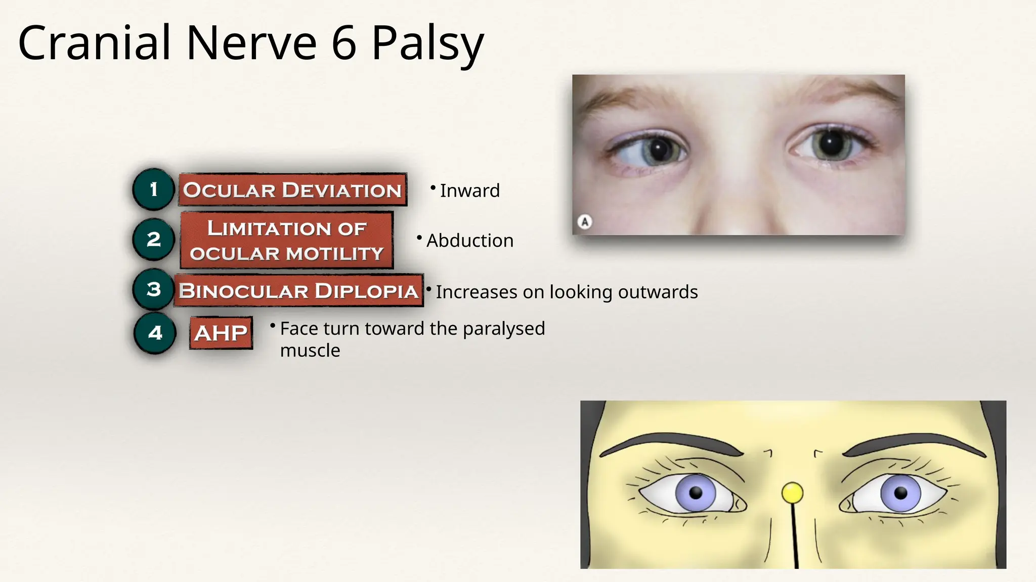
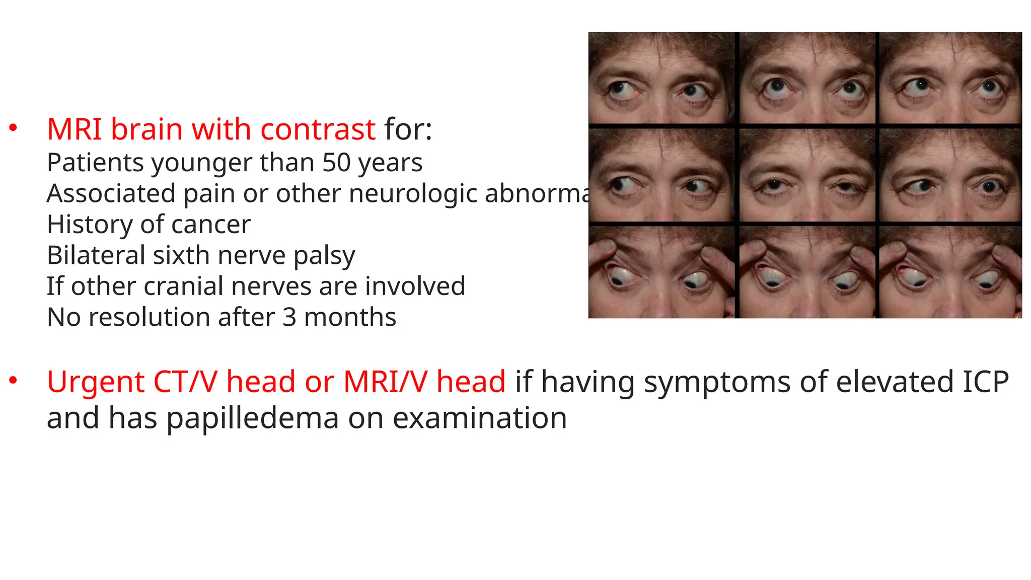
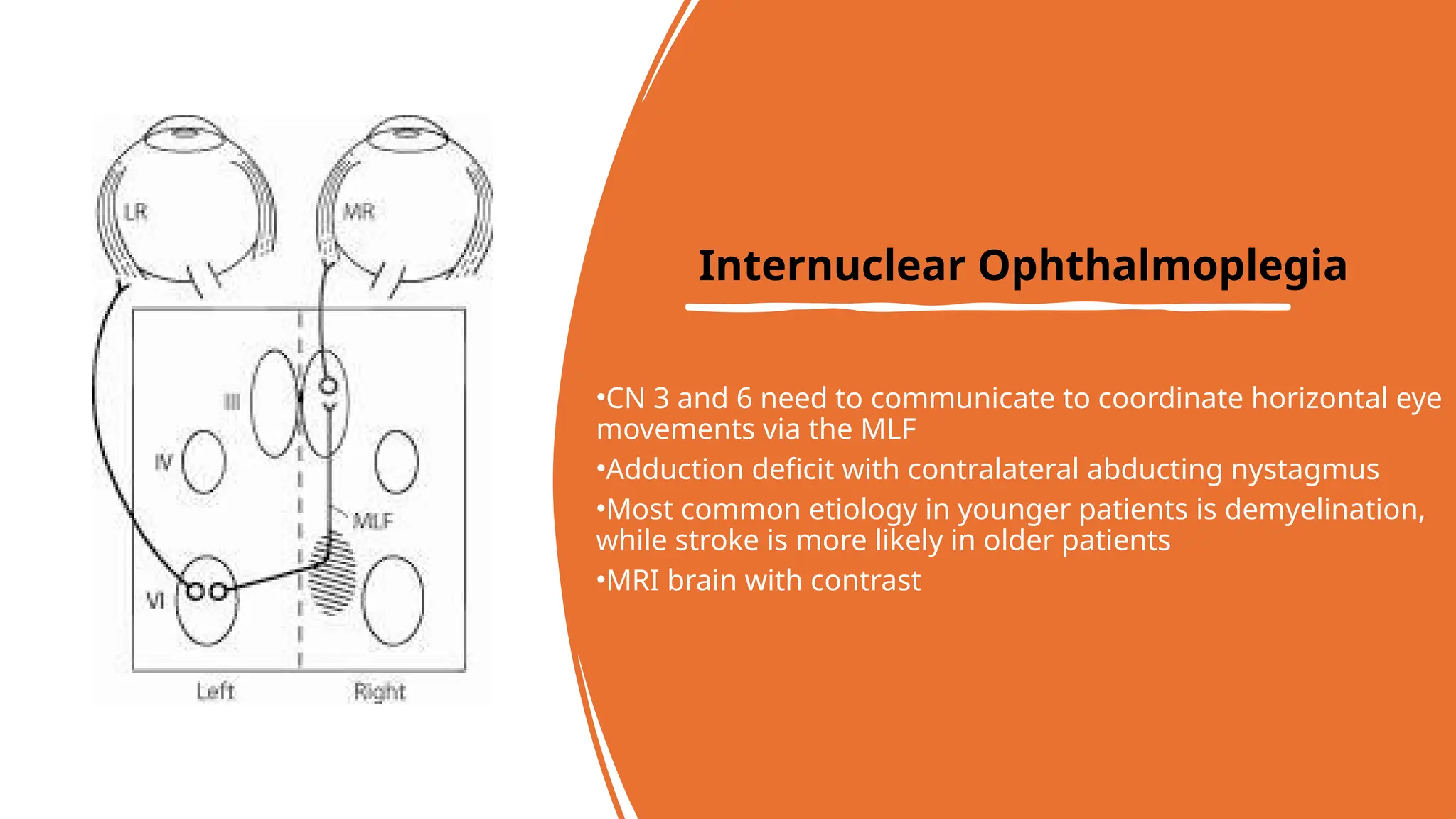
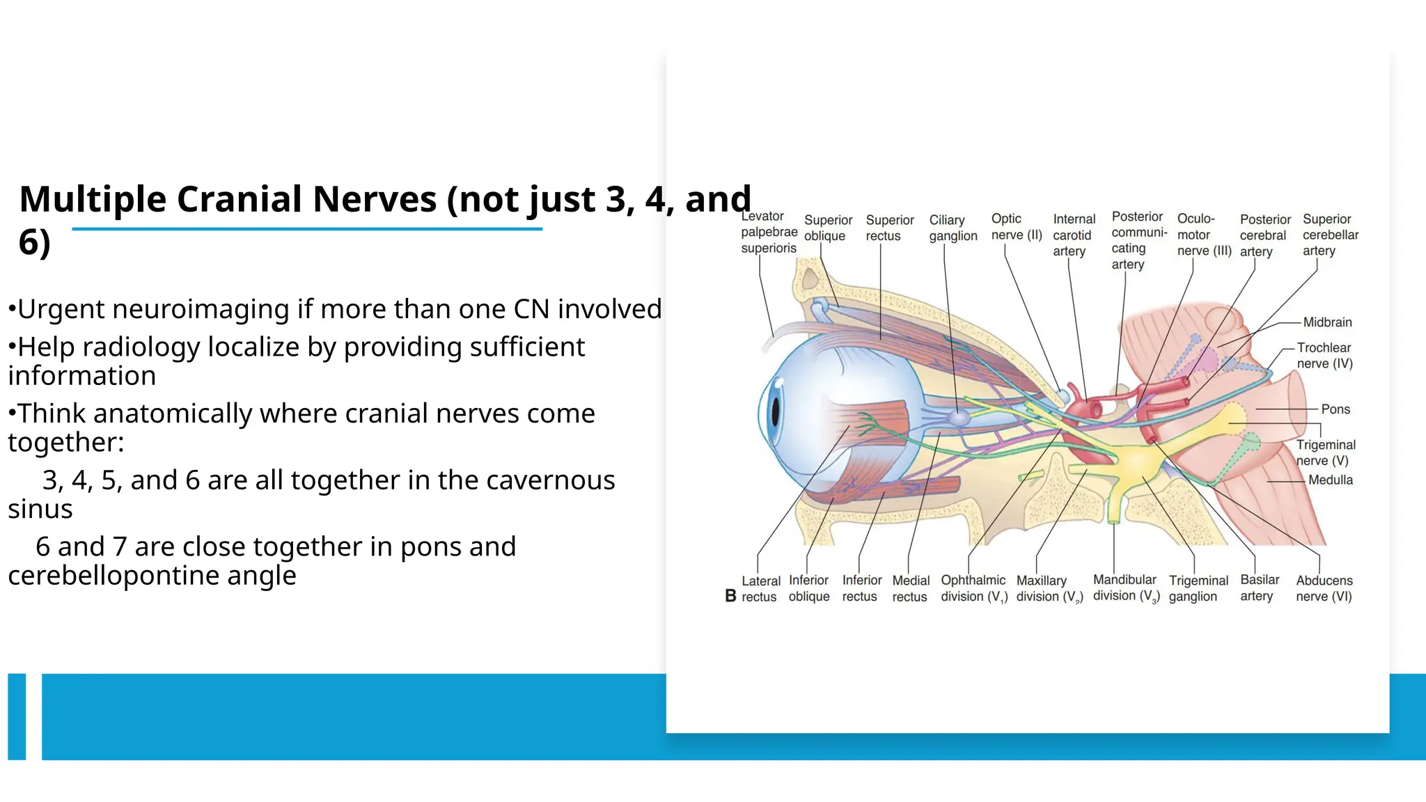
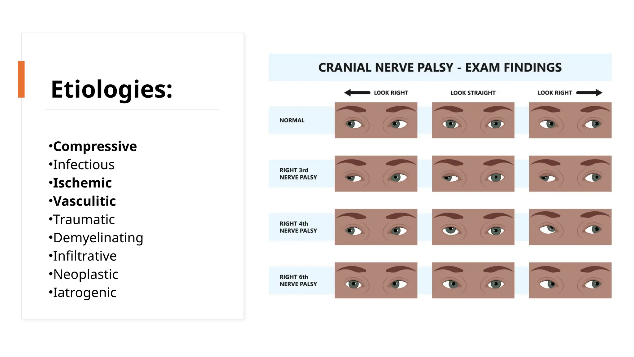
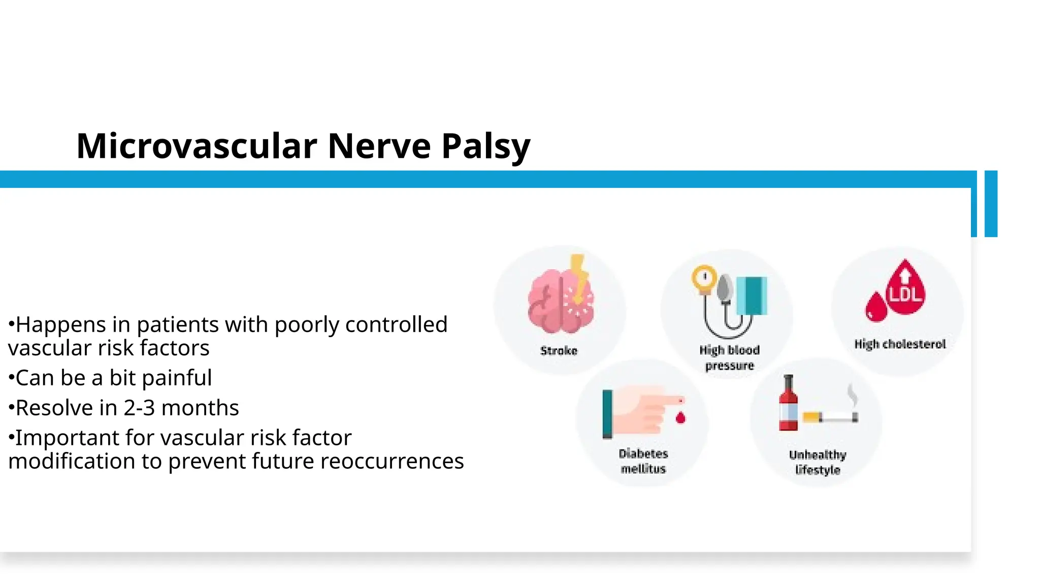
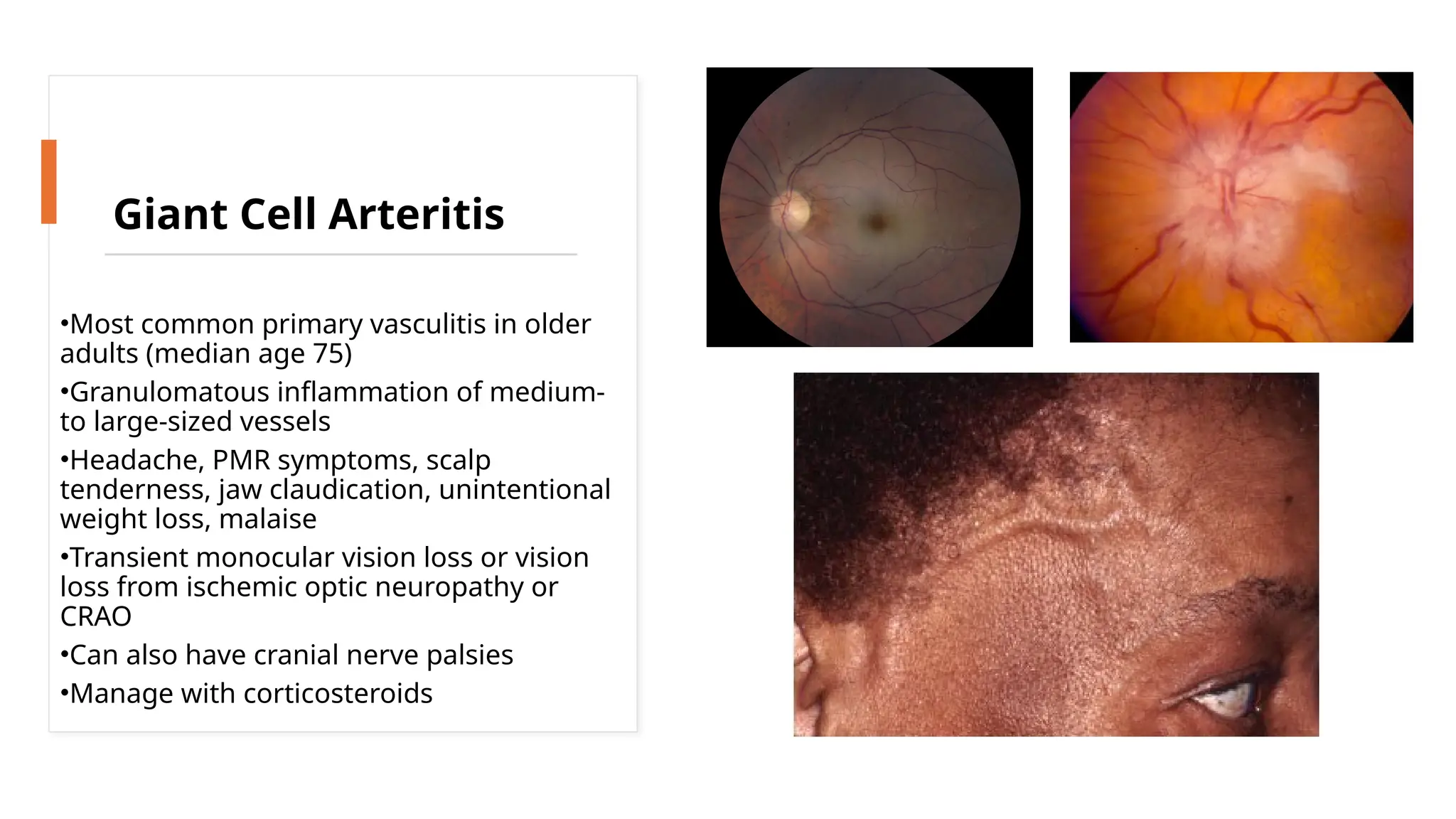
Mohamed Ahmed ELShafie
MD, HMS alumni
LR6SO4
Oculomotor nerve
(CN 3)
• Passes between the SCA below and the PCA above
• runs slightly oblique to the tentorial edge, parallel
and lateral to the posterior communicating artery
•Pupillary fibers usually found on the
dorsomedial surface of the nerve - vulnerable to
compression:
Posterior communicating artery aneurysm
Uncal herniation - most medial aspect of the temporal lobe is forced
through the tentorial notch by a supratentorial mass lesion
•Enters its own dural canal at the back edge of the
dura of the clivus just above CN 4
•Runs in the superior lateral wall of the cavernous
sinus and separates into the superior and inferior
divisions before entering the orbit
Trochlear nerve (CN 4)
• Nucleus in midbrain just below the aqueduct
Decussates before exiting
• Only CN that exits on the dorsal surface of
the brainstem
• the longest unprotected intracranial course –
vulnerable to closed-head trauma
• Swings around the midbrain, paralleling the
tentorium just under the tentorial edge
(vulnerable during neurosurgical procedures
that involve the tentorium)
• Enters the posterior lateral aspect of the
cavernous sinus underneath CN 3 and within
the lateral wall of the cavernous sinus.
Abducens nerve (CN
6)
• Nucleus in dorsal caudal pons surrounded by looping fibers of
CN 7
• Runs within the subarachnoid space along the clivus
• Enters dura approximately 1 cm below the petrous apex and
travels beneath petroclinoid ligament (Gruber ligament -
connects the petrous pyramid to the posterior clinoid)
• Enters Dorello’s canal and travels with the inferior petrosal
sinus
• Exits dura into cavernous sinus parallel to the horizontal
segment of carotid artery and V1
• Enters orbit through superior orbital fissure
 Vulnerable to elevated ICP due to its position between
brainstem and clivus in subarachnoid space (stretching of
nerve) and it’s tethering to Dorello’s canal
Cranial Nerve Palsy
Etiologies:
• Compressive
• Infectious
• Ischemic
• Vasculitic
• Traumatic
• Demyelinating
• Infiltrative
• Neoplastic
• Iatrogenic
• Outward
• In all directions except outward
Cranial Nerve 3 Palsy
Cranial Nerve 3 Palsy –
Imaging?
Urgent CTA head or MRA head:
• screen for posterior communicating artery aneurysm or
compressive lesion
• Presence of aberrant regeneration highly suggestive of
compressive or traumatic etiology
• Upward
• Down & in
• Increases on looking down & in
• Head tilt to the
opposite shoulder
• Chin depression
• Face turn to the same
side
Cranial Nerve 4 Palsy
Park’s 3-step test:
Which eye is hypertropic in primary position? Impacted muscle will be a
depressor of the higher eye (IR or SO) or an elevator of the lower eye (SR or IO)
Is the hypertropia worse in right or left gaze? If worse in left gaze, for a
paretic muscle, the SO in the right eye or the SR in the left eye is affected
If worse in right gaze, for a paretic muscle, the SO in the left eye or SR muscle in the
right eye is affected
Is the hypertropia worse on left head tilt or right head tilt. If worse in
right tilt, the right eye intorters (SO and SR) or left eye extorters (IO and IR) are
affected
If worse in left tilt, the left eye intorters (SO and SR) or right eye extorters (IO and IR)
are affected
Cranial Nerve 4 Palsy
• Longest and thinnest of the cranial nerves – most susceptible to
trauma
• Can be congenital, which can decompensate later in life or from
illness:
Longstanding head tilt (look at old photographs)
Facial asymmetry
Large vertical fusional amplitudes (>2 prism diopters)
• MRI brain with contrast – recommended if acute
Specify interest in CN 4 to neuro-radiology for best sequences (e.g. FIESTA)
• Inward
• Abduction
• Increases on looking outwards
• Face turn toward the paralysed
muscle
Cranial Nerve 6 Palsy
• MRI brain with contrast for:
Patients younger than 50 years
Associated pain or other neurologic abnormality
History of cancer
Bilateral sixth nerve palsy
If other cranial nerves are involved
No resolution after 3 months
• Urgent CT/V head or MRI/V head if having symptoms of elevated ICP
and has papilledema on examination
•CN 3 and 6 need to communicate to coordinate horizontal eye
movements via the MLF
•Adduction deficit with contralateral abducting nystagmus
•Most common etiology in younger patients is demyelination,
while stroke is more likely in older patients
•MRI brain with contrast
Internuclear Ophthalmoplegia
•Urgent neuroimaging if more than one CN involved
•Help radiology localize by providing sufficient
information
•Think anatomically where cranial nerves come
together:
3, 4, 5, and 6 are all together in the cavernous
sinus
6 and 7 are close together in pons and
cerebellopontine angle
Multiple Cranial Nerves (not just 3, 4, and
6)
•Compressive
•Infectious
•Ischemic
•Vasculitic
•Traumatic
•Demyelinating
•Infiltrative
•Neoplastic
•Iatrogenic
Etiologies:
•Happens in patients with poorly controlled
vascular risk factors
•Can be a bit painful
•Resolve in 2-3 months
•Important for vascular risk factor
modification to prevent future reoccurrences
Microvascular Nerve Palsy
•Most common primary vasculitis in older
adults (median age 75)
•Granulomatous inflammation of medium-
to large-sized vessels
•Headache, PMR symptoms, scalp
tenderness, jaw claudication, unintentional
weight loss, malaise
•Transient monocular vision loss or vision
loss from ischemic optic neuropathy or
CRAO
•Can also have cranial nerve palsies
•Manage with corticosteroids
Giant Cell Arteritis
The oculomotor nerve (CN 3):
A. Innervates the medial rectus, superior rectus, inferior rectus, and
inferior oblique.
B.Is responsible for eyelid closure.
C. Innervates the levator palpebrae superioris and the pupillary
sphincter.
D.A and C are true.
The trochlear nerve (CN 4):
A.Exits ventrally from the brainstem, like the other cranial nerves.
B.Can cause a hypertropia if damaged
C.Is hardly ever affected by head trauma
D.Only has a short segment that is unprotected in the subarachnoid
space
The abducens nerve (CN 6):
A. Nucleus is located in the pons and is surrounded by fibers of CN 8.
B. Innervates the lateral rectus
C.B and D are true
D. Communicates with CN 3 through the medial longitudinal fasciculus.
27
Dr. Ahmad Taher Azar 27

How to deal with 3,4 and 6 cranial nerve palsy

  • 1.
  • 2.
  • 4.
    Oculomotor nerve (CN 3) •Passes between the SCA below and the PCA above • runs slightly oblique to the tentorial edge, parallel and lateral to the posterior communicating artery •Pupillary fibers usually found on the dorsomedial surface of the nerve - vulnerable to compression: Posterior communicating artery aneurysm Uncal herniation - most medial aspect of the temporal lobe is forced through the tentorial notch by a supratentorial mass lesion •Enters its own dural canal at the back edge of the dura of the clivus just above CN 4 •Runs in the superior lateral wall of the cavernous sinus and separates into the superior and inferior divisions before entering the orbit
  • 6.
    Trochlear nerve (CN4) • Nucleus in midbrain just below the aqueduct Decussates before exiting • Only CN that exits on the dorsal surface of the brainstem • the longest unprotected intracranial course – vulnerable to closed-head trauma • Swings around the midbrain, paralleling the tentorium just under the tentorial edge (vulnerable during neurosurgical procedures that involve the tentorium) • Enters the posterior lateral aspect of the cavernous sinus underneath CN 3 and within the lateral wall of the cavernous sinus.
  • 7.
    Abducens nerve (CN 6) •Nucleus in dorsal caudal pons surrounded by looping fibers of CN 7 • Runs within the subarachnoid space along the clivus • Enters dura approximately 1 cm below the petrous apex and travels beneath petroclinoid ligament (Gruber ligament - connects the petrous pyramid to the posterior clinoid) • Enters Dorello’s canal and travels with the inferior petrosal sinus • Exits dura into cavernous sinus parallel to the horizontal segment of carotid artery and V1 • Enters orbit through superior orbital fissure  Vulnerable to elevated ICP due to its position between brainstem and clivus in subarachnoid space (stretching of nerve) and it’s tethering to Dorello’s canal
  • 8.
    Cranial Nerve Palsy Etiologies: •Compressive • Infectious • Ischemic • Vasculitic • Traumatic • Demyelinating • Infiltrative • Neoplastic • Iatrogenic
  • 9.
    • Outward • Inall directions except outward Cranial Nerve 3 Palsy
  • 10.
    Cranial Nerve 3Palsy – Imaging?
  • 11.
    Urgent CTA heador MRA head: • screen for posterior communicating artery aneurysm or compressive lesion • Presence of aberrant regeneration highly suggestive of compressive or traumatic etiology
  • 12.
    • Upward • Down& in • Increases on looking down & in • Head tilt to the opposite shoulder • Chin depression • Face turn to the same side Cranial Nerve 4 Palsy
  • 13.
    Park’s 3-step test: Whicheye is hypertropic in primary position? Impacted muscle will be a depressor of the higher eye (IR or SO) or an elevator of the lower eye (SR or IO) Is the hypertropia worse in right or left gaze? If worse in left gaze, for a paretic muscle, the SO in the right eye or the SR in the left eye is affected If worse in right gaze, for a paretic muscle, the SO in the left eye or SR muscle in the right eye is affected Is the hypertropia worse on left head tilt or right head tilt. If worse in right tilt, the right eye intorters (SO and SR) or left eye extorters (IO and IR) are affected If worse in left tilt, the left eye intorters (SO and SR) or right eye extorters (IO and IR) are affected
  • 15.
    Cranial Nerve 4Palsy • Longest and thinnest of the cranial nerves – most susceptible to trauma • Can be congenital, which can decompensate later in life or from illness: Longstanding head tilt (look at old photographs) Facial asymmetry Large vertical fusional amplitudes (>2 prism diopters) • MRI brain with contrast – recommended if acute Specify interest in CN 4 to neuro-radiology for best sequences (e.g. FIESTA)
  • 16.
    • Inward • Abduction •Increases on looking outwards • Face turn toward the paralysed muscle Cranial Nerve 6 Palsy
  • 17.
    • MRI brainwith contrast for: Patients younger than 50 years Associated pain or other neurologic abnormality History of cancer Bilateral sixth nerve palsy If other cranial nerves are involved No resolution after 3 months • Urgent CT/V head or MRI/V head if having symptoms of elevated ICP and has papilledema on examination
  • 18.
    •CN 3 and6 need to communicate to coordinate horizontal eye movements via the MLF •Adduction deficit with contralateral abducting nystagmus •Most common etiology in younger patients is demyelination, while stroke is more likely in older patients •MRI brain with contrast Internuclear Ophthalmoplegia
  • 19.
    •Urgent neuroimaging ifmore than one CN involved •Help radiology localize by providing sufficient information •Think anatomically where cranial nerves come together: 3, 4, 5, and 6 are all together in the cavernous sinus 6 and 7 are close together in pons and cerebellopontine angle Multiple Cranial Nerves (not just 3, 4, and 6)
  • 20.
  • 21.
    •Happens in patientswith poorly controlled vascular risk factors •Can be a bit painful •Resolve in 2-3 months •Important for vascular risk factor modification to prevent future reoccurrences Microvascular Nerve Palsy
  • 22.
    •Most common primaryvasculitis in older adults (median age 75) •Granulomatous inflammation of medium- to large-sized vessels •Headache, PMR symptoms, scalp tenderness, jaw claudication, unintentional weight loss, malaise •Transient monocular vision loss or vision loss from ischemic optic neuropathy or CRAO •Can also have cranial nerve palsies •Manage with corticosteroids Giant Cell Arteritis
  • 24.
    The oculomotor nerve(CN 3): A. Innervates the medial rectus, superior rectus, inferior rectus, and inferior oblique. B.Is responsible for eyelid closure. C. Innervates the levator palpebrae superioris and the pupillary sphincter. D.A and C are true.
  • 25.
    The trochlear nerve(CN 4): A.Exits ventrally from the brainstem, like the other cranial nerves. B.Can cause a hypertropia if damaged C.Is hardly ever affected by head trauma D.Only has a short segment that is unprotected in the subarachnoid space
  • 26.
    The abducens nerve(CN 6): A. Nucleus is located in the pons and is surrounded by fibers of CN 8. B. Innervates the lateral rectus C.B and D are true D. Communicates with CN 3 through the medial longitudinal fasciculus.
  • 27.

Editor's Notes

  • #11 Aberrant regeneration for CN 3 : muscle to muscle (on elevation lt eye move inward), muscle to lid (No ptosis in 1ry position, elevation of lid on looking down), and muscle to pupil
  • #15 No diplopia he can fuse
  • #20 Compressive etiologies are usually the most worrisome and often need to be referred to neurosurgery
  • #22 polymyalgia rheumatica order CBC, ESR, CRP
  • #24 d
  • #25 b
  • #26 c