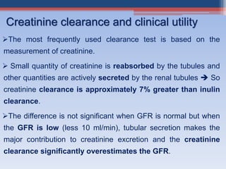
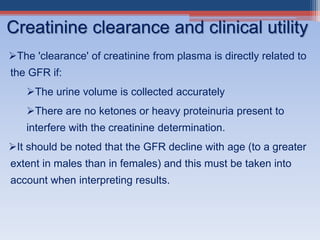
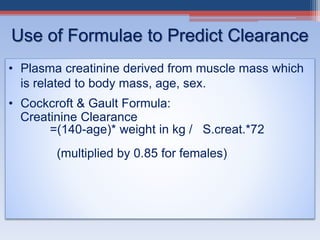
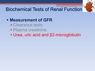
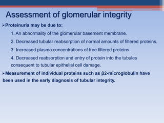
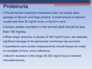
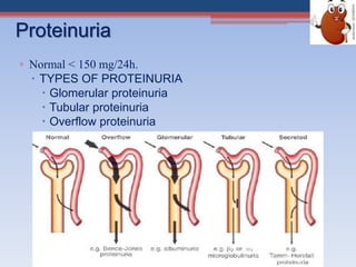
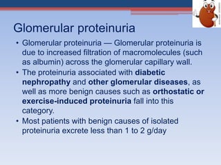
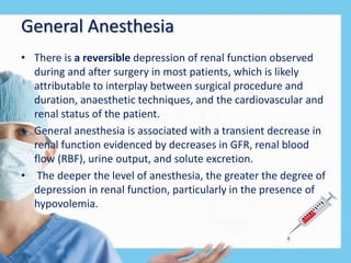
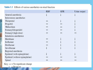
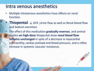
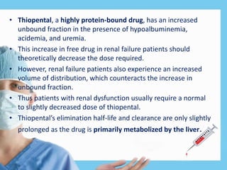
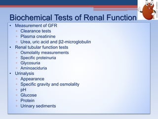
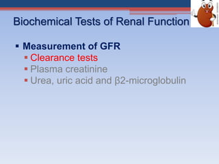
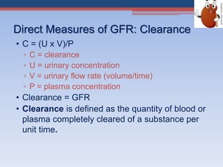
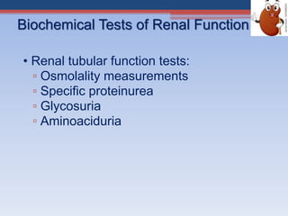
Renal function tests assess kidney function and detect impairment. They include tests of glomerular filtration rate (GFR) using creatinine, urea and inulin clearance. GFR is calculated using creatinine levels and equations factoring age, weight and sex. Other tests evaluate tubular function through urine concentration, osmolality, and checking for proteins, glucose and amino acids in urine. Together these tests provide valuable information about both glomerular and tubular integrity and kidney health.
KFT are used for evaluating kidney functions. there are several routine tests such as urea, creatinine and uric acid. Calculation of eGFR is recommended by national kidney organization whenever creatinine serum is measured.
Renal function test (RFT), also known as kidney function test is a group of tests used to assess the functions of kidney.
It is used screen for, detect, evaluate and monitor acute and chronic kidney diseases.
These are simple blood and urine tests that are used identify kidneys problems.
Tests of renal function have utility in-
Identifying the presence of renal disease
Monitoring the response of kidneys to treatment
Determining the progression of renal disease
RFT is ordered, if your doctor
thinks your kidneys may not be working properly which is known from signs and symptoms
and if you have other conditions that can harm the kidneys, such as diabetes or high blood pressure
The liver is the largest organ in the body
It is located below the diaphragm in the right upper quadrant of the abdominal cavity and extended approximately from the right 5th rib to the lower border of the rib cage.
rft is described in detail . function of kidney, objectives of doing the test. the various test available for assessing the renal function with clinical interpretation is available.
The main function of the kidney is excretion of water soluble waste products from our body.
Derangement of any of these function would result in either decreased excretion of waste products and hence their accumulation in the body or loss of some vital nutrient from the body.
A test in which blood or urine samples are checked for the amounts of certain substances released by the kidneys. A higher- or lower-than-normal amount of a substance can be a sign that the kidneys are not working the way they should. Also called kidney function test.
Best Ayurvedic medicine for Gas and IndigestionSwastikAyurveda
Here is the updated list of Top Best Ayurvedic medicine for Gas and Indigestion and those are Gas-O-Go Syp for Dyspepsia | Lavizyme Syrup for Acidity | Yumzyme Hepatoprotective Capsules etc
Title: Sense of Taste
Presenter: Dr. Faiza, Assistant Professor of Physiology
Qualifications:
MBBS (Best Graduate, AIMC Lahore)
FCPS Physiology
ICMT, CHPE, DHPE (STMU)
MPH (GC University, Faisalabad)
MBA (Virtual University of Pakistan)
Learning Objectives:
Describe the structure and function of taste buds.
Describe the relationship between the taste threshold and taste index of common substances.
Explain the chemical basis and signal transduction of taste perception for each type of primary taste sensation.
Recognize different abnormalities of taste perception and their causes.
Key Topics:
Significance of Taste Sensation:
Differentiation between pleasant and harmful food
Influence on behavior
Selection of food based on metabolic needs
Receptors of Taste:
Taste buds on the tongue
Influence of sense of smell, texture of food, and pain stimulation (e.g., by pepper)
Primary and Secondary Taste Sensations:
Primary taste sensations: Sweet, Sour, Salty, Bitter, Umami
Chemical basis and signal transduction mechanisms for each taste
Taste Threshold and Index:
Taste threshold values for Sweet (sucrose), Salty (NaCl), Sour (HCl), and Bitter (Quinine)
Taste index relationship: Inversely proportional to taste threshold
Taste Blindness:
Inability to taste certain substances, particularly thiourea compounds
Example: Phenylthiocarbamide
Structure and Function of Taste Buds:
Composition: Epithelial cells, Sustentacular/Supporting cells, Taste cells, Basal cells
Features: Taste pores, Taste hairs/microvilli, and Taste nerve fibers
Location of Taste Buds:
Found in papillae of the tongue (Fungiform, Circumvallate, Foliate)
Also present on the palate, tonsillar pillars, epiglottis, and proximal esophagus
Mechanism of Taste Stimulation:
Interaction of taste substances with receptors on microvilli
Signal transduction pathways for Umami, Sweet, Bitter, Sour, and Salty tastes
Taste Sensitivity and Adaptation:
Decrease in sensitivity with age
Rapid adaptation of taste sensation
Role of Saliva in Taste:
Dissolution of tastants to reach receptors
Washing away the stimulus
Taste Preferences and Aversions:
Mechanisms behind taste preference and aversion
Influence of receptors and neural pathways
Impact of Sensory Nerve Damage:
Degeneration of taste buds if the sensory nerve fiber is cut
Abnormalities of Taste Detection:
Conditions: Ageusia, Hypogeusia, Dysgeusia (parageusia)
Causes: Nerve damage, neurological disorders, infections, poor oral hygiene, adverse drug effects, deficiencies, aging, tobacco use, altered neurotransmitter levels
Neurotransmitters and Taste Threshold:
Effects of serotonin (5-HT) and norepinephrine (NE) on taste sensitivity
Supertasters:
25% of the population with heightened sensitivity to taste, especially bitterness
Increased number of fungiform papillae
- Video recording of this lecture in English language: https://youtu.be/kqbnxVAZs-0
- Video recording of this lecture in Arabic language: https://youtu.be/SINlygW1Mpc
- Link to download the book free: https://nephrotube.blogspot.com/p/nephrotube-nephrology-books.html
- Link to NephroTube website: www.NephroTube.com
- Link to NephroTube social media accounts: https://nephrotube.blogspot.com/p/join-nephrotube-on-social-media.html
These simplified slides by Dr. Sidra Arshad present an overview of the non-respiratory functions of the respiratory tract.
Learning objectives:
1. Enlist the non-respiratory functions of the respiratory tract
2. Briefly explain how these functions are carried out
3. Discuss the significance of dead space
4. Differentiate between minute ventilation and alveolar ventilation
5. Describe the cough and sneeze reflexes
Study Resources:
1. Chapter 39, Guyton and Hall Textbook of Medical Physiology, 14th edition
2. Chapter 34, Ganong’s Review of Medical Physiology, 26th edition
3. Chapter 17, Human Physiology by Lauralee Sherwood, 9th edition
4. Non-respiratory functions of the lungs https://academic.oup.com/bjaed/article/13/3/98/278874
Recomendações da OMS sobre cuidados maternos e neonatais para uma experiência pós-natal positiva.
Em consonância com os ODS – Objetivos do Desenvolvimento Sustentável e a Estratégia Global para a Saúde das Mulheres, Crianças e Adolescentes, e aplicando uma abordagem baseada nos direitos humanos, os esforços de cuidados pós-natais devem expandir-se para além da cobertura e da simples sobrevivência, de modo a incluir cuidados de qualidade.
Estas diretrizes visam melhorar a qualidade dos cuidados pós-natais essenciais e de rotina prestados às mulheres e aos recém-nascidos, com o objetivo final de melhorar a saúde e o bem-estar materno e neonatal.
Uma “experiência pós-natal positiva” é um resultado importante para todas as mulheres que dão à luz e para os seus recém-nascidos, estabelecendo as bases para a melhoria da saúde e do bem-estar a curto e longo prazo. Uma experiência pós-natal positiva é definida como aquela em que as mulheres, pessoas que gestam, os recém-nascidos, os casais, os pais, os cuidadores e as famílias recebem informação consistente, garantia e apoio de profissionais de saúde motivados; e onde um sistema de saúde flexível e com recursos reconheça as necessidades das mulheres e dos bebês e respeite o seu contexto cultural.
Estas diretrizes consolidadas apresentam algumas recomendações novas e já bem fundamentadas sobre cuidados pós-natais de rotina para mulheres e neonatos que recebem cuidados no pós-parto em unidades de saúde ou na comunidade, independentemente dos recursos disponíveis.
É fornecido um conjunto abrangente de recomendações para cuidados durante o período puerperal, com ênfase nos cuidados essenciais que todas as mulheres e recém-nascidos devem receber, e com a devida atenção à qualidade dos cuidados; isto é, a entrega e a experiência do cuidado recebido. Estas diretrizes atualizam e ampliam as recomendações da OMS de 2014 sobre cuidados pós-natais da mãe e do recém-nascido e complementam as atuais diretrizes da OMS sobre a gestão de complicações pós-natais.
O estabelecimento da amamentação e o manejo das principais intercorrências é contemplada.
Recomendamos muito.
Vamos discutir essas recomendações no nosso curso de pós-graduação em Aleitamento no Instituto Ciclos.
Esta publicação só está disponível em inglês até o momento.
Prof. Marcus Renato de Carvalho
www.agostodourado.com
2. Why test renal function?
To assess the functional capacity of kidney
Early detection of possible renal impairment.
Severity and progression of the impairment.
Monitor response to treatment
Monitor the safe and effective use of drugs which
are excreted in the urine.
3. When should we assess renal function?
Older age
Family history of Chronic Kidney disease (CKD)
Decreased renal mass
Low birth weight
Diabetes Mellitus (DM)
Hypertension (HTN)
Autoimmune disease
Systemic infections
Urinary tract infections (UTI)
Nephrolithiasis
Obstruction to the lower urinary tract
Drug toxicity
4. Biochemical Tests of Renal Function
• Measurement of GFR
▫ Clearance tests
▫ Plasma creatinine
▫ Urea, uric acid and β2-microglobulin
• Renal tubular function tests
▫ Osmolality measurements
▫ Specific proteinuria
▫ Glycosuria
▫ Aminoaciduria
• Urinalysis
▫ Appearance
▫ Specific gravity and osmolality
▫ pH
▫ Glucose
▫ Protein
▫ Urinary sediments
5. Glomerular Filtration Rate (GFR) Affected by:
1). Total filtration surface area
2). Membrane permeability
3). Net Filtration Pressure (as NFP goes up so does the GFR)
In the normal adult, this rate is about 120 ml/min; about 180 liters/Day
Glomerular filtration rate (GFR) :
▫GFR = rate (mL/min) at which substances in plasma are filtered through
the glomerulus
▫Best indicator of overall kidney function
▫Can be measured or calculated using a variety of markers
Glomerular Filtration Rate
6. Markers of GFR
• Ideal characteristics:
▫ Freely filtered at the glomerulus
▫ No tubular secretion or reabsorption
▫ No renal/tubular metabolism
• Exogenous or endogenous
▫ Exogenous – not normally present in the body
Inulin
▫ Endogenous – normally present in the body
Creatinine .
• Radiolabeled or non-radiolabeled .
7. Biochemical Tests of Renal Function
Measurement of GFR
Clearance tests
Plasma creatinine
Urea, uric acid and β2-microglobulin
8. Direct Measures of GFR: Clearance
• C = (U x V)/P
▫ C = clearance
▫ U = urinary concentration
▫ V = urinary flow rate (volume/time)
▫ P = plasma concentration
• Clearance = GFR
• Clearance is defined as the quantity of blood or
plasma completely cleared of a substance per
unit time.
9. Inulin clearance
• The Volume of blood from which inulin is cleared or completely
removed in one min is known as the inulin clearance and is equal to
the GFR.
• Gold standard for renal clearance
▫ Freely filtered at glomerulus
▫ No tubular metabolism
▫ No tubular reabsorption or secretion
• Protocol
▫ IV infusion
▫ Blood samples
▫ Urine catheter
• Limitations
▫ Expensive, hard to obtain
▫ Difficult to assay
▫ Invasive
10. Biochemical Tests of Renal Function
Measurement of GFR
Clearance tests
Plasma creatinine
Urea, uric acid and β2-microglobulin
11. Creatinine to Calculate GFR
• Creatinine clearance in adults is normally about of
120 ml/min.
• Advantages
▫ Endogenous
▫ Produced at a constant rate per day
▫ Routinely measured
▫ Freely filtered at glomerulus
-Inversely related to GFR
• Disadvantages
▫ Estimate of GFR
▫ 10% is secreted by renal tubules
▫ Secretion increases as kidney function decreases.
12. 1 to 2% of muscle creatine spontaneously converts to
creatinine daily and released into body fluids at a constant
rate.
Endogenous creatinine produced is proportional to muscle
mass, it is a function of total muscle mass the production
varies with age and sex
Dietary fluctuations of creatinine intake cause only minor
variation in daily creatinine excretion of the same person.
Creatinine released into body fluids at a constant rate and
its plasma levels maintained within narrow limits Creatinine
clearance may be measured as an indicator of GFR.
Creatinine
13. The most frequently used clearance test is based on the
measurement of creatinine.
Small quantity of creatinine is reabsorbed by the tubules and
other quantities are actively secreted by the renal tubules So
creatinine clearance is approximately 7% greater than inulin
clearance.
The difference is not significant when GFR is normal but when
the GFR is low (less 10 ml/min), tubular secretion makes the
major contribution to creatinine excretion and the creatinine
clearance significantly overestimates the GFR.
Creatinine clearance and clinical utility
14. The 'clearance' of creatinine from plasma is directly related to
the GFR if:
The urine volume is collected accurately
There are no ketones or heavy proteinuria present to
interfere with the creatinine determination.
It should be noted that the GFR decline with age (to a greater
extent in males than in females) and this must be taken into
account when interpreting results.
Creatinine clearance and clinical utility
15. Use of Formulae to Predict Clearance
• Plasma creatinine derived from muscle mass which
is related to body mass, age, sex.
• Cockcroft & Gault Formula:
Creatinine Clearance
=(140-age)* weight in kg / S.creat.*72
(multiplied by 0.85 for females)
16. Biochemical Tests of Renal Function
Measurement of GFR
Clearance tests
Plasma creatinine
Urea, uric acid and β2-microglobulin
17. Catabolism of proteins and nucleic acids results in formation
of so called nonprotein nitrogenous compounds.
Protein
Proteolysis, principally enzymatic
Amino acids
Transamination and oxidative deamination
Ammonia
Enzymatic synthesis in the “urea cycle”
Urea
Measurement of nonprotein nitrogen-containing
compounds
18. Urea is the major nitrogen-containing metabolic product of protein
catabolism in humans,
Its elimination in the urine represents the major route for
nitrogen excretion.
More than 90% of urea is excreted through the kidneys, with
losses through the GIT and skin
Urea is filtered freely by the glomeruli
Plasma urea concentration is often used as an index of renal
glomerular function
Urea production is increased by a high protein intake and it is
decreased in patients with a low protein intake or in patients with
liver disease.
Plasma Urea
19. Many renal diseases with various glomerular, tubular, interstitial or vascular
damage can cause an increase in plasma urea concentration.
The reference interval is 8-20 mg/dl.
Plasma concentrations also tend to be slightly higher in males than females.
Measurement of plasma creatinine provides a more accurate assessment
than urea because there are many factors that affect urea level.
Nonrenal factors can affect the urea level (normal adults is level 8-20 mg/dl)
like:
Mild dehydration,
high protein diet,
increased protein catabolism, muscle wasting as in starvation,
GIT haemorrhage,
treatment with cortisol or its synthetic analogues.
Plasma Urea
20. Clinical Significance
• States associated with elevated levels of urea in
blood are referred to as uremia or azotemia.
• Causes of urea plasma elevations:
Prerenal: renal hypoperfusion
Renal: acute tubular necrosis
Postrenal: obstruction of urinary flow.
21. In human, uric acid is the major product of the catabolism of the
purine nucleosides, adenosine and guanosine.
Purines are derived from catabolism of dietary nucleic acid
(nucleated cells, like meat) and from degradation of endogenous
nucleic acids.
Overproduction of uric acid may result from increased synthesis
of purine precursors.
In humans, approximately 75% of uric acid excreted is lost in the
urine; most of the reminder is secreted into the GIT .
Uric acid
22. Uric acid
Renal handling of uric acid is complex and involves four sequential steps:
Glomerular filtration of virtually all the uric acid in capillary plasma
entering the glomerulus.
Reabsorption in the proximal convoluted tubule of about 98 to 100%
of filtered uric acid.
Subsequent secretion of uric acid into the lumen of the distal portion
of the proximal tubule.
Further reabsorption in the distal tubule.
Hyperuricemia is defined by serum or plasma uric acid concentrations
higher than 7.0 mg/dl (0.42mmol/L) in men or greater than 6.0 mg/dl
(0.36mmol/L) in women.
23. It is present on the surface of most cells and in low concentrations
in the plasma.
It is completely filtered by the glomeruli and is reabsorbed and
catabolized by proximal tubular cells.
The plasma concentration of β2-microglobulin is a good index of
GFR in normal people, being unaffected by diet or muscle mass.
It is increased in certain malignancies and inflammatory diseases.
Since it is normally reabsorbed and catabolized in the tubules,
measurement of β2-microglobulin excretion provides a sensitive
method of assessing tubular integrity.
Plasma β2-microglobulin
24. Biochemical Tests of Renal Function
• Renal tubular function tests:
▫ Osmolality measurements
▫ Specific proteinurea
▫ Glycosuria
▫ Aminoaciduria
25. Renal tubular function tests
• To ensure that important constituents such as water,
sodium, glucose and a.a. are not lost from the body,
tubular reabsorption must be equally efficient
• Compared with the GFR as an assessment of glomerular
function, there are no easily performed tests which
measure tubular function in quantitative manner
• Osmolality measurements in plasma and urine; normal
urine : plasma osmolality ratio is usually between 1.0-3.0
26. Tubular function tests
Urine Concentration Test
The ability of the kidney to concentrate urine is a test of
tubular function that can be carried out readily with only
minor inconvenience to the patient.
This test requires a water deprivation for 14 hrs in
healthy individuals.
A specific gravity of > 1.02 indicates normal
concentrating power.
Specific gravity of 1.008 to 1.010 is isotonic with plasma
and indicates no work done by kidneys.
The test should not be performed on a dehydrated
patient.
27. Vasopressin Test
More patient friendly than water deprivation test.
The subject has nothing to drink after 6 p.m. At 8 p.m. five
units of vasopressin tannate is injected
subcutaneously. All urine samples are collected
separately until 9 a.m. the next morning.
Satisfactory concentration is shown by at least one
sample having a specific gravity above 1.020, or an
osmolality above 800 m osm/kg.
The urine/plasma osmolality ratio should reach 3 and
values less than 2 are abnormal.
28. Urine Dilution (Water Load) Test
After an overnight fast the subject empties his bladder
completely and is given 1000 ml of water to drink.
Urine specimens are collected for the next 4 hours, the
patient emptying bladder completely on each occasion.
Normally the patient will excrete at least 700 ml of urine in
the 4 hours, and at least one specimen will have a specific
gravity less than 1.004.
Kidneys which are severely damaged cannot excrete a
urine of lower specific gravity than 1.010 or a volume
above 400 ml in this time.
The test should not be done if there is oedema or renal
failure; water intoxication may result.
29. Proteinuria may be due to:
1. An abnormality of the glomerular basement membrane.
2. Decreased tubular reabsorption of normal amounts of filtered proteins.
3. Increased plasma concentrations of free filtered proteins.
4. Decreased reabsorption and entry of protein into the tubules
consequent to tubular epithelial cell damage.
Measurement of individual proteins such as β2-microglobulin have
been used in the early diagnosis of tubular integrity.
Assessment of glomerular integrity
30. The glomerular basement membrane does not usually allow
passage of albumin and large proteins. A small amount of albumin,
usually less than 30 mg/24 hours, is found in urine.
Urinary protein excretion in the normal adult should be less
than 150 mg/day.
When larger amounts, in excess of 300 mg/24 hours, are detected,
significant damage to the glomerular membrane has occurred.
Quantitative urine protein measurements should always be made
on complete 24-hour urine collections.
Albumin excretion in the range 30-300 mg/24 hours is termed
microalbuminuria.
Proteinuria
31. ▫ Normal < 150 mg/24h.
TYPES OF PROTEINURIA
Glomerular proteinuria
Tubular proteinuria
Overflow proteinuria
Proteinuria
32. Glomerular proteinuria
• Glomerular proteinuria — Glomerular proteinuria is
due to increased filtration of macromolecules (such
as albumin) across the glomerular capillary wall.
• The proteinuria associated with diabetic
nephropathy and other glomerular diseases, as
well as more benign causes such as orthostatic or
exercise-induced proteinuria fall into this
category.
• Most patients with benign causes of isolated
proteinuria excrete less than 1 to 2 g/day
33. Tubular proteinuria
• Low molecular weight proteins — have a
molecular weight ≤ 25,000 in comparison to the
69,000 molecular weight of albumin.
1. ß2-microglobulin,
2. immunoglobulin light chains,
3.retinol-binding protein, and amino acids —
• These smaller proteins can be filtered across the
glomerulus and are then almost completely
reabsorbed in the proximal tubule.
• Interference with proximal tubular reabsorption, due
to a variety of tubulointerstitial diseases or even
some primary glomerular diseases, can lead to
increased excretion of these smaller proteins.
34. Overflow proteinuria
• Increased excretion of low molecular weight
proteins can occur with marked overproduction
of a particular protein, leading to increased
glomerular filtration and excretion.
• Due to 1. immunoglobulin light chains in
multiple myeloma(most common)
2. lysozyme (in acute myelomonocytic
leukemia),
3.myoglobin (in rhabdomyolysis), or
4.hemoglobin (in intravascular hemolysis).
35. Biochemical Tests of Renal Function
• Urinalysis
▫ Appearance
▫ Specific gravity and osmolality
▫ pH
▫ Glucose
▫ Protein
▫ Urinary sediments
36. Urine Analysis
Urine examination is an extremely valuable and
most easily performed test for the evaluation of
renal functions.
It includes physical or macroscopic examination,
chemical examination and microscopic
examination of the sediment.
37. Macroscopic examination
Colour:
Normal- pale yellow in colour due to pigments
urochrome, urobilin and uroerythrin.
Cloudiness-caused by excessive cellular
material or protein, crystallization or precipitation
of salts upon standing at room temperature or in
the refrigerator.
Red colour -If the sample contains many red
blood cells.
38. To maintain water homeostasis, the kidneys must produce urine in a
volume precisely balances water intake and production to equal water
loss through extra renal routes.
Minimum urine volume is determined by the solute load to be excreted
whereas maximum urine volume is determined by the amount of excess
water that must be excreted.
Urine volume
39. Volume
Normal- 1-2.5 L/day
Oliguria- Urine Output < 400ml/day
Seen in
▫ Acute glomerulonephritis
▫ Renal Failure
Polyuria- Urine Output > 2.5 L/day
Seen in
▫ Increased water ingestion
▫ Diabetes mellitus and insipidus.
Anuria- Urine output < 100ml/day
Seen in renal shut down.
40. Specific Gravity
Measured by urinometer or refractometer.
It is measurement of urine density which reflects the ability
of the kidney to concentrate or dilute the urine relative to
the plasma from which it is filtered.
Normal :- 1.000- 1.030.
S.G Osmolality (mosm/kg)
1.001 100
1.010 300
1.020 800
1.025 1000
1.030 1200
1.040 1400
41. Increase in Specific Gravity seen in
Low water intake
Diabetes mellitus
Albuminuria
Acute nephritis.
Decrease in Specific Gravity is seen in
Absence of ADH
Renal Tubular damage.
Isosthenuria-Persistent production of fixed low
Specific gravity urine isoosmolar with plasma
despite variation in water intake.
42. Urinalysis: Osmolality measurements in
plasma and urine
– Osmolality serves as general marker of tubular function. Because the
ability to concentrate the urine is highly affected by renal diseases.
– This is conveniently done by determining the osmolality, and then
comparing this to the plasma.
– If the urine osmolality is 600mosm/kg or more, tubular function is
usually regarded as intact
– When the urine osmolality does not differ greatly from plasma (urine:
plasma osmolality ratio=1), the renal tubules are not reabsorbing water
43. pH
Urine pH ranges from 4.5 to 8
Normally it is slightly acidic lying between 6 – 6.5.
On exposure to atmosphere, urea in urine splits
causing NH4
+ release resulting in alkaline
reaction.
44. Biochemical testing of urine involves the use of commercially available disposable
strips When the strip is manually immersed in the urine specimen, the reagents
react with a specific component of urine in such a way that to form color
Colour change produced is proportional to the concentration of the component
being tested for.
To test a urine sample:
fresh urine is collected into a clean dry container
the sample is not centrifuged
the disposable strip is briefly immersed in the urine specimen;
The colour of the test areas are compared with those provided on a colour chart.
Urinalysis using disposable strips
46. Urine Sediments
- Microscopic examination of sediment from freshly
passed urine involves looking for cells, casts, fat
droplets
- Blood: haematuria is consistent with various
possibilities ranging from malignancy through urinary
tract infection to contamination from menstruation.
- Red Cell casts could indicate glomerular disease
- Crystals
- Leucocytes in the urine suggests acute inflammation
and the presence of a urinary tract infection.
47. -are cylindrical structures produced by the kidney and present in the
urine in certain disease states.
- They form in the distal convoluted tubule and collecting ducts of
nephrons, then dislodge and pass into the urine, where they can be
detected by microscope.
- They form via precipitation of Tamm-Hrsfall mucoprotein which is
secreted by renal tubule cells, and sometimes also by albumin.
Urinary casts
48. Red blood cell cast in urine
White blood cell cast in
urine
Urinary casts. (A) Hyaline cast
(200 X); (B) erythrocyte cast
(100 X); (C) leukocyte cast
(100 X); (D) granular cast (100
X)
50. Clinical significance of RFT in AKI
The RIFLE criteria, proposed by the Acute Dialysis
Quality Initiative (ADQI) group, aid in the staging of
patients with AKI (previously ARF).
51. Cystatin C
Novel biomarker for non-invasive estimation of Glomerular
Filtration Rate and Early Renal Impairment.
• Cystatin C is a nonglycosylated basic protein produced at
a constant rate by all nucleated cells.
• It is freely filtered by the renal glomeruli and primarily
catabolized in the tubule (not secreted or reabsorbed as
an intact molecule).
• As serum cystatin C concentration is independent of age,
sex, and muscle mass, it has been postulated to be an
improved marker of glomerular filtration rate (GFR)
compared with serum creatinine level.
53. • The National Kidney Foundation Kidney Disease Outcomes
Quality Initiative (K/DOQI) definition of chronic kidney disease
is the presence of kidney damage or a reduction in the
glomerular filtration rate (GFR) for three months or longer.
• The K/DOQI chronic kidney disease staging system is based on
GFR.
• Inappropriate dosing in patients with chronic kidney disease
can cause toxicity or ineffective therapy.
• Older patients are at a higher risk of developing advanced
disease and related adverse events caused by age-related
decline in renal function and the use of multiple medications
to treat comorbid conditions.
54.
55. • Chronic kidney disease can affect glomerular blood
flow and filtration, tubular secretion and
reabsorption, and renal bioactivation and
metabolism.
• Drug absorption, bioavailability, protein binding,
distribution volume, and non-renal clearance
(metabolism) can also be altered in these patients.
• In patients with a GFR < 60 mL/min/1.73 m2, the
MDRD equation has been shown to be superior to
the Cockcroft-Gault equation.
56. • The MDRD equation has been shown to be the best
method for detecting a GFR < 90 ml /min/ 1.73 m2 in
older patients.
• Because the production and excretion of creatinine
declines with age, normal serum creatinine values
may not represent normal renal function in older
patients.
57. Dosing Adjustments
• Loading doses usually do not need to be adjusted in patients
with chronic kidney disease.
• For Maintenance dosing adjustments:
1.dose reduction,
2.lengthening the dosing interval, or both.
• Dose reduction involves reducing each dose while
maintaining the normal dosing interval.
• This approach maintains more constant drug concentrations,
but associated with a higher risk of toxicities if the dosing
interval is inadequate to allow for drug elimination.
58. Dosing Adjustments
• Lengthening of dose interval-
Normal doses are maintained with the extended
interval method, but the dosing interval is lengthened to
allow time for drug elimination before re-dosing.
• Lengthening the dosing interval has been associated with a
lower risk of toxicities but a higher risk of subtherapeutic
drug concentrations.
59. Anti Hypertensives
• Thiazide diuretics -first-line agents for treating uncomplicated
hypertension, but they are not recommended if the serum
creatinine level is higher than 2.5 mg per dL or if the
creatinine clearance is lower than 30 mL per minute.
• Loop diuretics -most commonly used to treat uncomplicated
hypertension in patients with chronic kidney disease.
• Potassium-sparing diuretics and aldosterone blockers -
avoided in patients with severe chronic kidney disease
because of the rise in serum potassium that typically
accompanies renal dysfunction.
61. ACE Inhibitors/ARB
• Angiotensin-converting enzyme (ACE) inhibitors and
angiotensin receptor blockers (ARBs) are first-line
hypertensive agents for patients with type 1 or 2 diabetes
mellitus and proteinuria or early chronic kidney disease.
• These 1.reduce blood pressure and proteinuria,
2.slow the progression of kidney disease, and provide
long-term cardiovascular protection.
• Inhibit the renin-angiotensin-aldosterone system in patients
with chronic kidney disease and in patients with normal
baseline serum creatinine levels, causing efferent arteriolar
dilation.
62.
63. • This causes –
• 1.Acute decline in GFR of > 15% from baseline with
• 2. proportional elevations in serum creatinine within the first week
of initiating therapy.
• This most commonly occurs in patients with congestive heart
failure, in patients using concomitant diuretics or nonsteroidal
anti-inflammatory drugs (NSAIDs), and in patients receiving high
doses of ACE inhibitors or ARBs.
• ACE inhibitors and ARBs can be continued safely if the rise in serum
creatinine is < 30%.
• Typically, the levels will return to baseline in four to six weeks.
• Because of long-term renoprotective and cardioprotective effects,
no patient should be denied an ACE-inhibitor or ARB trial without
careful evaluation.
64. • Hydrophilic beta blockers - Atenolol, Bisoprolol, Nadolol,
Acebutolol-eliminated renally -dosing adjustments are needed
in patients with chronic kidney failure.
• Metoprolol tartrate , Metoprolol succinate, Propranolol,and
Labetalol are metabolized by the liver and adjustments are not
required.
• Other AHA’s that do not require dosing adjustments include
calcium channel blockers, clonidine and alpha blockers.
65. Oral hypoglycemics
• METFORMIN-is 90 to 100 percent renally excreted,
• Not recommended when the serum creatinine level is higher
than 1.5 mg/dL in men or higher than 1.4 mg/dl in women,
• In patients older than 80 years, or
• In patients with chronic heart failure.
• The primary concern about the use of metformin in patients
with renal insufficiency is that other hypoxemic conditions
(e.g., acute myocardial infarction, severe infection, respiratory
disease, liver disease) increase the risk of lactic acidosis.
66.
67. OHA
• Sulfonylureas -Chlorpropamide , Glyburide
• Should be avoided in patients with stages 3 to 5 chronic
kidney disease.
• The half-life of chlorpropamide is significantly increased in
these patients, which can cause severe hypoglycemia.
• Glyburide has an active metabolite that is eliminated renally,
and accumulation of this metabolite can cause prolonged
hypoglycemia in patients with chronic kidney disease.
• Glipizide, however, does not have an active metabolite and is
safe in these patients.
68. Antibiotics
• Penicillins-Excessive serum levels of injectable penicillinG or
carbenicillin may be associated with neuromuscular toxicity,
myoclonus, seizures,or coma.
• Carbapenems- Imipenem/cilastatin can accumulate in
patients with chronic kidney disease, causing seizures if doses
are not reduced.
• Patients with advanced disease should receive a different
carbapenem, such as meropenem.
• Tetracyclines- with the exception of doxycycline , have an
antianabolic effect that may significantly worsen the uremic
state in patients with severe disease.
• Nitrofurantoin - has a toxic metabolite that can
accumulate in patients with chronic kidney disease, causing
peripheral neuritis.
69. Antibiotics
• Aminoglycosides -avoided in patients with chronic
kidney disease when possible.
• If used, initial doses should be based on an accurate GFR
estimate.
70. Opioids
• Patients with stage 5 kidney disease are more likely to experience
adverse effects from opioid use.
• Metabolites of meperidine ,dextropropoxyphene(propoxyphene ),
morphine , tramadol , and codeine can accumulate in patients
with chronic kidney disease, causing central nervous system and
respiratory adverse effects.
• These agents are not recommended in patients with stage 4 or 5
disease.
• A 50 to 75 percent dose reduction for morphine and codeine is
recommended in patients with a creatinine clearance less than
50 mL per minute.
71. Opioids
• Extended-release tramadol should be avoided in patients with
chronic kidney disease. The dosing interval of tramadol (regular
release) may need to be increased to every 12 hours in patients
with a creatinine clearance < 30 mL/ min .
72. Opioids
• Morphine is metabolized primarily by hepatic glucuronidation
to form morphine-6-glucuronide and morphine-3-
glucuronide, both of which are excreted renally .
• Morphine-6-glucuronide is more potent than morphine and
may accumulate in renal patients causing prolonged
respiratory depression.
• Meperidine is metabolized by the liver to normeperidine,
which is eliminated both renally and hepatically.
• Accumulation of high levels of normeperidine can produce
excitatory central nervous symptoms including seizures in
extreme cases.
• More appropriate narcotics in renal patients include fentanyl ,
sufentanil, alfentanil, and remifentanil that do not undergo
transformation to long-acting renally excreted metabolites.
73. • At doses of 1–2 mg/kg, Morphine does not decrease blood
pressure or urine flow.
• Fentanyl may decrease GFR, urine flow, and mean arterial
pressure (MAP), though with conflicting data regarding renal
blood flow .
74. NSAIDS
• Adverse renal effects - 1.Acute renal failure;
2.Nephrotic syndrome with interstitial nephritis; and
3.chronic renal failure with or without glomerulopathy,
4.interstitial nephritis, and
5. papillary necrosis.
• The risk of ARF is three times higher in NSAID users .
• Other adverse effects - include decreased potassium excretion -
hyperkalemia, and decreased sodium excretion-peripheral
edema, elevated blood pressure, and decompensation of heart
failure.
• NSAIDs can blunt antihypertensive treatment, especially if beta
blockers, ACE inhibitors, or ARBs are Used.
75. NSAIDS
• Although selective cyclooxygenase-2 (COX-2) inhibitors may
cause slightly fewer adverse gastrointestinal effects, adverse
renal effects are similar to traditional NSAIDs.
• Short-term use - generally safe in patients who are well
hydrated; who have good renal function; and who do not have
heart failure, diabetes, or hypertension.
• Long-term use and high daily dosages of COX-2 inhibitors and
other NSAIDs should be avoided if possible.
76. NSAIDS
• Patients at high risk of NSAID-induced kidney disease
should receive serum creatinine measurements
every two to four weeks for several weeks after
initiation of therapy because renal insufficiency may
occur early in the course of therapy.
• Acetaminophen can be used safely in patients with
renal impairment.
78. • While multiple anaesthetic drugs have direct effects on the
kidneys and their function due to hemodynamic effects, they
are often also dependent on the kidney for renal excretion of
either the drug itself or of its metabolites.
• Hydrophilic and ionized drugs depend primarily on renal
excretion.
• Mechanisms of renal excretion depend on renal blood flow.
• Thus renal blood flow decreases due to surgery, anaesthesia,
or pre-existing conditions may result in decreased renal
excretion by the kidneys.
• In addition to accumulation of drugs and their metabolites,
renal failure patients may also have an altered volume of
distribution, hypoalbuminemia, anaemia, hyperkalemia, and
metabolic acidosis.
79. General Anesthesia
• There is a reversible depression of renal function observed
during and after surgery in most patients, which is likely
attributable to interplay between surgical procedure and
duration, anaesthetic techniques, and the cardiovascular and
renal status of the patient.
• General anesthesia is associated with a transient decrease in
renal function evidenced by decreases in GFR, renal blood
flow (RBF), urine output, and solute excretion.
• The deeper the level of anesthesia, the greater the degree of
depression in renal function, particularly in the presence of
hypovolemia.
80.
81. Intra venous anesthetics
• Multiple intravenous anesthetics have effects on renal
function.
• Thiopental- ↓ GFR ,Urine flow as well as Renal blood flow
and Sodium excretion.
• The effect of this medication gradually reverses, and animal
studies on high-dose thiopental show renal blood flow
remains unchanged in spite of a decrease in myocardial
contractility, cardiac preload and blood pressure, and a reflex
increase in systemic vascular resistance.
82. • Thiopental, a highly protein-bound drug, has an increased
unbound fraction in the presence of hypoalbuminemia,
acidemia, and uremia.
• This increase in free drug in renal failure patients should
theoretically decrease the dose required.
• However, renal failure patients also experience an increased
volume of distribution, which counteracts the increase in
unbound fraction.
• Thus patients with renal dysfunction usually require a normal
to slightly decreased dose of thiopental.
• Thiopental’s elimination half-life and clearance are only slightly
prolonged as the drug is primarily metabolized by the liver.
83. • The effects of Propofol on renal injury remain controversial.
Recent rat studies suggest propofol may have a protective
effect in acute kidney injury .
• Midazolam, in induction doses, decreases urine flow but does
not significantly affect renal blood flow, renal vascular
resistance, or sodium excretion.
• Ketamine has been shown in dogs to increase blood pressure,
renal blood flow, and renal vascular resistance though studies
are conflicting.
• The more protein-bound barbiturates, ketamine, propofol,
and benzodiazepines require no alteration in induction doses
in patients with renal failure.
84. Inhalational anesthetics
• Elimination of these drugs is not dependent on renal function.
• Volatile anesthetics are variably metabolized by the liver to
metabolites including inorganic fluoride, which is dependent
on renal excretion and is nephrotoxic.
• This metabolization is highest in halothane (12–20%) and
followed by sevoflurane, enflurane, isoflurane, and desflurane
(3%,2%,0.2%, and 0.02%, respectively).
• Sevoflurane has not been reported to cause renal toxicity in
patients despite this laboratory data.
85. • Halothane -
Most studies show a decrease in GFR, sodium excretion, and
urine output with a variable effect on renal blood flow during
halothane administration. Data suggest that halothane may
not decrease renal blood flow .
• Enflurane decreases GFR, RBF, and urine flow in humans.
• Isoflurane decreases GFR and urine output in pigs with little
change in renal blood flow.
86. • Sevoflurane metabolism to inorganic fluoride has been
implicated in experimental studies of renal toxicity; however,
no human studies are available to indicate this effect.
• Desflurane decreases renal vascular resistance as well as
RBF, thus maintaining renal blood flow .
87. Depolarizing muscle relaxants
• Succinylcholine increases serum potassium by 0.5 meq/l.
• This increase is no larger in renal patients than in nonrenal
patients: however, the baseline potassium must be taken into
consideration.
• Succinylcholine is metabolized by the hepatically produced
plasma cholinesterase.
• This cholinesterase may be decreased in uremic renal patients,
but this does not usually lead to any clinically significant effect.
• A metabolite of succinylcholine, succinylmonocholine, is
excreted by the kidney and may be active as nondepolarizing
neuromuscular blocker. Thus continuous infusions of
succinylcholine should be avoided in patients with renal failure.
88. Non depolarizing muscle relaxants
• Pancuronium, metocurine, gallamine, doxacurium, and
pipercurium are renally excreted and will exhibit prolonged
elimination half-lives in patients with renal failure.
• Atracurium, vecuronium, and cisatracurium are the
paralytics of choice for intermediate duration as their
pharmacodynamics are minimally affected.
• Atracurium metabolism depends on ester hydrolysis and
Hoffman’s elimination, which do not require renal function.
However, a metabolite of atracurium, laudanosine, is a central
nervous system excitatory agent, which may accumulate in
renal patients, though has not been documented to reach
clinical significance.
89. • Cisatracurium is metabolized by Hoffman’s elimination and is
safe in renal failure.
• Vecuronium is metabolized by the liver; however, the clinical
duration of the drug may be increased in renal failure due to an
increase in elimination half-life and decrease in clearance.
• The elimination half-life of Rocuronium is increased in renal
dysfunction due to the increased volume of distribution;
however, there is no clinical difference noted in terms of onset,
duration, and recovery of neuromuscular blockade.
• Mivacurium is hydrolyzed by plasma cholinesterase and shows
a prolonged duration of action in renal failure.
90. Anticholinesterases
• Neostigmine, pyridostigmine, and edrophonium .
• All highly dependent on renal excretion .
• As a result they have prolonged durations of action in patients
with renal failure.
• Anticholinergics such as atropine and glycopyrrolate also
have prolonged durations of actions in these patients.
91. Regional Anesthesia
• A spinal block as high as T1 produces only slight depressions in
GFR and RBF in humans as long as systemic blood pressure is
maintained .
• Likewise, epidural blocks to thoracic levels with epinephrine-
free local anesthetics produce minimal decreases in GFR and
renal blood flow .
• However, epidural blocks to the thoracic region with
epinephrine containing local anesthetics induce moderate
reductions in GRF and RBF that parallel reductions in mean
arterial pressure.
• Most likely the effects of neuraxial blockade on renal function
depend on the hemodynamic effects induced by the
sympathetic blockade.