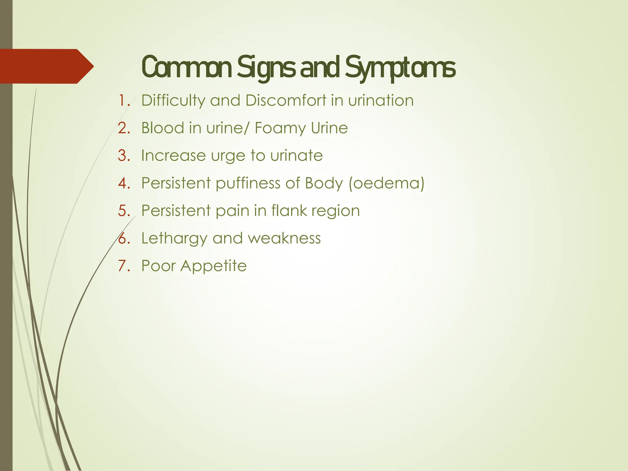
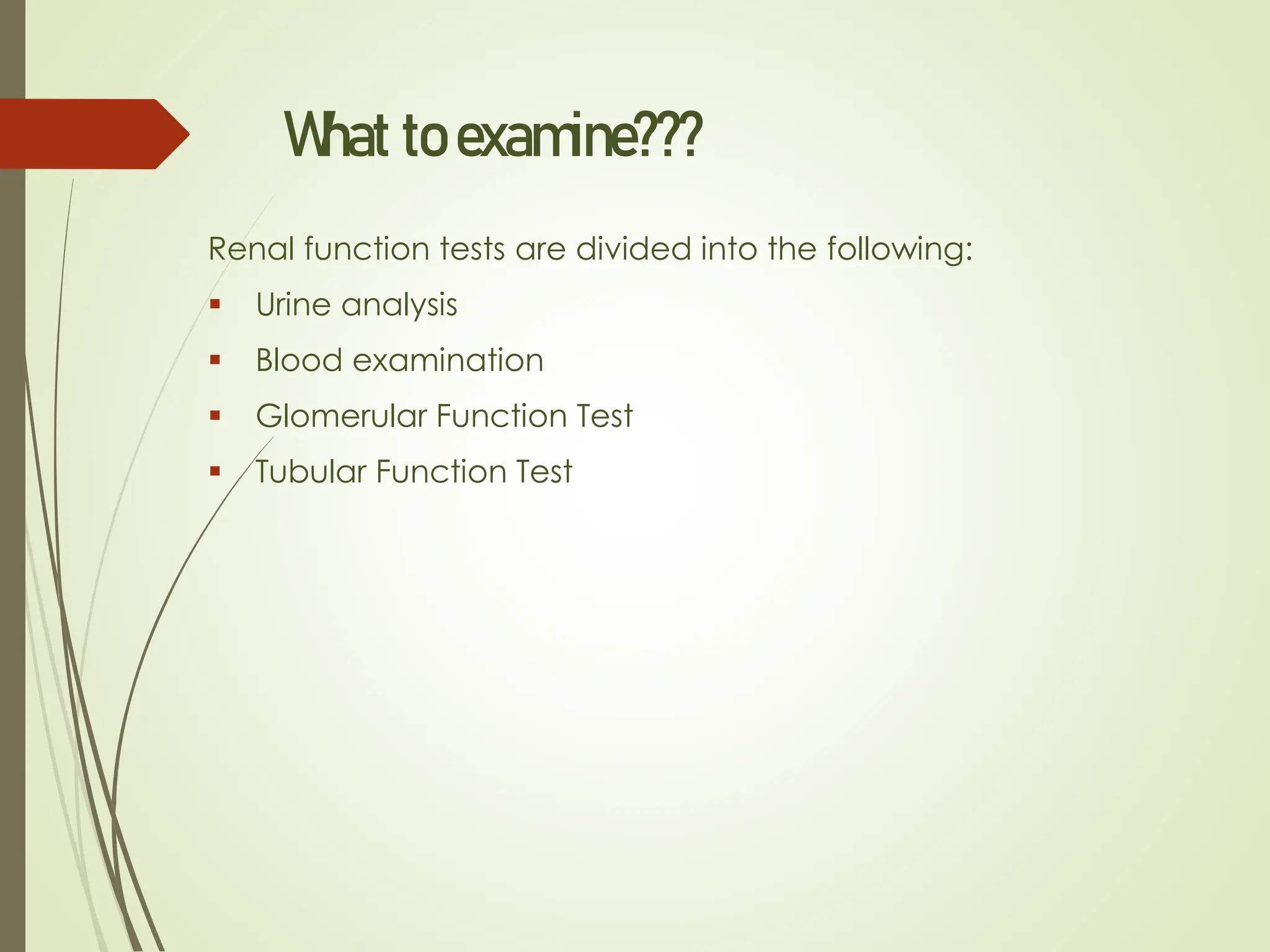
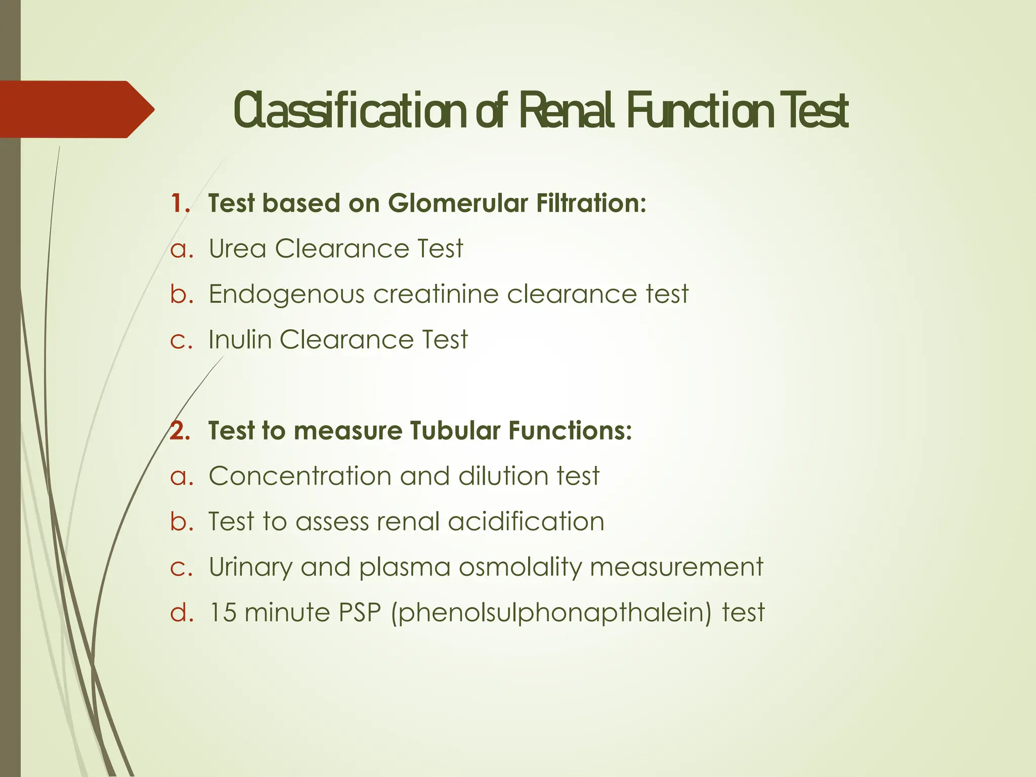
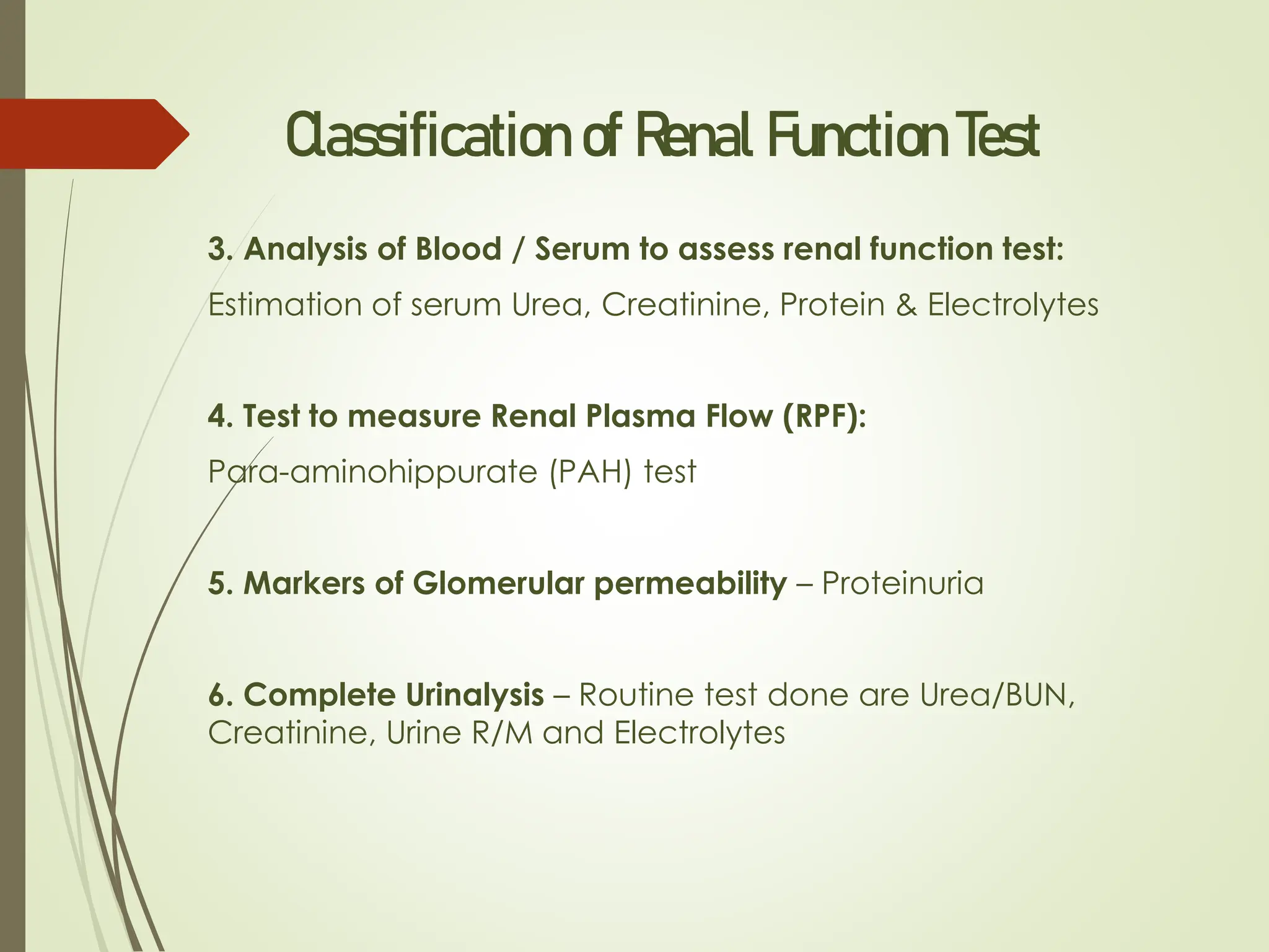
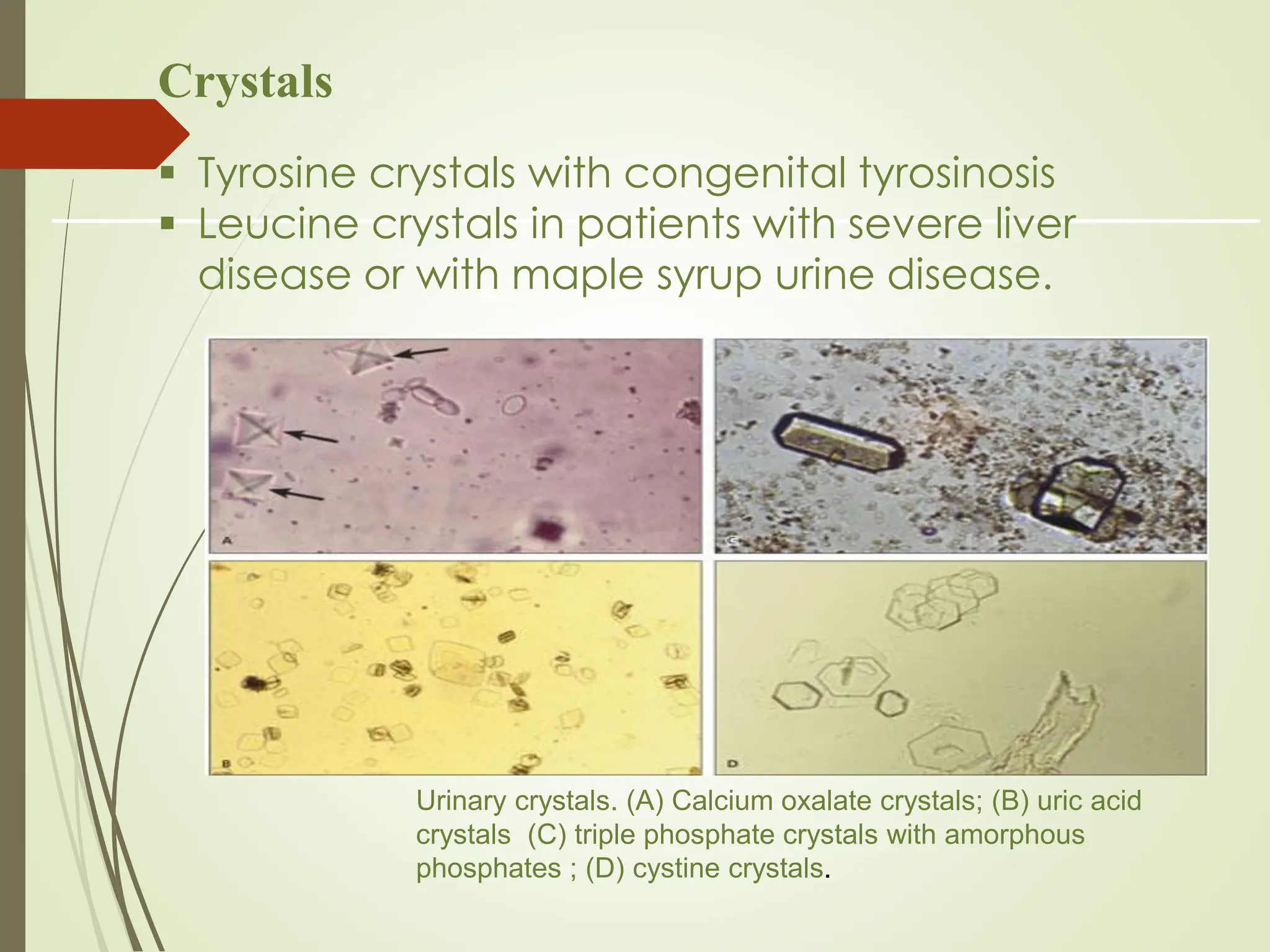
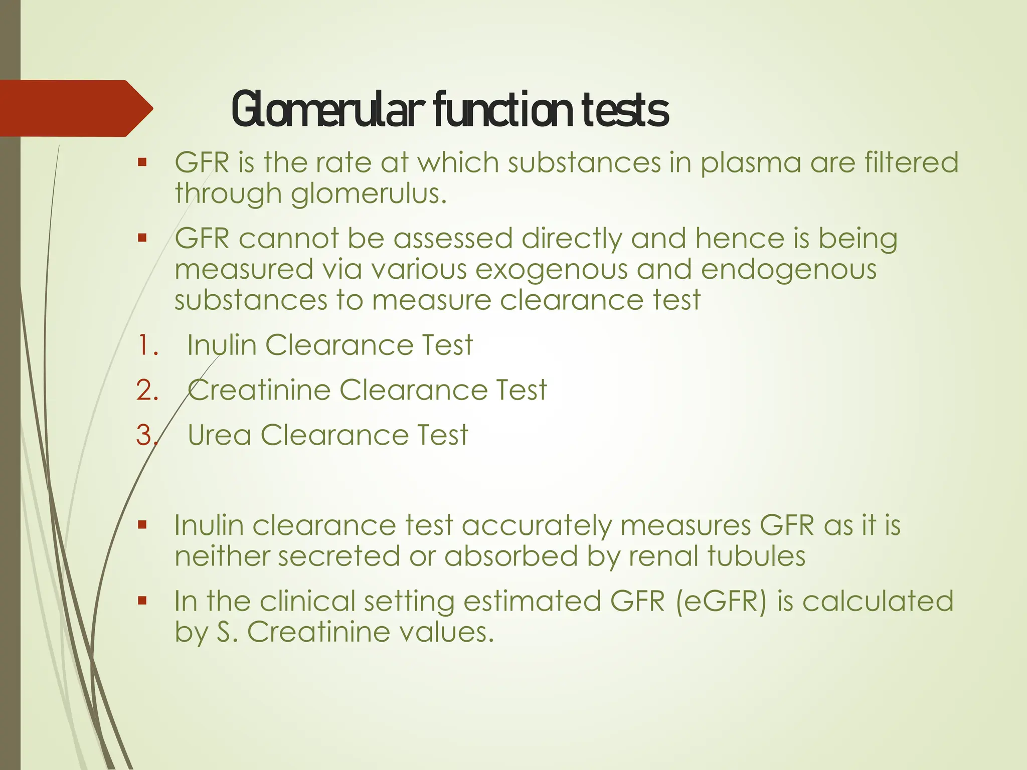
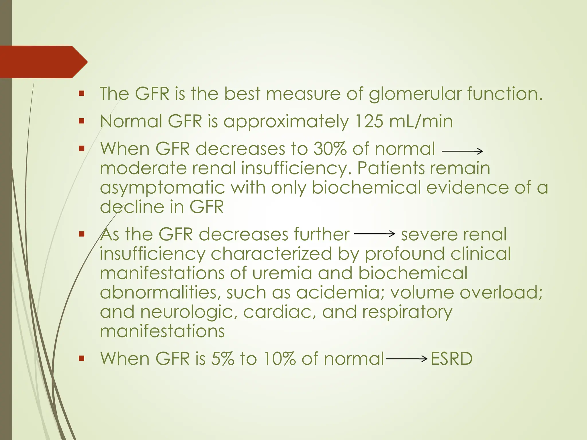
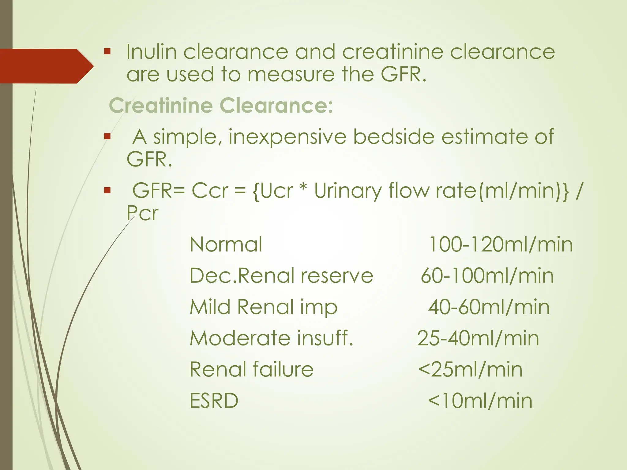
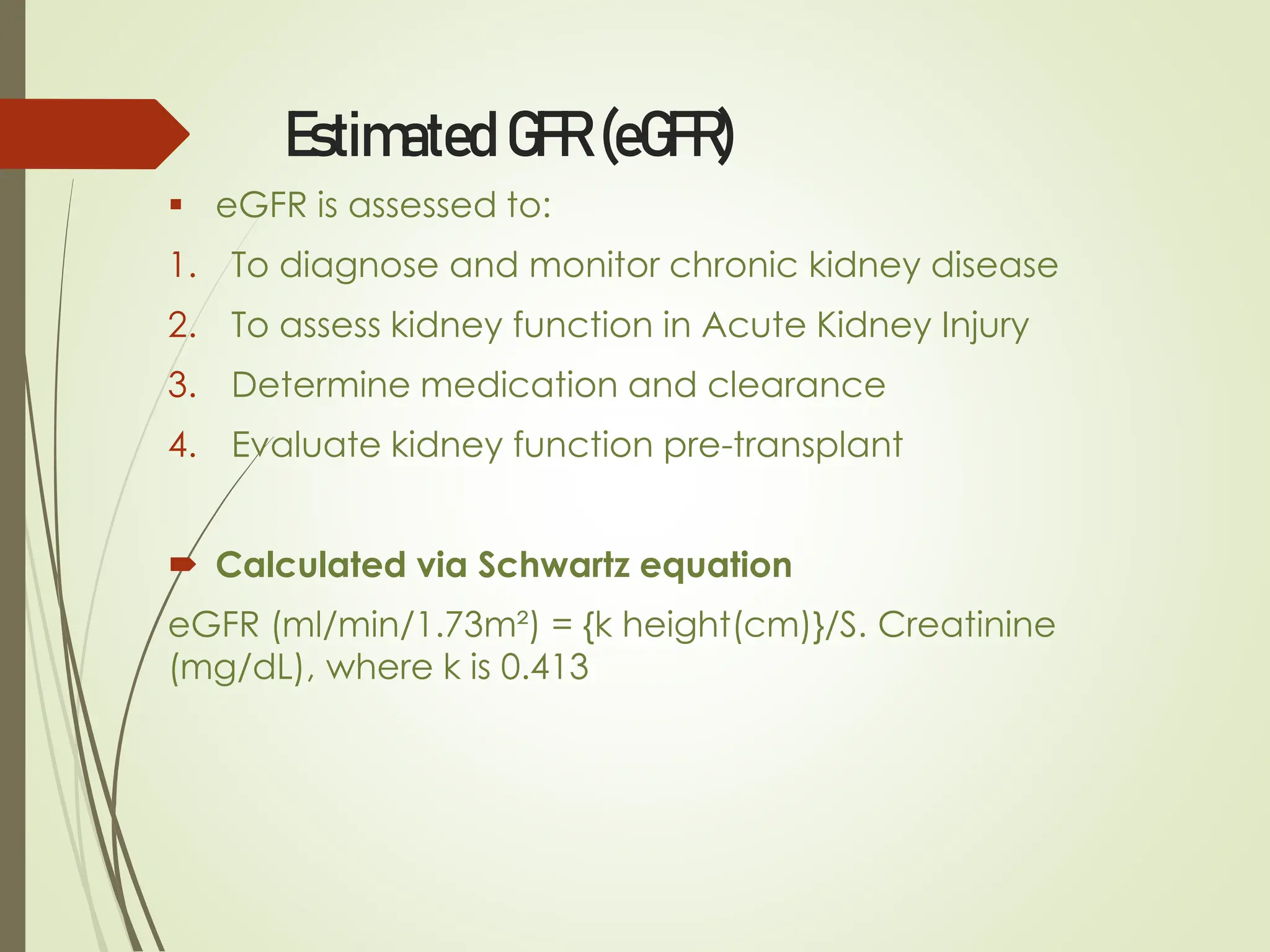
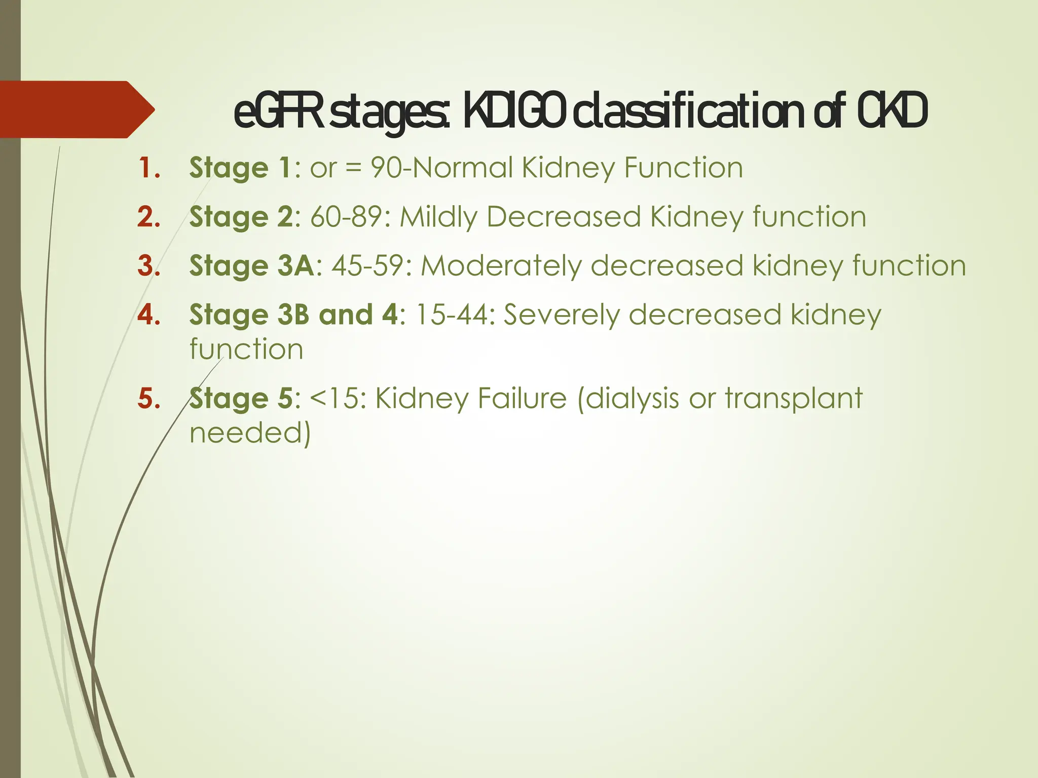
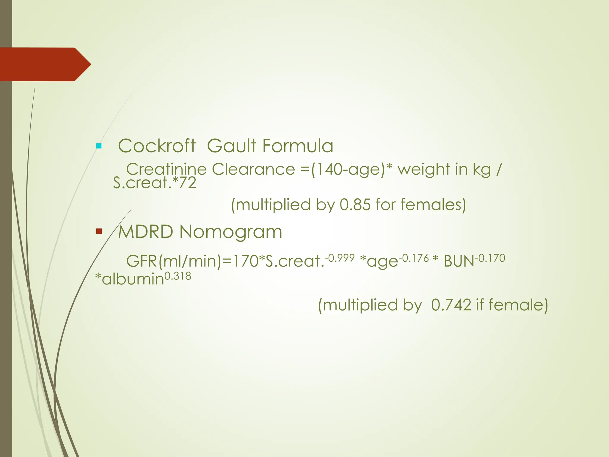
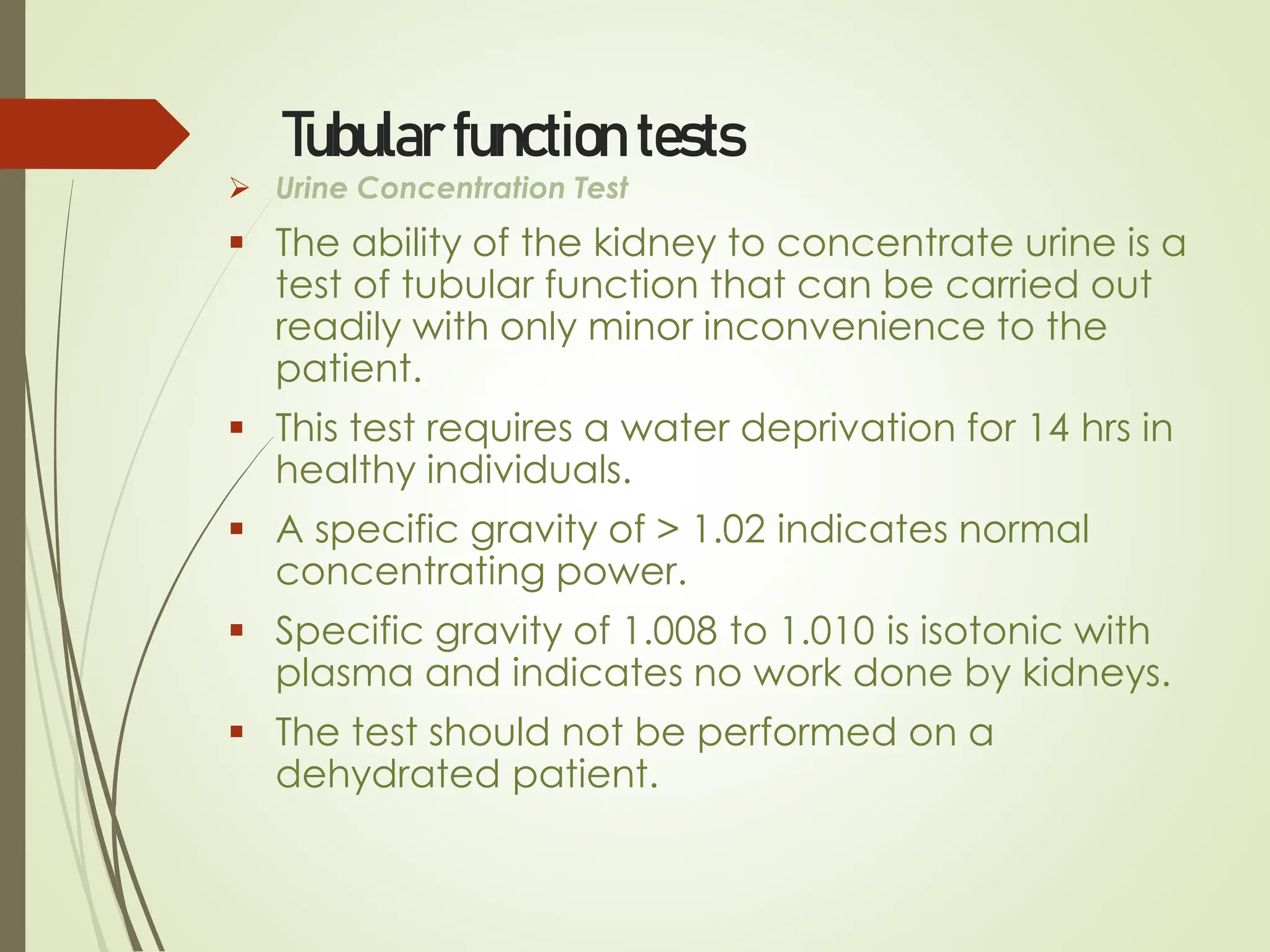
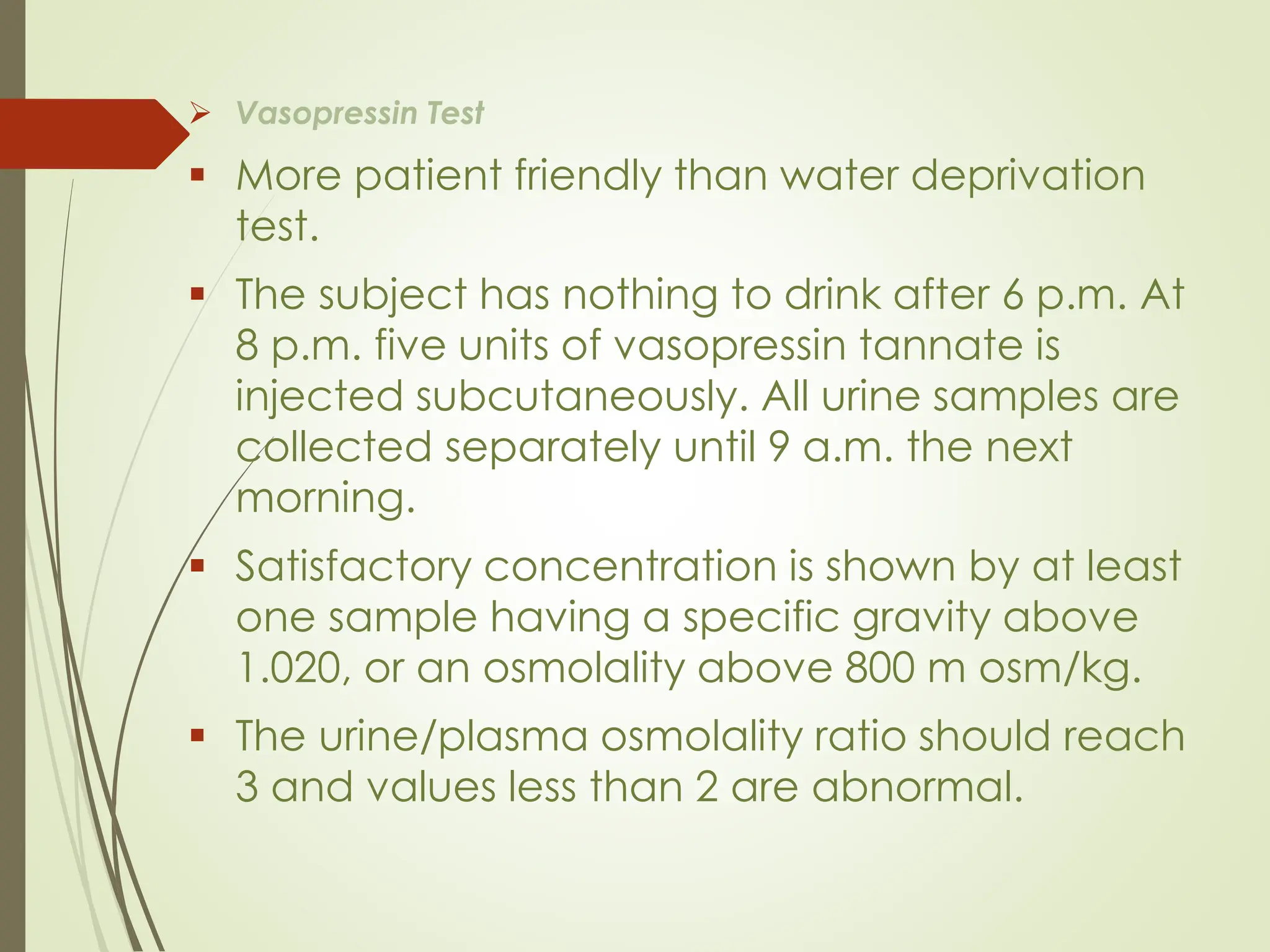
RENAL
FUNCTION
TESTS
Functions of Kidney
▪ Regulation of inorganic ions
▪ Regulation of water balance and
osmolality
▪ Excretion of nitrogenous waste (urea &
creatinine)
▪ Regulation of Ph and HCO3-
▪ Synthesis of Renin
▪ Synthesis of EPO; activation of VitD3
Why test renal function?
▪ To asses the functional capacity of
kidney
▪ Early detection of possible renal
impairment.
▪ Severity and progression of the
impairment.
▪ Monitor response to treatment
▪ Monitor the safe and effective use of
drugs which are excreted in the urine
When should we assess renal function?
▪ Older age
▪ Family history of Chronic Kidney disease (CKD)
▪ Decreased renal mass
▪ Low birth weight
▪ Diabetes Mellitus (DM)
▪ Hypertension (HTN)
▪ Autoimmune disease
▪ Systemic infections
▪ Urinary tract infections (UTI)
▪ Nephrolithiasis
▪ Obstruction to the lower urinary tract
▪ Drug toxicity
Common Signs and Symptoms
1. Difficulty and Discomfort in urination
2. Blood in urine/ Foamy Urine
3. Increase urge to urinate
4. Persistent puffiness of Body (oedema)
5. Persistent pain in flank region
6. Lethargy and weakness
7. Poor Appetite
What to examine???
Renal function tests are divided into the following:
▪ Urine analysis
▪ Blood examination
▪ Glomerular Function Test
▪ Tubular Function Test
Classification of Renal Function Test
1. Test based on Glomerular Filtration:
a. Urea Clearance Test
b. Endogenous creatinine clearance test
c. Inulin Clearance Test
2. Test to measure Tubular Functions:
a. Concentration and dilution test
b. Test to assess renal acidification
c. Urinary and plasma osmolality measurement
d. 15 minute PSP (phenolsulphonapthalein) test
Classification of Renal Function Test
3. Analysis of Blood / Serum to assess renal function test:
Estimation of serum Urea, Creatinine, Protein & Electrolytes
4. Test to measure Renal Plasma Flow (RPF):
Para-aminohippurate (PAH) test
5. Markers of Glomerular permeability – Proteinuria
6. Complete Urinalysis – Routine test done are Urea/BUN,
Creatinine, Urine R/M and Electrolytes
Urine Analysis
▪ Urine examination is an extremely
valuable and most easily performed test
for the evaluation of renal functions.
▪ It includes physical or macroscopic
examination, chemical examination and
microscopic examination of the sediment.
Macroscopic examination
✓ Colour
▪ Normal- pale yellow in colour due to pigments
urochrome,urobilin and uroerythrin.
▪ Cloudiness may be caused by excessive cellular
material or protein, crystallization or precipitation of
salts upon standing at room temperature or in the
refrigerator.
▪ If the sample contains many red blood cells, it would
be cloudy as well as red.
Blue Green Pink-Orange-
Red
Red-brown-black
Methylene Blue Haemoglobin Haemoglobin
Pseudomonas Myoglobin Myoglobin
Riboflavin Phenolpthalein Red blood cells
Porphyrins Homogentisic Acid
Rifampicin L -DOPA
Melanin
Methyldopa
▪ Colour of urine depending upon it’s constituents.
✓ Volume
▪ Normal- 1-2.5 L/day
▪ Oliguria- Urine Output < 400ml/day
Seen in
 Acute glomerulonephritis
 Renal Failure
▪ Polyuria- Urine Output > 2.5 L/day
Seen in
 Increased water ingestion
 Diabetes mellitus and insipidus.
▪ Anuria- Urine output < 100ml/day
Seen in renal shut down
✓Specific Gravity
▪ Measured by urinometer or refractometer.
▪ It is measurement of urine density which
reflects the ability of the kidney to
concentrate or dilute the urine relative to the
plasma from which it is filtered.
▪ Normal :- 1.001- 1.040.
▪ Increase in Specific Gravity seen in
▪ Low water intake
▪ Diabetes mellitus
▪ Albuminuruia
▪ Acute nephritis.
▪ Decrease in Specific Gravity is seen in
▪ Absence of ADH
▪ Renal Tubular damage.
▪ Isosthenuria-Persistent production of fixed low
Specific gravity urine isoosmolar with plasma
despite variation in water intake.
✓ pH
▪ Urine pH ranges from 4.5 to 8
▪ Normally it is slightly acidic lying between 6 – 6.5.
▪ After meal it becomes alkaline.
▪ On exposure to atmosphere,urea in urine splits
causing NH4
+ release resulting in alkaline reaction.
Microscopic examination
▪ A sample of well-mixed urine (usually 10-15 ml) is
centrifuged in a test tube at relatively low speed
(about 2000-3,000 rpm) for 5-10 minutes which
produces a concentration of sediment (cellular
matter) at the bottom of the tube.
▪ A drop of sediment is poured onto a glass slide
and a thin slice of glass (a coverslip) is place over
it.
▪ The sediment is first examined under low power to
identify crystals, casts, squamous cells, and other
large objects. "Casts" are plugs of material which
came from individual tubules.
▪ The numbers of casts seen are usually reported as
number of each type found per low power field
(LPF). For an example: "5-10 hyaline casts/L
casts/LPF."
▪ Then, examination is carried out at high power to
identify crystals, cells, and bacteria.
▪ The various types of cells are usually described as
the number of each type found per average
high power field (HPF). For example: "1-5
WBC/HPF."
▪ Epithelial cells and 1-2 WBC or pus cell/HPF is
normally seen
▪ If more leukocytes per each high power field appear in
non-contaminated urine, the specimen is probably
abnormal showing pyuria. Leukocytes have lobed
nuclei and granular cytoplasm.
▪ Usually, the WBC's are granulocytes. White cells from the
vagina, in the presence of vaginal and cervical
infections, or the external urethral meatus in men and
women may contaminate the urine.
▪ Presence of Granular Casts, RBC, bacteria, Glucose,
Albumin and Ketone bodies is abnormal.
▪ Hematuria is the presence of abnormal numbers of red
cells in urine due to any of several possible causes.
 glomerular damage,
 tumors which erode the urinary tract anywhere along its length,
 kidney trauma,
 urinary tract stones,
 acute tubular necrosis,
 upper and lower urinary tract infections,
 nephrotoxins
▪ Red blood cells may stick together and form red blood cell
casts. Such casts are indicative of glomerulonephritis, with
leakage of RBC's from glomeruli, or severe tubular damage.
▪ White blood cell casts are most typical for acute
pyelonephritis, but they may also be present with
glomerulonephritis. Their presence indicates inflammation
of the kidney.
Urinary crystals. (A) Calcium oxalate crystals; (B) uric acid
crystals (C) triple phosphate crystals with amorphous
phosphates ; (D) cystine crystals.
Crystals
▪ Tyrosine crystals with congenital tyrosinosis
▪ Leucine crystals in patients with severe liver
disease or with maple syrup urine disease.
Blood examination
▪ Done to measure substance in blood
that are normally excreted by kidney.
▪ Their level in blood increases in kidney
dysfunction.
▪ As markers of renal function creatinine,
urea,uric acid and electrolytes are done
for routine analysis
➢ Serum creatinine
▪ Creatinine is a breakdown product of creatine
phosphate in muscle, and is usually produced at a
fairly constant rate by the body depending on
muscle mass
▪ Creatinine is filtered but not reabsorbed in kidney.
▪ Normal range is 0.8-1.3 mg/dl in men and 0.6-1
mg/dl in women.
▪ Not increased above normal until GFR<50 ml/min .
▪ The methods most widely used for serum
creatinine are based on the Jaffe reaction. This
reaction occurs between creatinine and the
picrate ion formed in alkaline medium (sodium
picrate); a red-orange solution develops which is
read colorimetrically at 520 nm .
 Increased serum creatinine:
 Impaired renal function
 Very high protein diet
 Anabolic steroid users
 Vary large muscle mass: body
builders, giants, acromegaly patients
 Rhabdomyolysis/crush injury
 Athletes taking oral creatine.
 Drugs:
• Probenecid
• Cimetidine
• Triamterene
• Trimethoprim
• Amiloride
➢ Blood urea
▪ Urea is major nitrogenous end product of protein
and amino acid catabolism, produced by liver
and distributed throughout intracellular and
extracellular fluid.
▪ Urea is filtered freely by the glomeruli .
▪ Many renal diseases with various glomerular,
tubular, interstitial or vascular damage can cause
an increase in plasma urea concentration.
▪ The reference interval for serum urea of healthy
adults is 10-40 mg/dl.
▪ Plasma concentrations also tend to be slightly
higher in males than females. High protein diet
causes significant increases in plasma urea
concentrations and urinary excretion.
▪ Measurement of plasma creatinine provides a more
accurate assessment than urea because there are
many non renal factors that affect urea level.
▪ Nonrenal factors can affect the urea level (normal
adults is level 10-40mg/dl) like:
▪ Mild dehydration,
▪ high protein diet,
▪ increased protein catabolism, muscle wasting as in
starvation,
▪ reabsorption of blood proteins after a GIT haemorrhage,
▪ treatment with cortisol or its synthetic analogous
▪ States associated with elevated levels of urea in blood
are referred to as uremia or azotemia.
▪ Causes of urea plasma elevations:
▪ Prerenal: renal hypoperfusion
▪ Renal: acute tubular necrosis
▪ Postrenal: obstruction of urinary flow
▪ Blood urea is normally doubled when the GFR is
halved.
▪ Parallel determination of urea and creatinine is
performed to differentiate between pre-renal and
post-renal azotemia.
▪ Pre-renal azotemia leads to increased urea levels,
while creatinine values remain within the reference
range. In post-renal azotemias both urea and
creatinine levels rise, but creatinine in a smaller
extent.
▪ Enzymatic Berthelot Method is used for blood urea
estimation:
▪ Principal:
▪ Urea + H2O Urease > Ammonia + CO2
▪ Ammonia + Phenolic Chromogen + Hypochlorite > Green
Colored Complex whoose absorbance is read at 570nm
➢ Serum Uric Acid
▪ In human, uric acid is the major product of
the catabolism of the purine nucleosides,
adenosine and guanosine.
▪ Purines are derived from catabolism of
dietary nucleic acid and from degradation of
endogenous nucleic acids.
▪ Overproduction of uric acid may result from
increased synthesis of purine precursors.
▪ In humans, approximately 75% of uric acid
excreted is lost in the urine; most of the
reminder is secreted into the GIT
▪ Renal handling of uric acid is complex and
involves four sequential steps:
Glomerular filtration of virtually all the uric acid in
capillary plasma entering the glomerulus.
Reabsorption in the proximal convoluted tubule
of about 98 to 100% of filtered uric acid.
Subsequent secretion of uric acid into the lumen
of the distal portion of the proximal tubule.
Further reabsorption in the distal tubule.
▪ Hyperuricemia is defined by serum or plasma
uric acid concentrations higher than 7.0 mg/dl
(0.42mmol/L) in men or greater than 6.0 mg/dl
(0.36mmol/L) in women
Glomerular function tests
▪ GFR is the rate at which substances in plasma are filtered
through glomerulus.
▪ GFR cannot be assessed directly and hence is being
measured via various exogenous and endogenous
substances to measure clearance test
1. Inulin Clearance Test
2. Creatinine Clearance Test
3. Urea Clearance Test
▪ Inulin clearance test accurately measures GFR as it is
neither secreted or absorbed by renal tubules
▪ In the clinical setting estimated GFR (eGFR) is calculated
by S. Creatinine values.
▪ The GFR is the best measure of glomerular function.
▪ Normal GFR is approximately 125 mL/min
▪ When GFR decreases to 30% of normal
moderate renal insufficiency. Patients remain
asymptomatic with only biochemical evidence of a
decline in GFR
▪ As the GFR decreases further severe renal
insufficiency characterized by profound clinical
manifestations of uremia and biochemical
abnormalities, such as acidemia; volume overload;
and neurologic, cardiac, and respiratory
manifestations
▪ When GFR is 5% to 10% of normal ESRD
▪ Inulin clearance and creatinine clearance
are used to measure the GFR.
Creatinine Clearance:
▪ A simple, inexpensive bedside estimate of
GFR.
▪ GFR= Ccr = {Ucr * Urinary flow rate(ml/min)} /
Pcr
Normal 100-120ml/min
Dec.Renal reserve 60-100ml/min
Mild Renal imp 40-60ml/min
Moderate insuff. 25-40ml/min
Renal failure <25ml/min
ESRD <10ml/min
Estimated GFR (eGFR)
▪ eGFR is assessed to:
1. To diagnose and monitor chronic kidney disease
2. To assess kidney function in Acute Kidney Injury
3. Determine medication and clearance
4. Evaluate kidney function pre-transplant
 Calculated via Schwartz equation
eGFR (ml/min/1.73m²) = {k height(cm)}/S. Creatinine
(mg/dL), where k is 0.413
eGFR stages: KDIGO classification of CKD
1. Stage 1: or = 90-Normal Kidney Function
2. Stage 2: 60-89: Mildly Decreased Kidney function
3. Stage 3A: 45-59: Moderately decreased kidney function
4. Stage 3B and 4: 15-44: Severely decreased kidney
function
5. Stage 5: <15: Kidney Failure (dialysis or transplant
needed)
▪ Cockroft Gault Formula
Creatinine Clearance =(140-age)* weight in kg /
S.creat.*72
(multiplied by 0.85 for females)
▪ MDRD Nomogram
GFR(ml/min)=170*S.creat.-0.999 *age-0.176 * BUN-0.170
*albumin0.318
(multiplied by 0.742 if female)
Tubular function tests
➢ Urine Concentration Test
▪ The ability of the kidney to concentrate urine is a
test of tubular function that can be carried out
readily with only minor inconvenience to the
patient.
▪ This test requires a water deprivation for 14 hrs in
healthy individuals.
▪ A specific gravity of > 1.02 indicates normal
concentrating power.
▪ Specific gravity of 1.008 to 1.010 is isotonic with
plasma and indicates no work done by kidneys.
▪ The test should not be performed on a
dehydrated patient.
➢ Vasopressin Test
▪ More patient friendly than water deprivation
test.
▪ The subject has nothing to drink after 6 p.m. At
8 p.m. five units of vasopressin tannate is
injected subcutaneously. All urine samples are
collected separately until 9 a.m. the next
morning.
▪ Satisfactory concentration is shown by at least
one sample having a specific gravity above
1.020, or an osmolality above 800 m osm/kg.
▪ The urine/plasma osmolality ratio should reach
3 and values less than 2 are abnormal.
➢ Urine Dilution (Water Load) Test
▪ After an overnight fast the subject empties his
bladder completely and is given 1000 ml of water
to drink.
▪ Urine specimens are collected for the next 4 hours,
the patient emptying bladder completely on
each occasion.
▪ Normally the patient will excrete at least 700 ml of
urine in the 4 hours, and at least one specimen will
have a specific gravity less than 1.004.
▪ Kidneys which are severely damaged cannot
excrete a urine of lower specific gravity than 1.010
or a volume above 400 ml in this time.
▪ The test should not be done if there is oedema or
renal failure; water intoxication may result.
➢ Para Aminohippuric Acid Clearance
▪ Maximum secretory capacity of tubules for
PAH is nearly constantat about 80mg/min.
▪ A decrease in the TmPAH indicates tubular
damage.
➢ Micropuncturing
▪ Micropuncturing various part of tubule and
analysis of fluid for volume and composition.
➢ Microcryoscopic study
▪ Studying slices of renal tissue at different
depths.
➢ Microelectrode study
▪ Measuring membrane potential of the tubular
cells
Serum Electrolytes
Normal Levels:
Sodium-135-145 meq/L
Potassium-3.5-5.5 meq/L
Chloride 96-108 meq/L
• Potassium is the most convincing electrolyte marker
for renal failure.
• Combination of decreased filtration and decreased
secretion of potassium in distal tube during renal
failure causes increased potassium.
• Hyperkalaemia is the most significant and life
threatening component of Renal failure.
POTASSIUM
 Serum potassium concentration normally ranges between 3.5
and 5 mEq/1The level is higher in newborns, ranging from 4-6
mEq/l in term babies, and 5.5-7mEq/l in preterm infants.
 Potassium is mostly an intracellular ion; the ECF contains only 2%
of the total body potassium.
 Even though extracellular potassium constitutes a fraction of the
total body content, changes of its levels in this compartment
have a potential for morbidity, in view of its effect on cardiac
conduction.
 The serum potassium concentration is influenced both by the
total body content of potassium and by movement of the lon
between the ECF and the intracellular fluid (ICF).
Hyperkalemia
 Hyperkalaemia, defined as serum potassium level above 5.5
mEq/L, is almost always secondary to inadequate renal
excretion of potassium.
 This may result from acute or chronic renal failure with a decline
in the glomerular filtration rate, a deficiency of aldosterone, or
from an intrinsic inability of the distal nephron to secrete
potassium into the tubular lumen.
Causes
 Spurious – Haemolysis, Leucocytosis thrombocytosis, Application
of tourniquet, clenching during blood draw
 Shift of potassium from intracellular to extracellular
compartment – Metabolic acidosis, Hyperosmolality with insulin
deficiency, beta-blocker therapy, Hyperkalaemic periodic
paralysis
 Increase intake, release from damaged cells – Exogenous
dietary administration, IV fluids with excess potassium,
Transfusion of old blood, Massive haemolysis, Rhabdomyolysis,
Severe exercise, Infection, Gastrointestinal bleeding
 Decreased excretion – Acute and chronic renal failure (Renal
tubular acidosis type IV)
 Mineralocorticoid deficiency, resistance
Clinical Features
• Mild hyperkalaemia is usually asymptomatic and is detected on
routine laboratory tests.
• Patients with severe hyperkalaemia with a serum potassium
level >6.5 mEq
• The earliest change is peaking of the T-waves.
• This is followed by flattening P-waves, prolongation of PR
Interval, and widening of the QRS complex. Events include a
sine-wave pattern on the EKG with ventricular fibrillation,
cardiac arrest.
• Cardiac toxicity is more evident if there is a rapid rise in the
serum, if there are underlying cardiac problems
Management
 Spurious hyperkalaemia should always be excluded, especially
if the clinical setting does not support the diagnosis.
 If the serum potassium level >7 mEq/l, or If there are EKG
changes, therapeutic measures should be Instituted
Immediately while awaiting confirmation of the abnormal result.
 The goals of emergency management are to counteract the
cardiac toxicity and to shift potassium into the Intracellular
space.
 Drugs used in management of hyperkalaemia
• Calcium gluconate (10%)
• Insulin
• Sodium bicarbonate
• Nebulized salbutamol
• Kayexalate (sodium polystyrene sulfonate)
 Dialysis (peritoneal/haemodialysis)
Interpretation of Renal Function Tests UG

Interpretation of Renal Function Tests UG

  • 1.
  • 2.
    Functions of Kidney ▪Regulation of inorganic ions ▪ Regulation of water balance and osmolality ▪ Excretion of nitrogenous waste (urea & creatinine) ▪ Regulation of Ph and HCO3- ▪ Synthesis of Renin ▪ Synthesis of EPO; activation of VitD3
  • 3.
    Why test renalfunction? ▪ To asses the functional capacity of kidney ▪ Early detection of possible renal impairment. ▪ Severity and progression of the impairment. ▪ Monitor response to treatment ▪ Monitor the safe and effective use of drugs which are excreted in the urine
  • 4.
    When should weassess renal function? ▪ Older age ▪ Family history of Chronic Kidney disease (CKD) ▪ Decreased renal mass ▪ Low birth weight ▪ Diabetes Mellitus (DM) ▪ Hypertension (HTN) ▪ Autoimmune disease ▪ Systemic infections ▪ Urinary tract infections (UTI) ▪ Nephrolithiasis ▪ Obstruction to the lower urinary tract ▪ Drug toxicity
  • 5.
    Common Signs andSymptoms 1. Difficulty and Discomfort in urination 2. Blood in urine/ Foamy Urine 3. Increase urge to urinate 4. Persistent puffiness of Body (oedema) 5. Persistent pain in flank region 6. Lethargy and weakness 7. Poor Appetite
  • 6.
    What to examine??? Renalfunction tests are divided into the following: ▪ Urine analysis ▪ Blood examination ▪ Glomerular Function Test ▪ Tubular Function Test
  • 7.
    Classification of RenalFunction Test 1. Test based on Glomerular Filtration: a. Urea Clearance Test b. Endogenous creatinine clearance test c. Inulin Clearance Test 2. Test to measure Tubular Functions: a. Concentration and dilution test b. Test to assess renal acidification c. Urinary and plasma osmolality measurement d. 15 minute PSP (phenolsulphonapthalein) test
  • 8.
    Classification of RenalFunction Test 3. Analysis of Blood / Serum to assess renal function test: Estimation of serum Urea, Creatinine, Protein & Electrolytes 4. Test to measure Renal Plasma Flow (RPF): Para-aminohippurate (PAH) test 5. Markers of Glomerular permeability – Proteinuria 6. Complete Urinalysis – Routine test done are Urea/BUN, Creatinine, Urine R/M and Electrolytes
  • 9.
    Urine Analysis ▪ Urineexamination is an extremely valuable and most easily performed test for the evaluation of renal functions. ▪ It includes physical or macroscopic examination, chemical examination and microscopic examination of the sediment.
  • 10.
    Macroscopic examination ✓ Colour ▪Normal- pale yellow in colour due to pigments urochrome,urobilin and uroerythrin. ▪ Cloudiness may be caused by excessive cellular material or protein, crystallization or precipitation of salts upon standing at room temperature or in the refrigerator. ▪ If the sample contains many red blood cells, it would be cloudy as well as red.
  • 11.
    Blue Green Pink-Orange- Red Red-brown-black MethyleneBlue Haemoglobin Haemoglobin Pseudomonas Myoglobin Myoglobin Riboflavin Phenolpthalein Red blood cells Porphyrins Homogentisic Acid Rifampicin L -DOPA Melanin Methyldopa ▪ Colour of urine depending upon it’s constituents.
  • 12.
    ✓ Volume ▪ Normal-1-2.5 L/day ▪ Oliguria- Urine Output < 400ml/day Seen in  Acute glomerulonephritis  Renal Failure ▪ Polyuria- Urine Output > 2.5 L/day Seen in  Increased water ingestion  Diabetes mellitus and insipidus. ▪ Anuria- Urine output < 100ml/day Seen in renal shut down
  • 13.
    ✓Specific Gravity ▪ Measuredby urinometer or refractometer. ▪ It is measurement of urine density which reflects the ability of the kidney to concentrate or dilute the urine relative to the plasma from which it is filtered. ▪ Normal :- 1.001- 1.040.
  • 14.
    ▪ Increase inSpecific Gravity seen in ▪ Low water intake ▪ Diabetes mellitus ▪ Albuminuruia ▪ Acute nephritis. ▪ Decrease in Specific Gravity is seen in ▪ Absence of ADH ▪ Renal Tubular damage. ▪ Isosthenuria-Persistent production of fixed low Specific gravity urine isoosmolar with plasma despite variation in water intake.
  • 15.
    ✓ pH ▪ UrinepH ranges from 4.5 to 8 ▪ Normally it is slightly acidic lying between 6 – 6.5. ▪ After meal it becomes alkaline. ▪ On exposure to atmosphere,urea in urine splits causing NH4 + release resulting in alkaline reaction.
  • 16.
    Microscopic examination ▪ Asample of well-mixed urine (usually 10-15 ml) is centrifuged in a test tube at relatively low speed (about 2000-3,000 rpm) for 5-10 minutes which produces a concentration of sediment (cellular matter) at the bottom of the tube. ▪ A drop of sediment is poured onto a glass slide and a thin slice of glass (a coverslip) is place over it. ▪ The sediment is first examined under low power to identify crystals, casts, squamous cells, and other large objects. "Casts" are plugs of material which came from individual tubules. ▪ The numbers of casts seen are usually reported as number of each type found per low power field (LPF). For an example: "5-10 hyaline casts/L casts/LPF."
  • 17.
    ▪ Then, examinationis carried out at high power to identify crystals, cells, and bacteria. ▪ The various types of cells are usually described as the number of each type found per average high power field (HPF). For example: "1-5 WBC/HPF." ▪ Epithelial cells and 1-2 WBC or pus cell/HPF is normally seen ▪ If more leukocytes per each high power field appear in non-contaminated urine, the specimen is probably abnormal showing pyuria. Leukocytes have lobed nuclei and granular cytoplasm. ▪ Usually, the WBC's are granulocytes. White cells from the vagina, in the presence of vaginal and cervical infections, or the external urethral meatus in men and women may contaminate the urine.
  • 18.
    ▪ Presence ofGranular Casts, RBC, bacteria, Glucose, Albumin and Ketone bodies is abnormal. ▪ Hematuria is the presence of abnormal numbers of red cells in urine due to any of several possible causes.  glomerular damage,  tumors which erode the urinary tract anywhere along its length,  kidney trauma,  urinary tract stones,  acute tubular necrosis,  upper and lower urinary tract infections,  nephrotoxins ▪ Red blood cells may stick together and form red blood cell casts. Such casts are indicative of glomerulonephritis, with leakage of RBC's from glomeruli, or severe tubular damage. ▪ White blood cell casts are most typical for acute pyelonephritis, but they may also be present with glomerulonephritis. Their presence indicates inflammation of the kidney.
  • 20.
    Urinary crystals. (A)Calcium oxalate crystals; (B) uric acid crystals (C) triple phosphate crystals with amorphous phosphates ; (D) cystine crystals. Crystals ▪ Tyrosine crystals with congenital tyrosinosis ▪ Leucine crystals in patients with severe liver disease or with maple syrup urine disease.
  • 21.
    Blood examination ▪ Doneto measure substance in blood that are normally excreted by kidney. ▪ Their level in blood increases in kidney dysfunction. ▪ As markers of renal function creatinine, urea,uric acid and electrolytes are done for routine analysis
  • 22.
    ➢ Serum creatinine ▪Creatinine is a breakdown product of creatine phosphate in muscle, and is usually produced at a fairly constant rate by the body depending on muscle mass ▪ Creatinine is filtered but not reabsorbed in kidney. ▪ Normal range is 0.8-1.3 mg/dl in men and 0.6-1 mg/dl in women. ▪ Not increased above normal until GFR<50 ml/min . ▪ The methods most widely used for serum creatinine are based on the Jaffe reaction. This reaction occurs between creatinine and the picrate ion formed in alkaline medium (sodium picrate); a red-orange solution develops which is read colorimetrically at 520 nm .
  • 23.
     Increased serumcreatinine:  Impaired renal function  Very high protein diet  Anabolic steroid users  Vary large muscle mass: body builders, giants, acromegaly patients  Rhabdomyolysis/crush injury  Athletes taking oral creatine.  Drugs: • Probenecid • Cimetidine • Triamterene • Trimethoprim • Amiloride
  • 24.
    ➢ Blood urea ▪Urea is major nitrogenous end product of protein and amino acid catabolism, produced by liver and distributed throughout intracellular and extracellular fluid. ▪ Urea is filtered freely by the glomeruli . ▪ Many renal diseases with various glomerular, tubular, interstitial or vascular damage can cause an increase in plasma urea concentration. ▪ The reference interval for serum urea of healthy adults is 10-40 mg/dl. ▪ Plasma concentrations also tend to be slightly higher in males than females. High protein diet causes significant increases in plasma urea concentrations and urinary excretion.
  • 25.
    ▪ Measurement ofplasma creatinine provides a more accurate assessment than urea because there are many non renal factors that affect urea level. ▪ Nonrenal factors can affect the urea level (normal adults is level 10-40mg/dl) like: ▪ Mild dehydration, ▪ high protein diet, ▪ increased protein catabolism, muscle wasting as in starvation, ▪ reabsorption of blood proteins after a GIT haemorrhage, ▪ treatment with cortisol or its synthetic analogous ▪ States associated with elevated levels of urea in blood are referred to as uremia or azotemia. ▪ Causes of urea plasma elevations: ▪ Prerenal: renal hypoperfusion ▪ Renal: acute tubular necrosis ▪ Postrenal: obstruction of urinary flow
  • 26.
    ▪ Blood ureais normally doubled when the GFR is halved. ▪ Parallel determination of urea and creatinine is performed to differentiate between pre-renal and post-renal azotemia. ▪ Pre-renal azotemia leads to increased urea levels, while creatinine values remain within the reference range. In post-renal azotemias both urea and creatinine levels rise, but creatinine in a smaller extent. ▪ Enzymatic Berthelot Method is used for blood urea estimation: ▪ Principal: ▪ Urea + H2O Urease > Ammonia + CO2 ▪ Ammonia + Phenolic Chromogen + Hypochlorite > Green Colored Complex whoose absorbance is read at 570nm
  • 27.
    ➢ Serum UricAcid ▪ In human, uric acid is the major product of the catabolism of the purine nucleosides, adenosine and guanosine. ▪ Purines are derived from catabolism of dietary nucleic acid and from degradation of endogenous nucleic acids. ▪ Overproduction of uric acid may result from increased synthesis of purine precursors. ▪ In humans, approximately 75% of uric acid excreted is lost in the urine; most of the reminder is secreted into the GIT
  • 28.
    ▪ Renal handlingof uric acid is complex and involves four sequential steps: Glomerular filtration of virtually all the uric acid in capillary plasma entering the glomerulus. Reabsorption in the proximal convoluted tubule of about 98 to 100% of filtered uric acid. Subsequent secretion of uric acid into the lumen of the distal portion of the proximal tubule. Further reabsorption in the distal tubule. ▪ Hyperuricemia is defined by serum or plasma uric acid concentrations higher than 7.0 mg/dl (0.42mmol/L) in men or greater than 6.0 mg/dl (0.36mmol/L) in women
  • 29.
    Glomerular function tests ▪GFR is the rate at which substances in plasma are filtered through glomerulus. ▪ GFR cannot be assessed directly and hence is being measured via various exogenous and endogenous substances to measure clearance test 1. Inulin Clearance Test 2. Creatinine Clearance Test 3. Urea Clearance Test ▪ Inulin clearance test accurately measures GFR as it is neither secreted or absorbed by renal tubules ▪ In the clinical setting estimated GFR (eGFR) is calculated by S. Creatinine values.
  • 30.
    ▪ The GFRis the best measure of glomerular function. ▪ Normal GFR is approximately 125 mL/min ▪ When GFR decreases to 30% of normal moderate renal insufficiency. Patients remain asymptomatic with only biochemical evidence of a decline in GFR ▪ As the GFR decreases further severe renal insufficiency characterized by profound clinical manifestations of uremia and biochemical abnormalities, such as acidemia; volume overload; and neurologic, cardiac, and respiratory manifestations ▪ When GFR is 5% to 10% of normal ESRD
  • 31.
    ▪ Inulin clearanceand creatinine clearance are used to measure the GFR. Creatinine Clearance: ▪ A simple, inexpensive bedside estimate of GFR. ▪ GFR= Ccr = {Ucr * Urinary flow rate(ml/min)} / Pcr Normal 100-120ml/min Dec.Renal reserve 60-100ml/min Mild Renal imp 40-60ml/min Moderate insuff. 25-40ml/min Renal failure <25ml/min ESRD <10ml/min
  • 32.
    Estimated GFR (eGFR) ▪eGFR is assessed to: 1. To diagnose and monitor chronic kidney disease 2. To assess kidney function in Acute Kidney Injury 3. Determine medication and clearance 4. Evaluate kidney function pre-transplant  Calculated via Schwartz equation eGFR (ml/min/1.73m²) = {k height(cm)}/S. Creatinine (mg/dL), where k is 0.413
  • 33.
    eGFR stages: KDIGOclassification of CKD 1. Stage 1: or = 90-Normal Kidney Function 2. Stage 2: 60-89: Mildly Decreased Kidney function 3. Stage 3A: 45-59: Moderately decreased kidney function 4. Stage 3B and 4: 15-44: Severely decreased kidney function 5. Stage 5: <15: Kidney Failure (dialysis or transplant needed)
  • 34.
    ▪ Cockroft GaultFormula Creatinine Clearance =(140-age)* weight in kg / S.creat.*72 (multiplied by 0.85 for females) ▪ MDRD Nomogram GFR(ml/min)=170*S.creat.-0.999 *age-0.176 * BUN-0.170 *albumin0.318 (multiplied by 0.742 if female)
  • 35.
    Tubular function tests ➢Urine Concentration Test ▪ The ability of the kidney to concentrate urine is a test of tubular function that can be carried out readily with only minor inconvenience to the patient. ▪ This test requires a water deprivation for 14 hrs in healthy individuals. ▪ A specific gravity of > 1.02 indicates normal concentrating power. ▪ Specific gravity of 1.008 to 1.010 is isotonic with plasma and indicates no work done by kidneys. ▪ The test should not be performed on a dehydrated patient.
  • 36.
    ➢ Vasopressin Test ▪More patient friendly than water deprivation test. ▪ The subject has nothing to drink after 6 p.m. At 8 p.m. five units of vasopressin tannate is injected subcutaneously. All urine samples are collected separately until 9 a.m. the next morning. ▪ Satisfactory concentration is shown by at least one sample having a specific gravity above 1.020, or an osmolality above 800 m osm/kg. ▪ The urine/plasma osmolality ratio should reach 3 and values less than 2 are abnormal.
  • 37.
    ➢ Urine Dilution(Water Load) Test ▪ After an overnight fast the subject empties his bladder completely and is given 1000 ml of water to drink. ▪ Urine specimens are collected for the next 4 hours, the patient emptying bladder completely on each occasion. ▪ Normally the patient will excrete at least 700 ml of urine in the 4 hours, and at least one specimen will have a specific gravity less than 1.004. ▪ Kidneys which are severely damaged cannot excrete a urine of lower specific gravity than 1.010 or a volume above 400 ml in this time. ▪ The test should not be done if there is oedema or renal failure; water intoxication may result.
  • 38.
    ➢ Para AminohippuricAcid Clearance ▪ Maximum secretory capacity of tubules for PAH is nearly constantat about 80mg/min. ▪ A decrease in the TmPAH indicates tubular damage. ➢ Micropuncturing ▪ Micropuncturing various part of tubule and analysis of fluid for volume and composition. ➢ Microcryoscopic study ▪ Studying slices of renal tissue at different depths. ➢ Microelectrode study ▪ Measuring membrane potential of the tubular cells
  • 39.
    Serum Electrolytes Normal Levels: Sodium-135-145meq/L Potassium-3.5-5.5 meq/L Chloride 96-108 meq/L • Potassium is the most convincing electrolyte marker for renal failure. • Combination of decreased filtration and decreased secretion of potassium in distal tube during renal failure causes increased potassium. • Hyperkalaemia is the most significant and life threatening component of Renal failure.
  • 40.
    POTASSIUM  Serum potassiumconcentration normally ranges between 3.5 and 5 mEq/1The level is higher in newborns, ranging from 4-6 mEq/l in term babies, and 5.5-7mEq/l in preterm infants.  Potassium is mostly an intracellular ion; the ECF contains only 2% of the total body potassium.  Even though extracellular potassium constitutes a fraction of the total body content, changes of its levels in this compartment have a potential for morbidity, in view of its effect on cardiac conduction.  The serum potassium concentration is influenced both by the total body content of potassium and by movement of the lon between the ECF and the intracellular fluid (ICF).
  • 41.
    Hyperkalemia  Hyperkalaemia, definedas serum potassium level above 5.5 mEq/L, is almost always secondary to inadequate renal excretion of potassium.  This may result from acute or chronic renal failure with a decline in the glomerular filtration rate, a deficiency of aldosterone, or from an intrinsic inability of the distal nephron to secrete potassium into the tubular lumen.
  • 42.
    Causes  Spurious –Haemolysis, Leucocytosis thrombocytosis, Application of tourniquet, clenching during blood draw  Shift of potassium from intracellular to extracellular compartment – Metabolic acidosis, Hyperosmolality with insulin deficiency, beta-blocker therapy, Hyperkalaemic periodic paralysis  Increase intake, release from damaged cells – Exogenous dietary administration, IV fluids with excess potassium, Transfusion of old blood, Massive haemolysis, Rhabdomyolysis, Severe exercise, Infection, Gastrointestinal bleeding  Decreased excretion – Acute and chronic renal failure (Renal tubular acidosis type IV)  Mineralocorticoid deficiency, resistance
  • 43.
    Clinical Features • Mildhyperkalaemia is usually asymptomatic and is detected on routine laboratory tests. • Patients with severe hyperkalaemia with a serum potassium level >6.5 mEq • The earliest change is peaking of the T-waves. • This is followed by flattening P-waves, prolongation of PR Interval, and widening of the QRS complex. Events include a sine-wave pattern on the EKG with ventricular fibrillation, cardiac arrest. • Cardiac toxicity is more evident if there is a rapid rise in the serum, if there are underlying cardiac problems
  • 44.
    Management  Spurious hyperkalaemiashould always be excluded, especially if the clinical setting does not support the diagnosis.  If the serum potassium level >7 mEq/l, or If there are EKG changes, therapeutic measures should be Instituted Immediately while awaiting confirmation of the abnormal result.  The goals of emergency management are to counteract the cardiac toxicity and to shift potassium into the Intracellular space.  Drugs used in management of hyperkalaemia • Calcium gluconate (10%) • Insulin • Sodium bicarbonate • Nebulized salbutamol • Kayexalate (sodium polystyrene sulfonate)  Dialysis (peritoneal/haemodialysis)