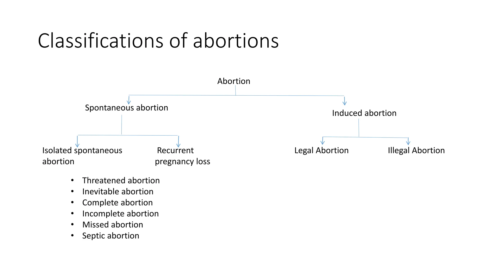
Abortion
Dr Aksshaya
1st year Post graduate
MS Obstetrics and Gynecology
Abortion
• Termination of pregnancy < 20 weeks of gestation.
• WHO criteria : Termination of pregnancy occurs in fetus weighing <
500g, at the time of termination
• Early pregnancy loss : < 12 weeks
• Still birth : Pregnancy loss > 20 weeks
• Anembryonic pregnancy : Non viable pregnancy with a gestational sac
that does not contain a yolk sac/ embryo. AKA blighted ovum.
Risk factors
• Increased maternal age ≥ 35 years
• Previous history of abortions
• Maternal infections like
Viral : Rubella, CMV, Variola, Vaccinia or HIV
Parascitic : Toxoplasma, Malaria
Bacterial : Ureplasma, Chlamyia, Brucella, Spirochetes.
• Maternal factors : Uncontrolled diabetes, Thyroid disorders, Obesity,
Stress, Pregnancy with IUCD in place, Substance abuse and radiation
exposure.
Classifications of abortions
Abortion
Recurrent
pregnancy loss
Isolated spontaneous
abortion
Induced abortion
Spontaneous abortion
Illegal Abortion
Legal Abortion
• Threatened abortion
• Inevitable abortion
• Complete abortion
• Incomplete abortion
• Missed abortion
• Septic abortion
Isolated abortions
• Can happen in 1st and 2nd trimester
• Most common cause : Chromosomal anamolies
Aneuploidy > Trisomy (group trisomy) > Monosomy X (20%) > Trisomy
(16%)
• Uterine anomalies : Fibroids, adhesions, septae, Cervical
incompetence
• Trauma
• Infections : Chorioamnionitis, maternal infections like TORCH
• Thrombophilias
Isolated abortions (Contd..)
• Environmental factors : Cigarette smoking, Contraceptive agents,
drugs, chemicals,noxious agents
Clinical classification of Spontaneous abortion
• Threatened abortion
• Inevitable abortion
• Complete abortion
• Incomplete abortion
• Missed abortion
• Septic abortion
Threatened abortion
• Clinical entity where the process of miscarriage has started but has not
progressed to a state from which recovery is impossible.
• History : Spotting PV +/- Pain abdomen
• C/F :
Bleeding PV > Pain
P/A : Height of the uterus = Period of gestation
P/S : Bleeding if any, escapes through os
Internal os - Closed (reversible)
D/D : Cervical ectopy, polyps or carcinoma, ectopic pregnancy and molar
pregnancy
USG : Cardiac activity +
Management : No defenitive treatment
Emperical treatment :
• Avoid heavy weight lifting
• Avoid intercourse
• Rest for 48 hours
Anti D injection to be given to the Rh negative pregnant women with
threatened abortion at ≥ 12 weeks(2nd trimester)
Inevitable abortion
• The process of abortion has reached the stage from where it is not
reversible
• History : Bleeding + pain abdomen
• No H/O expulsion of products of conception
• P/A : Height of the uterus = POG
• Internal os - Open (Not reversible)
• USG : Cardiac activity is absent
• Management : Complete the process of abortion by Medical/ Surgical
methods (MTP)
• Anti D injection to be given to the all Rh negative pregnant women with
inevitable abortion whether it is < 12 weeks or ≥ 12 weeks due to increased
chance of fetomaternal hemorrhage
(< 12 weeks - 50 mcg, ≥ 12 weeks - 300 mcg)
Incomplete abortion
• The process of abortion begins and POC starts coming out
• 2nd MC type of abortion which leads to shock
• History : POC coming out, bleeding, pain abdomen
• P/A : Height of the uterus < POG
• Internal os : Open + POC coming out
• Management : Complete the process of abortion by Medical/ Surgical
methods (MTP)
• Anti D injection to be given to the all Rh negative pregnant women
with incomplete abortion whether it is < 12 weeks or ≥ 12 weeks
Complete abortion
• The entire process of abortion is completed on its own
• History : Initial H/O bleeding, pain abdomen, expulsion of POC with
stoppage of bleeding later
• P/A : Height of the uterus < POG
• Internal os - Closed
• USG : Empty uterus
• Management : Reassurance
• Anti D injection to be given if complete abortion occurs at ≥ 12 weeks
Missed abortion
• Cardiac activity of the fetus has stopped and patient is unaware about
the abortion
• No H/O bleeding
• P/A : Height of the uterus < POG
• Internal os - Closed
Anti D injection to be given to the all Rh negative pregnant women with
missed abortion whether it is < 12 weeks or ≥ 12 weeks
Diagnosis of Missed abortion
• Mean sac diameter (MSD) ≥ 25mm and no embryo is seen
• ≥ 11 days after the sac showing gestational sac with yolk sac but no
embryo
• ≥ 2 weeks after scan showing gestational sac without yolk sac or
embryo
• If CRL ≥ 7mm with no cardiac activity
• Most common time for abortion : First trimester - more commonly <8
weeks
Approach to a case of abortion
Approach
Open
No H/O POC coming out
POC came out
Open
Close Close
Incomplete
abortion
Complete
abortion
Inevitable
abortion
Height of the
uterus < POG
Height of the
uterus = POG
Threatened
abortion
Missed abortion
Septic abortion
• Any abortion associated with clinical evidence of infection of uterus
and its contents
• Criteria :
• Rise of temprature of atleast 100.4o F for 24 hours or more
• Offensive or purulent vaginal discharge
• Other evidence of pelvic infection such as lower abdominal pain and
tenderness
• Majority of cases, the infection occurs following illegal induced
abortion
Mode of infection
• Anaerobic : Bacteriodes,anaerobic streptococcus, clostridium welchii
and tetanus
• Aerobic : E coli, Klebsiella, Staphylococcus, Pseudomonas, Beta
hemolytic strep, MRSA
• Mixed infection is more common
Clinical grading
• Grade 1 : Infection localized inside the uterus
• Grade 2 : Infection spreading beyond Uterus and parametrium, tubes
and ovaries or pelvic peritoneum
• Grade 3 : Generalised peritonitis and/ or endotoxic shock or jaundice
or acute renal failure
Management
General management :
• Hospitalization
• High vaginal or cervical swab
• Vaginal examination
• Overall assessment
• Investigations
Grade 1
Drugs :
• Antibiotics
• Prophylactic antigas gangrene serum of 8,000 units and 3,000 units of
anti tetanus serum IM
• Analgesics and sedatives
• Evacuation of the uterus : Should be performed at a convinent time
within 24 hours following antibiotic therapy
Grade 2
• Antibiotics
• Analgesics and ATS
• TPR/BP/Urine I/O charting
• Look for pain progression, tenderness in lower abdomen.
• Evacuation of the uterus : Withheld for atleast 48 hours when the
infection is controlled or localised, only exception is excessive
bleeding
• Posterior colpotomy : Infection in POD, pelvic abscess
Grade 3
• Along with antibiotics and clinical monitoring
• Supportive therapy to treat generalised peritonitis by gastric suction
• Laparotomy : Uterus should be removed irrespective of parity. Adnexa
is to be removed or preserved according to pathology
• Even when nothing is found on laparotomy, simple drainage of pus is
effective
Recurrent pregnancy loss
• ≥ 3 consecutive pregnancy losses at < 20 weeks.
• Investigations should begin ≥ 2 abortions
• M/C cause : Idiopathic
• M/C group among other causes : Endocrinopathies > Uterine causes >
Immunological causes > Chromosomal abnormalities
Endocrinopathies (15-
60%)
Uterine causes (10-50%) Immunological causes Chromosomal
abnormalities
• Hypothyroidism
• Hyperthyroidism
• Uncontrolled DM
• Increased Prolactin
levels
• Leuteal Phase defect
• PCOS
• Cervical Incompetence
• Uterine malformations
• Submucosal fibroid
• Asherman’s syndrome
APLA syndrome (Anti
Phospholipid antibody)
Balanced translocations
of chromosomes
Endocrinopathies
• Leuteal phase defect :Decreased progesterone in the 2nd half of the
cycle leading to abortion
• Overt hypothyroidism or hyperthyroidism associated with increased
fetal loss
• Uncontrolled diabetes leads to increased miscarriage
Infections
• Viral : Rubella, CMV, Variola, Vaccinia and HIV
• Parasites : Toxoplasma, Malaria
• Bacterial : Ureplasma, Chlamydia, brucella, Spirochetes
• Infections are not reason for Recurrent pregnancy loss
• Syphilis follows Kassowitz’s law : As the number of pregnancy losses
increases the period of pregnancy at which the loss occurs also
increases
• 1st pregnancy : Abortion (<20 wks)
• 2nd Pregnancy : Stillbirth (>20 wks)
• 3rd pregnancy : Preterm labour
APLA syndrome
Antibodies against :
• Lupus anticoagulant : Most common
• Anti cardiolipin antibody
• Beta 2 glycoprotein antibody
All these cause thrombosis : Arterial, Venous or placntal
Placental thrombosis :
Complete cut off of blood supply < 20 weeks - abortion (RPL)
Complete cut off blood supply > 20 weeks - Still birth
• Incomplete cut off blood supply - IUGR and PIH in mother
Diagnosis of APLA syndrome
• Modifies Sapporo criteria/ Sydney Criteria : 1 clinical criteria with 1 lab
criteria should be present
• Clinical criteria :
≥ 3 pregnancy losses at < 10 weeks with maternal anatomic or hormonal
abnormalities and paternal and maternal chromosomal causes excluded
≥ 1 pregnancy loss at > 10 weeks morphologically normal fetus
≥ 1 preterm labour at < 34 weeks due to early onset eclampsia or IUGR
morphologically normal neonate
Vascular thrombosis
Lab criteria :
Presence of LAC/ Anti cardiolipin antibody/ Beta 2 glycoprotein : Presence of
any of these in medium to high titres on 2 occasions 12 weeks apart
Management
Non pregnant women : Warfarin (Drug of choice)
Pregnant women : Low dose Aspirin
• Given in all case of APLA syndrome
• Started as soon as pregnancy is diagnosed
• Ideally should be started before conception
Low molecular weight heparin :
• Started after confirmation of intrauterine pregnancy
• Given only if there is H/O thrombosis or abortions
Pregnant female with persistent LAC
antibodies
• Low dose Aspirin started as antibodies are present
• If H/O thrombosis : Low dose aspirin and LMWH
• If no H/O thrombosis : Check for pregnancy complications related to
APLA syndrome
• If complications present along with H/O abortion : Low dose aspirin +
LMWH
• If complications present along with H/O pre term labour due to PIH/
Uteroplacental insufficiency : Low dose Aspirin
Uterine anomalies
• Can be either congenital/ acquired
• Congenital : M/C due to Mullerian anamolies ( Septate > Bicornuate)
• Acquired causes :
Cervical incompetence : M/C uterine anamolies causing RPL
Submucous fibroid
Polyps
Adenomyosis
Asherman’s syndrome
Investigations for Uterine anamolies
• Pregnant female : TVS
• Non pregnant female : Saline infusion sonography
• 3D USG : IOC for mullerian anamolies
• MRI : Gold standard investigation for mullerian anamolies
• Hysteroscopy
• Laparoscopy
Cervical incompetence
• Causes mid trimester pregnancy loss
• History based diagnosis of cervical incompetence ≥ 2 painless 2nd
trimester pregnancy losses
• Spontaneous dilation of cervix which is painless leading to expulsion
of POC spontaneously
• With every subsequent pregnancy, the time of pregnancy loss
decreases
Risk factors
• Past H/O surgeries on cervix : Conization/ LEEP (past H/O CIN) or
amputation of cervix in fothergill’s surgery for vaginal prolapse
• Cervical trauma during labour, instrumental delivery
• Congenital abnormality is rare
• Mostly Incompetence is a acquired defect
Diagnosis
• H/O pregnant female with ≥ 2 painless 2nd trimester pregnancy
losses managed with cervical encercalage and progesterone
• H/O 1 painless 2nd trimester abortion : TVS done between 18-24
weeks (ideal time) can be done as early as 14 weeks
• Principle for diagnosis : As the cervix dilates, length of the cervix
shortens
• Cervical length ≤ 2.5 cms with one 2nd trimester pregnancy losses is
taken as cervical incompetence
Normal length : 3-4 cms
• Diameter of internal os : ≥ 2 cm
• Shape of cervix becomes U shaped
Normal shape of cervix in TVS : T shaped
As the cervix shortens, Cx becomes Y shaped , then V shaped and
then U shaped.
Os is completely dilated
USG based diagnosis
• H/O of one or more 2nd trimester abortions and TVS showing cervical
length as ≤ 2.5 cm
• Management : Cervical encercalage and progesterone
• Diagnosis of cervical incompetence in a non pregnant female :
If Hegar dilator no 8 can be passed through the internal os without any
resistence from the female
Management
• Surgery : Cervical encercalage (Transvaginal > Transabdominal)
• M/C : Transvaginal (McDonalds and Shirodkars Cercalage)
• McDonalds cercalage : Attempt is made to reach as close as possible
to the internal os
Sutures are applied at cervicovaginal junction
Purse string sutures with non absorbable suture material
2’O clock --> 10’ O Clock --> 8’ O Clock --> 2 ‘O clock (Anti clockwise
direction)
• Ideal time for cercalage : 12 - 14 weeks
• Can be done up till 24 weeks --> not to be done beyond 24 weeks
• In all patient who has undergone cervical cercalage : Supplemental
progesterone given up to 36 weeks + 6 days of gestation
• Shirodkar’s cercalage :
The Cervicovaginal junction is out
Suture applied at the internal Os
Non absorbable sutures used
Less failure rate
• Transabdominal cercalage : Only done if transvaginal fails
A Mersilene tape is placed at the level of isthmus between uterine
walls and uterine vessels
Done at 11 and 13 weeks following laparotomy
Cervical encercalage
Indications
• If history based or USG based
criteria for incompetence is met
Contraindications
• Absolute : Gross Congenital
anomalies, Current pelvic
infections, ruptured membranes
• Relative : Placenta previa
Cervical stitch removal
• Time to remove : 37 weeks
• Cervical stitch should be removed irrespective of period of gestation
in case of :
• Ruptured membranes
• Patient goes in preterm labour
• Chorioamnionitis
As per 2022 amedment
• MTP can be done upto 24 weeks
• If pregnancy due to contraceptive failure can be done upto 20 weeks
• Fetal anamoly : 24 weeks, If severe fetal anamolies - No upper limit
• Single doctor’s opinion is needed uptil 20 weeks
• 2 doctors opinion needed for 20-24 weeks
Medical Termination of pregnancy
• MTP act : 1971
• Ammendment : 2022
Consent :
• Only female’s consent needed
• If the female is < 18 years or mentally ill : Gaurdian’s consent
Qualifications for MTP :
• RMP who has assisted in 25 MTPs (atleast 5 as primary surgeon)
• RMP who has done 6 months as house surgeon in OBG
• Diploma/Degree in OBG
Methods of doing MTP
1st trimester :
• Medical abortion
• Suction evacuation
• Manual vaccum aspiration
2nd trimester
• Prostaglandins
• Oxytocin
• Dilatation and evacuation
Medical abortion
Indian guidelines WHO
Upper limit 7 weeks 9 weeks
Done As a OPD procedure if 7-9 weeks , IP basis
Day 1 T. Mife 200 mg orally T. Mife 200 mg orally
Day 3 T. Misoprostol 400 mcg oral/
buccal/PV/Sublingual/PR
T. Misoprostol 800 mcg oral/
buccal/PV/Sublingual/PR
Day 15 To ensure that the process is
complete
To ensure that the process is
complete
Suction evacuation
• Done using Karman’s canula
• Number of Karman’s canula corresponds to the size of uterus
• Dilatation : Hegar’s dilator
• Pressure generated : 600 mmHg
End point of suction :
• Decreased blood loss
• Gripping sensation
• Grating sensation
• Air bubbles in the cannula
Dilatation and evacuation
• Similar to suction and evacuation till the dilatation of internal os
• Sponge holding forceps/ Ovum forceps is used to carry out abortions
• Check curettage with blunt curette is done
MVA syringe :
Used in rural areas where electricity is not available
References
• Williams Obstetrics - 26th edition
• DC dutta’s text book of obstetrics - 9th edition
• Comprehensive Abortion care by Ministry of health and family
welfare - Third editin (2023)
THANK YOU

Abortion PG Seminar Power point presentation

  • 1.
    Abortion Dr Aksshaya 1st yearPost graduate MS Obstetrics and Gynecology
  • 2.
    Abortion • Termination ofpregnancy < 20 weeks of gestation. • WHO criteria : Termination of pregnancy occurs in fetus weighing < 500g, at the time of termination • Early pregnancy loss : < 12 weeks • Still birth : Pregnancy loss > 20 weeks • Anembryonic pregnancy : Non viable pregnancy with a gestational sac that does not contain a yolk sac/ embryo. AKA blighted ovum.
  • 3.
    Risk factors • Increasedmaternal age ≥ 35 years • Previous history of abortions • Maternal infections like Viral : Rubella, CMV, Variola, Vaccinia or HIV Parascitic : Toxoplasma, Malaria Bacterial : Ureplasma, Chlamyia, Brucella, Spirochetes. • Maternal factors : Uncontrolled diabetes, Thyroid disorders, Obesity, Stress, Pregnancy with IUCD in place, Substance abuse and radiation exposure.
  • 4.
    Classifications of abortions Abortion Recurrent pregnancyloss Isolated spontaneous abortion Induced abortion Spontaneous abortion Illegal Abortion Legal Abortion • Threatened abortion • Inevitable abortion • Complete abortion • Incomplete abortion • Missed abortion • Septic abortion
  • 5.
    Isolated abortions • Canhappen in 1st and 2nd trimester • Most common cause : Chromosomal anamolies Aneuploidy > Trisomy (group trisomy) > Monosomy X (20%) > Trisomy (16%) • Uterine anomalies : Fibroids, adhesions, septae, Cervical incompetence • Trauma • Infections : Chorioamnionitis, maternal infections like TORCH • Thrombophilias
  • 6.
    Isolated abortions (Contd..) •Environmental factors : Cigarette smoking, Contraceptive agents, drugs, chemicals,noxious agents
  • 7.
    Clinical classification ofSpontaneous abortion • Threatened abortion • Inevitable abortion • Complete abortion • Incomplete abortion • Missed abortion • Septic abortion
  • 8.
    Threatened abortion • Clinicalentity where the process of miscarriage has started but has not progressed to a state from which recovery is impossible. • History : Spotting PV +/- Pain abdomen • C/F : Bleeding PV > Pain P/A : Height of the uterus = Period of gestation P/S : Bleeding if any, escapes through os Internal os - Closed (reversible) D/D : Cervical ectopy, polyps or carcinoma, ectopic pregnancy and molar pregnancy USG : Cardiac activity +
  • 9.
    Management : Nodefenitive treatment Emperical treatment : • Avoid heavy weight lifting • Avoid intercourse • Rest for 48 hours Anti D injection to be given to the Rh negative pregnant women with threatened abortion at ≥ 12 weeks(2nd trimester)
  • 10.
    Inevitable abortion • Theprocess of abortion has reached the stage from where it is not reversible • History : Bleeding + pain abdomen • No H/O expulsion of products of conception • P/A : Height of the uterus = POG • Internal os - Open (Not reversible) • USG : Cardiac activity is absent • Management : Complete the process of abortion by Medical/ Surgical methods (MTP) • Anti D injection to be given to the all Rh negative pregnant women with inevitable abortion whether it is < 12 weeks or ≥ 12 weeks due to increased chance of fetomaternal hemorrhage (< 12 weeks - 50 mcg, ≥ 12 weeks - 300 mcg)
  • 11.
    Incomplete abortion • Theprocess of abortion begins and POC starts coming out • 2nd MC type of abortion which leads to shock • History : POC coming out, bleeding, pain abdomen • P/A : Height of the uterus < POG • Internal os : Open + POC coming out • Management : Complete the process of abortion by Medical/ Surgical methods (MTP) • Anti D injection to be given to the all Rh negative pregnant women with incomplete abortion whether it is < 12 weeks or ≥ 12 weeks
  • 12.
    Complete abortion • Theentire process of abortion is completed on its own • History : Initial H/O bleeding, pain abdomen, expulsion of POC with stoppage of bleeding later • P/A : Height of the uterus < POG • Internal os - Closed • USG : Empty uterus • Management : Reassurance • Anti D injection to be given if complete abortion occurs at ≥ 12 weeks
  • 13.
    Missed abortion • Cardiacactivity of the fetus has stopped and patient is unaware about the abortion • No H/O bleeding • P/A : Height of the uterus < POG • Internal os - Closed Anti D injection to be given to the all Rh negative pregnant women with missed abortion whether it is < 12 weeks or ≥ 12 weeks
  • 14.
    Diagnosis of Missedabortion • Mean sac diameter (MSD) ≥ 25mm and no embryo is seen • ≥ 11 days after the sac showing gestational sac with yolk sac but no embryo • ≥ 2 weeks after scan showing gestational sac without yolk sac or embryo • If CRL ≥ 7mm with no cardiac activity • Most common time for abortion : First trimester - more commonly <8 weeks
  • 15.
    Approach to acase of abortion Approach Open No H/O POC coming out POC came out Open Close Close Incomplete abortion Complete abortion Inevitable abortion Height of the uterus < POG Height of the uterus = POG Threatened abortion Missed abortion
  • 16.
    Septic abortion • Anyabortion associated with clinical evidence of infection of uterus and its contents • Criteria : • Rise of temprature of atleast 100.4o F for 24 hours or more • Offensive or purulent vaginal discharge • Other evidence of pelvic infection such as lower abdominal pain and tenderness • Majority of cases, the infection occurs following illegal induced abortion
  • 17.
    Mode of infection •Anaerobic : Bacteriodes,anaerobic streptococcus, clostridium welchii and tetanus • Aerobic : E coli, Klebsiella, Staphylococcus, Pseudomonas, Beta hemolytic strep, MRSA • Mixed infection is more common
  • 18.
    Clinical grading • Grade1 : Infection localized inside the uterus • Grade 2 : Infection spreading beyond Uterus and parametrium, tubes and ovaries or pelvic peritoneum • Grade 3 : Generalised peritonitis and/ or endotoxic shock or jaundice or acute renal failure
  • 19.
    Management General management : •Hospitalization • High vaginal or cervical swab • Vaginal examination • Overall assessment • Investigations
  • 20.
    Grade 1 Drugs : •Antibiotics • Prophylactic antigas gangrene serum of 8,000 units and 3,000 units of anti tetanus serum IM • Analgesics and sedatives • Evacuation of the uterus : Should be performed at a convinent time within 24 hours following antibiotic therapy
  • 21.
    Grade 2 • Antibiotics •Analgesics and ATS • TPR/BP/Urine I/O charting • Look for pain progression, tenderness in lower abdomen. • Evacuation of the uterus : Withheld for atleast 48 hours when the infection is controlled or localised, only exception is excessive bleeding • Posterior colpotomy : Infection in POD, pelvic abscess
  • 22.
    Grade 3 • Alongwith antibiotics and clinical monitoring • Supportive therapy to treat generalised peritonitis by gastric suction • Laparotomy : Uterus should be removed irrespective of parity. Adnexa is to be removed or preserved according to pathology • Even when nothing is found on laparotomy, simple drainage of pus is effective
  • 23.
    Recurrent pregnancy loss •≥ 3 consecutive pregnancy losses at < 20 weeks. • Investigations should begin ≥ 2 abortions • M/C cause : Idiopathic • M/C group among other causes : Endocrinopathies > Uterine causes > Immunological causes > Chromosomal abnormalities
  • 24.
    Endocrinopathies (15- 60%) Uterine causes(10-50%) Immunological causes Chromosomal abnormalities • Hypothyroidism • Hyperthyroidism • Uncontrolled DM • Increased Prolactin levels • Leuteal Phase defect • PCOS • Cervical Incompetence • Uterine malformations • Submucosal fibroid • Asherman’s syndrome APLA syndrome (Anti Phospholipid antibody) Balanced translocations of chromosomes
  • 25.
    Endocrinopathies • Leuteal phasedefect :Decreased progesterone in the 2nd half of the cycle leading to abortion • Overt hypothyroidism or hyperthyroidism associated with increased fetal loss • Uncontrolled diabetes leads to increased miscarriage
  • 26.
    Infections • Viral :Rubella, CMV, Variola, Vaccinia and HIV • Parasites : Toxoplasma, Malaria • Bacterial : Ureplasma, Chlamydia, brucella, Spirochetes • Infections are not reason for Recurrent pregnancy loss • Syphilis follows Kassowitz’s law : As the number of pregnancy losses increases the period of pregnancy at which the loss occurs also increases • 1st pregnancy : Abortion (<20 wks) • 2nd Pregnancy : Stillbirth (>20 wks) • 3rd pregnancy : Preterm labour
  • 27.
    APLA syndrome Antibodies against: • Lupus anticoagulant : Most common • Anti cardiolipin antibody • Beta 2 glycoprotein antibody All these cause thrombosis : Arterial, Venous or placntal
  • 28.
    Placental thrombosis : Completecut off of blood supply < 20 weeks - abortion (RPL) Complete cut off blood supply > 20 weeks - Still birth • Incomplete cut off blood supply - IUGR and PIH in mother
  • 29.
    Diagnosis of APLAsyndrome • Modifies Sapporo criteria/ Sydney Criteria : 1 clinical criteria with 1 lab criteria should be present • Clinical criteria : ≥ 3 pregnancy losses at < 10 weeks with maternal anatomic or hormonal abnormalities and paternal and maternal chromosomal causes excluded ≥ 1 pregnancy loss at > 10 weeks morphologically normal fetus ≥ 1 preterm labour at < 34 weeks due to early onset eclampsia or IUGR morphologically normal neonate Vascular thrombosis Lab criteria : Presence of LAC/ Anti cardiolipin antibody/ Beta 2 glycoprotein : Presence of any of these in medium to high titres on 2 occasions 12 weeks apart
  • 30.
    Management Non pregnant women: Warfarin (Drug of choice) Pregnant women : Low dose Aspirin • Given in all case of APLA syndrome • Started as soon as pregnancy is diagnosed • Ideally should be started before conception Low molecular weight heparin : • Started after confirmation of intrauterine pregnancy • Given only if there is H/O thrombosis or abortions
  • 31.
    Pregnant female withpersistent LAC antibodies • Low dose Aspirin started as antibodies are present • If H/O thrombosis : Low dose aspirin and LMWH • If no H/O thrombosis : Check for pregnancy complications related to APLA syndrome • If complications present along with H/O abortion : Low dose aspirin + LMWH • If complications present along with H/O pre term labour due to PIH/ Uteroplacental insufficiency : Low dose Aspirin
  • 32.
    Uterine anomalies • Canbe either congenital/ acquired • Congenital : M/C due to Mullerian anamolies ( Septate > Bicornuate) • Acquired causes : Cervical incompetence : M/C uterine anamolies causing RPL Submucous fibroid Polyps Adenomyosis Asherman’s syndrome
  • 33.
    Investigations for Uterineanamolies • Pregnant female : TVS • Non pregnant female : Saline infusion sonography • 3D USG : IOC for mullerian anamolies • MRI : Gold standard investigation for mullerian anamolies • Hysteroscopy • Laparoscopy
  • 34.
    Cervical incompetence • Causesmid trimester pregnancy loss • History based diagnosis of cervical incompetence ≥ 2 painless 2nd trimester pregnancy losses • Spontaneous dilation of cervix which is painless leading to expulsion of POC spontaneously • With every subsequent pregnancy, the time of pregnancy loss decreases
  • 35.
    Risk factors • PastH/O surgeries on cervix : Conization/ LEEP (past H/O CIN) or amputation of cervix in fothergill’s surgery for vaginal prolapse • Cervical trauma during labour, instrumental delivery • Congenital abnormality is rare • Mostly Incompetence is a acquired defect
  • 36.
    Diagnosis • H/O pregnantfemale with ≥ 2 painless 2nd trimester pregnancy losses managed with cervical encercalage and progesterone • H/O 1 painless 2nd trimester abortion : TVS done between 18-24 weeks (ideal time) can be done as early as 14 weeks • Principle for diagnosis : As the cervix dilates, length of the cervix shortens
  • 37.
    • Cervical length≤ 2.5 cms with one 2nd trimester pregnancy losses is taken as cervical incompetence Normal length : 3-4 cms • Diameter of internal os : ≥ 2 cm • Shape of cervix becomes U shaped Normal shape of cervix in TVS : T shaped As the cervix shortens, Cx becomes Y shaped , then V shaped and then U shaped. Os is completely dilated
  • 38.
    USG based diagnosis •H/O of one or more 2nd trimester abortions and TVS showing cervical length as ≤ 2.5 cm • Management : Cervical encercalage and progesterone • Diagnosis of cervical incompetence in a non pregnant female : If Hegar dilator no 8 can be passed through the internal os without any resistence from the female
  • 39.
    Management • Surgery :Cervical encercalage (Transvaginal > Transabdominal) • M/C : Transvaginal (McDonalds and Shirodkars Cercalage) • McDonalds cercalage : Attempt is made to reach as close as possible to the internal os Sutures are applied at cervicovaginal junction Purse string sutures with non absorbable suture material 2’O clock --> 10’ O Clock --> 8’ O Clock --> 2 ‘O clock (Anti clockwise direction)
  • 40.
    • Ideal timefor cercalage : 12 - 14 weeks • Can be done up till 24 weeks --> not to be done beyond 24 weeks • In all patient who has undergone cervical cercalage : Supplemental progesterone given up to 36 weeks + 6 days of gestation
  • 41.
    • Shirodkar’s cercalage: The Cervicovaginal junction is out Suture applied at the internal Os Non absorbable sutures used Less failure rate • Transabdominal cercalage : Only done if transvaginal fails A Mersilene tape is placed at the level of isthmus between uterine walls and uterine vessels Done at 11 and 13 weeks following laparotomy
  • 42.
    Cervical encercalage Indications • Ifhistory based or USG based criteria for incompetence is met Contraindications • Absolute : Gross Congenital anomalies, Current pelvic infections, ruptured membranes • Relative : Placenta previa
  • 43.
    Cervical stitch removal •Time to remove : 37 weeks • Cervical stitch should be removed irrespective of period of gestation in case of : • Ruptured membranes • Patient goes in preterm labour • Chorioamnionitis
  • 44.
    As per 2022amedment • MTP can be done upto 24 weeks • If pregnancy due to contraceptive failure can be done upto 20 weeks • Fetal anamoly : 24 weeks, If severe fetal anamolies - No upper limit • Single doctor’s opinion is needed uptil 20 weeks • 2 doctors opinion needed for 20-24 weeks
  • 45.
    Medical Termination ofpregnancy • MTP act : 1971 • Ammendment : 2022 Consent : • Only female’s consent needed • If the female is < 18 years or mentally ill : Gaurdian’s consent Qualifications for MTP : • RMP who has assisted in 25 MTPs (atleast 5 as primary surgeon) • RMP who has done 6 months as house surgeon in OBG • Diploma/Degree in OBG
  • 46.
    Methods of doingMTP 1st trimester : • Medical abortion • Suction evacuation • Manual vaccum aspiration 2nd trimester • Prostaglandins • Oxytocin • Dilatation and evacuation
  • 47.
    Medical abortion Indian guidelinesWHO Upper limit 7 weeks 9 weeks Done As a OPD procedure if 7-9 weeks , IP basis Day 1 T. Mife 200 mg orally T. Mife 200 mg orally Day 3 T. Misoprostol 400 mcg oral/ buccal/PV/Sublingual/PR T. Misoprostol 800 mcg oral/ buccal/PV/Sublingual/PR Day 15 To ensure that the process is complete To ensure that the process is complete
  • 48.
    Suction evacuation • Doneusing Karman’s canula • Number of Karman’s canula corresponds to the size of uterus • Dilatation : Hegar’s dilator • Pressure generated : 600 mmHg End point of suction : • Decreased blood loss • Gripping sensation • Grating sensation • Air bubbles in the cannula
  • 49.
    Dilatation and evacuation •Similar to suction and evacuation till the dilatation of internal os • Sponge holding forceps/ Ovum forceps is used to carry out abortions • Check curettage with blunt curette is done MVA syringe : Used in rural areas where electricity is not available
  • 50.
    References • Williams Obstetrics- 26th edition • DC dutta’s text book of obstetrics - 9th edition • Comprehensive Abortion care by Ministry of health and family welfare - Third editin (2023)
  • 51.