Download to read offline


![International Journal of Keratoconus and Ectatic Corneal Diseases, September-December 2016;5(3):114-127 119
IJKECD
Ferrara Intrastromal Corneal Ring Segments
showed an increase. The UDVA changed from 0.14 lines
preoperatively to 0.32 lines postoperatively, while the
best-corrected visual acuity (BCVA) changing from 0.40
to 0.59. The spherical equivalent changed from –8.34 D
before the operation to –2.83 D after the operation.
In 2014, we published a paper19
with the longest follow-
up ever described, after ICRS implantation. The mean
UDVA (logMAR) improved from 1.01 ± 0.28 to 0.71° ± °0.38
at 5 years (p = 0.000) and 0.67° ± °0.25 at 10 years (p = 0.735).
The mean CDVA (logMAR) improved from 0.45° ± °0.45 to
0.24° ± °0.19 at 5 years (p = 0.004) and 0.29° ± °0.09 at 10 years
(p = 0.292). The mean maximum K value decreased from
54.99° ± °6.33 to 50.58° ± °5.11 D at 5 years (p = 0.000) and
50.65° ± °5.17 D at 10 years (p = 0.854). The mean minimum
K value decreased from 48.85° ± °5.70 to 46.90° ± °5.08
D at 5 years (p = 0.000) and 47.12° ± °4.22 D at 10 years
(p = 0.945). The central corneal thickness decreased from
457.42° ± °58.21 to 421.34° ± °74.12 μm at 5 years (p = 0.039)
and 434.32° ± °77.65 μm at 10 years (p = 0.427).
These studies showed that the Ferrara ICRS could be
a valuable tool to provide topographic and visual stabil-
ity, delay the progression of keratoconus, and postpone a
corneal grafting surgery to more physiological position.4,18
THE 140 FERRARA ICRS
There are different models of Ferrara ICRS with varying
sizes and arc thickness. These segments induce an arch
shortening effect in the lamella, leading to central flat-
tening of the cornea. There are three main corneal ring
arc diameters, 140, 160, and 210°. The shorter the segment,
the greater the astigmatic correction, the lesser the asphe-
ricity change.27
A new Ferrara intrastromal ring model
has a short arc length of 140° (140-ICRS) and has been
recently used. The main advantage of this 140 arc length
is its effect in astigmatism reduction. That is why, they
have as a main indication, pellucid marginal degenera-
tion,45
which leads to high astigmatism, besides corneal
deformity. It has also been used in cases of astigmatic
cones. These are central cones with high astigmatism
and high keratometry.
We retrospectively reviewed the chart records of 65
consecutive patients implanted with Ferrara ICRS, which
has a 140° arch length (unpublished study). Patients
included in the study presented with clear corneas and
minimum corneal thickness of 300 μm at the ring track.
Patients were contact lens intolerant and/or showed pro-
gression of ectasia. The average follow-up was 16 months.
The average age was 33.38 (±13.25), 54 females (80.6%)
and 13 males (19.4%).
The UDVA improved from 0.22 (decimal) preopera-
tively (±0.15) to 0.42 postoperatively (±0.42) (p 0.001).
The CDVA improved from 0.38 (±0.20) to 0.59 (±0.21)
(p 0.001). The mean minimum keratometry (K1) did
not change significantly; it reduced from 45.49 (±6.38) to
45.14 (±5.10) (p = 0.354). The mean K2 decreased from 54.11
(±8.40) to 49.54 (±5.11) (p 0.001). The average keratometry
reduced from 49.87 (±7.018) to 47.34 (±4.90) (p 0.001). The
average asphericity changed from –0.60 (±0.86) to –0.23
(±0.67) (p 0.001). The refractive astigmatism decreased
from –4.95 (±1.61) to –2.55 D (±1.31) (p 0.001). The mean
preoperative astigmatism topography decreased from
–8.00 (±3.45) to –4.53 (±2.52) (p 0.001).
In selected cases of keratoconus with high astigma-
tism, the short arch segments (140 arch) seem to be more
effective, in order to get a significant astigmatic reduction.
THE 210 FERRARA ICRS
The 210° of arc Ferrara intrastromal ICRS have three
major advantages over the conventional ICRS (160°):
(1) Minimal astigmatic induction; (2) corneal flattening,
and (3) implantation of a single segment. These ICRS are
especially useful for the nipple type of keratoconus. The
210-ICRS are an efficient method for keratoconus correc-
tion, significantly decreasing the keratometric values and
spherical equivalent and improving UDVA and CDVA.
We retrospectively reviewed patient records of
80 eyes of 76 patients, which were consecutively oper-
ated, in which the 210-ICRS were implanted.24
Statistical
analysis included preoperative and postoperative UDVA,
BCVA, spherical equivalent, and keratometry.
The mean follow-up time was 6.65 months. The mean
UDVA increased from 20/350 to 20/136 (p° = °0.001). The
mean CDVA increased from 20/125 to 20/55 (p° = °0.0001).
Themeanpreoperativesphericalequivalentdecreasedfrom
–5.22D,preoperative,to–2.26D(p° = °0.050),postoperative.
Corneal tomography (Pentacam®
) showed corneal flat-
tening in all eyes. The mean K1 decreased from 51.49 D to
47.40 D (p° = °0.00014), and the mean K2 decreased from
54.33 D to 49.14 D (p° = °0.00022). The mean keratometric
astigmatismdecreasedfrom3.65D(preoperative)to2.69D
(postoperative) (p° = °0.0001).
IMPLANTATION OF FERRARA ICRS
IN POSTREFRACTIVE SURGERY
CORNEAL ECTASIA
We evaluated 25 eyes of 20 patients with corneal ectasia
(13 males [15 eyes] and 7 females [10 eyes]) who under-
went Ferrara intracorneal ICRS implantation.46
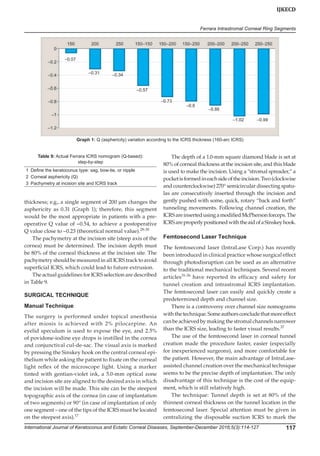
The mean follow-up time was 39.8° ± °21.1 months
(Table 1). All patients had implanted only one segment of
ICRS. A 160° arc ICRS (160-ICRS) was implanted in 18 eyes,
and the 210° arc ICRS (210-ICRS) was implanted in 7 eyes.
The mean UDVA increased from 20/185 to 20/66
(p = 0.005). The mean CDVA increased from 20/125 to
20/40 (p = 0.008) (Graph 1). The mean asphericity values](https://image.slidesharecdn.com/ferrararingreview2017-180820152145/85/Ferrara-ring-review-2017-6-320.jpg)
![120
Leonardo Torquetti et al
decreased from –0.95, preoperatively, to –0.23 (p = 0.006),
postoperatively.
The mean pachymetry at the apex of the cornea
increased from (mean) 457.7 ± 48.7 μm (361–542) to 466.2 ±
49.8 μm (381–559) (p = 0.025), and the pachymetry at the
thinnest point of the cornea increased from 436.3 ± 46.2 μm
(348–533) to 453.9 ± 49.3 μm (370–548) (p = 0.000). A signifi-
cant reduction in keratometric values was found at the last
follow-up examination; mean preoperative keratometry
was decreased from 45.41 ± 5.63 D (37.3–55.5) and changed
significantly to 42.88 ± 4.44 D (31.2–54.1) (p = 0.000).
Our postoperative results show a significant improve-
ment in UDVA and CDVA. Moreover, there was signifi-
cant increase in corneal thickness. This can be explained
by a theoretical cornea collagen remodeling induced by
the implantation of ICRS.
We found a significant increase in asphericity values
after the implantation of ICRS in this study. Interest-
ingly, the mean postoperative value was –0.23, which is
considered the “normal” value for the general popula-
tion.25,28-30,47
This value means that the normal physiologic
asphericity of the cornea shows a significant individual
variation ranging from mild oblate to moderate prolate.
In an unpublished study, where we evaluated the corneal
asphericity changes induced by the ICRS in keratoconus,
we found that the Ferrara intrastromal ICRS implanta-
tion significantly increased the mean corneal asphericity
from –0.85 to –0.32. It is well known that most corneas
after ablation laser procedures tend to become oblate,
and when ectasia develops these corneas usually become
prolate. However, the excess of prolatism usually found
in keratoconus (primary) is usually of a much larger
amount that that found in postrefractive surgery ectasia.
The probable reason is that the Q value after Ferrara ICRS
becomes much closer to “normal” values than when the
ICRS is used for keratoconus. As asphericity is one of the
markers of visual quality, turning it “normal” can be a
predictor of improvement of visual quality.
Thekeratometryvaluesreducedsignificantlyinalleyes.
It can be realized that the mean preoperative values are
usually lower than ones found in keratoconus (primary).
This can be explained somewhat by the corneal flattening
induced by the refractive procedure, usually in an optic
zone of greater extent than the location of the ectasia.
Most of the implanted ICRS were 160-ICRS, the “con-
ventional” ICRS. The remainder of eyes received the 210-
ICRS. The latter is usually reserved for central cones of
nipple type.24
Some ectasia assume the same topographi-
cal pattern of nipple cones, in which we usually use the
210-ICRS with excellent results. These ICRS are reserved
for cases with low astigmatism, in which we desire to
flatten the cornea with minimal astigmatic induction.
The potential advantages of ICRS implantation
over keratoplasty in eyes with post-LASIK ectasia are
many.23,48
First, it avoids further laser treatment, eliminat-
ing central corneal wound healing. This leaves the optical
center of the cornea untouched, enhancing the refractive
outcome. Second, the technique is reversible in cases of an
unsatisfactory refractive or clinical outcome, and minimal
postoperative care is required. Third, adjustment can
be performed using thinner or thicker ICRS. In cases of
unexpected corneal shape changes, one segment can be
removed or exchanged.49
Fourth, it avoids the complica-
tions of intraocular surgery.
These data are confirmed by several studies.9,50-52
Some long-term studies (ICRS in ectasia after LASIK)
showed that ICRS yielded improvements in visual acuity,
refractive status, and keratometric values without any
progression in cases with post-LASIK corneal ectasia.53
ENDOTHELIUM EVALUATION AFTER
FERRARA ICRS IMPLANTATION
We retrospectively reviewed patient records of 102 eyes
of 81 patients, which were followed for a period of at least
1 year (mean follow-up: 45.7 months, standard deviation:
16.4 months; range: 13–71 months).54
All patients had the
diagnosis of keratoconus, post-LASIK ectasia, or pellucid
degeneration.Statisticalanalysisincludedpreoperativeand
postoperative keratometry and endothelial characteristics
(cell count, average cell size, and coefficient of variation).
All patients completed at least 1 year of follow-up
(13–71 months). Mean age was 30.5 ± 8 years. The mean
cell count decreased from (mean ± standard deviation
[SD]) 2714 ± 372 to 2562 ± 406 cells/mm2
(p 0.001). The
calculated exponential cell loss rate over the mean inter-
val of follow-up (4 years) was 1.4% per year. The average
cell size increased from (mean ± SD) 375 ± 56 to –399 ± 61 µ2
(p 0.001). The coefficient of variation increased from
(mean ± SD) 0.22 ± 0.075 to 0.26 ± 0.010 (p = 0.001).
The mean maximum cell size increased from (mean ±
SD)529 ± 116to639 ± 225µ2
(p 0.001).Themeanminimum
cell size varied from (mean ± SD) 225 ± 36 to 226 ± 54 µ2
(p = 0.936).
There was significant corneal flattening as shown
by keratometry changes. The mean K decreased from
47.70 ± 2.29 (43.70–53.80) to 44.86 ± 2.02 (41.20–51.20)
(p = 0.0001).
In our study, we found a 1.4% loss of endothelial cells
per year. Considering that most of the studied patients
were young, the rate of endothelial cell loss was slightly
higher than in normal eyes (1.1%).55,56
Moreover, there is no
study in the current literature that shows the profile of the
“normal” endothelial loss in keratoconus corneas, which
could be higher than in normal corneas. The only report
in the literature regarding the endothelium profile of
keratoconus is nonprospective and studied only 12 eyes.57](https://image.slidesharecdn.com/ferrararingreview2017-180820152145/85/Ferrara-ring-review-2017-7-320.jpg)


The document discusses the use of Ferrara intrastromal corneal ring segments (ICRS) for treating ectatic corneal disorders such as keratoconus, highlighting their efficacy in reshaping the cornea to improve visual acuity and delay the need for more invasive procedures like keratoplasty. It details the history, mechanism of action, indications, contraindications, and selection criteria for ICRS, emphasizing the importance of surgical technique and nomograms in achieving optimal outcomes. Clinical findings and advancements in ICRS technology are also reviewed, focusing on patient tolerance and visual correction results.


