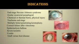
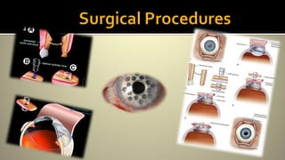
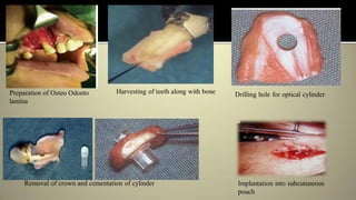
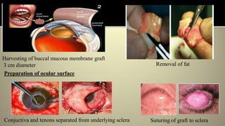
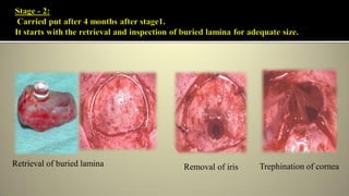
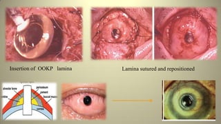
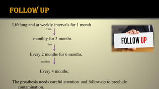
 Introduction
 History
 Ideal Keratoprosthesis
 Indication
 Contraindication
 Preoperative Assessment
 Surgical Procedure
 Complication
 Conclusion
OSTEO – Bone
ODONTO – Tooth
KERATO - Eye
PROSTHESIS - Artificial body part
Eyes are the “Windows to the soul;” the cornea is the “ Window of the
eye.” and most commonly occurring loss out of all sensory organs.
Corneal diseases are the major cause of blindness in the world today
remains second only to cataract.
Keratoprosthesis - only viable option for
restoring vision in end-stage inflammatory
corneal diseases.
EXAMPLE:
I. For eyes with a good ocular surface:
Boston Kpro type 1 .
II. Designed to treat severe dry eyes and
damaged ocular surfaces : osteo-odonto
keratoprosthesis (OOKP ) and Boston
KPro type 2.
 1789-Pellier de quengs glass lens in silver ring for leukomatous
cornea
 1853-Nussbaum collar-stud glass device consisting of two
plates sandwiching the cornea and connected by an optical cylinder, 2
with trials in rabbit eyes.
 1859-Heusser first to implant a keratoprosthesis in a human
eye;this was retained for 3 months.
 Other attempts made in Later half of 19th century (Von Hippel 1877,
Dimmer 1889, Baker1889, van Millingen 1895, Salzer 1895) but almost all
the implants failed and were extruded.
During the Second World War, the incidental discovery of corneal tissue
tolerance to plexi-glass fragments from aeroplane canopies suggested a new
direction for future research.
Surgical approach was developed by Stampelli in 1963 using a plastic acrylic
implant cemented to Patient’s tooth.
Falcinelli gave a Stepwise technique by Modifying Strampelli technique
 Able to surpass the natural cornea by having an improved optical quality
 Decreased aberrations.
 Excellent Biointegration.
 Replicate properties of the cornea such as drug penetration and intraocular
pressure measurement
 Provide resistance against infection .
 Enhance comfort and reduce mechanical shearing forces.
 Inexpensive
The properties of the PMMA optical cylinder are:
 Intraocular diameter, 4.1 mm
 Extraocular diameter, 3.65 mm
 Length, 7.75 mm
 Extraocular surface, 16 mm
 Intraocular surface, 6.5 mm
 Refractive index, 1.49;
 Equivalent power, 50.8 diopters
Ideal OOKP lamina size : 12 mm x 6 mm x 3 mm
 End-stage Stevens–Johnson syndrome
 Ocular cicatricial pemphigoid
 Chemical or thermal burns, physical injury
 Trachoma end-stage
 Multiple failed penetrating keratoplasty.
 Corneal failure after vitrectomy
 Epidermolysis bullosa
 Severe keratitis
 Uveitis
 Graft versus host disease
OOKP surgery is absolutely contraindicated in :
 Children under the age of 17.
 Eyes with no perception of light.
 Advanced glaucoma.
 Irreparable retinal detachment.
 Smoking and betel nut chewing etc.
Relative Contraindication:
 Defective light perception.
 Mentally unstable patient
 Unable to commit lifelong follow up
 Multidisciplinary approach .
 Surgical team comprises
a) Ophthalmologist,
b) Oral surgeon, and
c) Radiologist.
 Preoperative assessment includes
a) Opthomological asessment.
b) Oral assessment.
c) Psychological assessment.
 Primary diagnosis and previous
surgical interventions (detailed
general ophthalmic history and
careful examination)
 Determining intact and
functional retina and optic nerve
using Echography, ultrasound
examinations
Buccal mucosa assessment
 Tissue condition (severe scaring)
Dental assessment
 Oral hygiene, periodontal status
 Orthopantomograms and intraoral periapical radiographs (to evaluate length of the
root, Pulpal, periapical status and periodontal status of tooth etc).
 Expectations (regarding sight and cosmesis)
 Financial, time, emotional stress
 Risk of complications
 Lifelong follow-up
Ookp is usually carried out in two stages separated by an average period of
12 weeks
Stage 1 :
Step 1a: Tooth ostectomy ,preparation of OOKP lamina and submuscular
implantation.
Step 1b: Removal of ocular surface ,harvesting a full thickness buccal
mucosal graft and coverage of ocular surface.
Preparation of Osteo Odonto
lamina
Harvesting of teeth along with bone Drilling hole for optical cylinder
Removal of crown and cementation of cylinder Implantation into subcutaneous
pouch
Preparation of ocular surface
Harvesting of buccal mucous membrane graft
3 cm diameter Removal of fat
Conjuctiva and tenons separated from underlying sclera Suturing of graft to sclera
Retrieval of buried lamina Removal of iris Trephination of cornea
Lamina sutured and repositionedInsertion of OOKP lamina
Lifelong and at weekly intervals for 1 month
Then
monthly for 3 months
then
Every 2 months for 6 months,
and then
Every 4 months.
The prosthesis needs careful attention and follow-up to preclude
contamination.
 Heterotrophic auto graft of living human tissue.
 Provide long term stability
 Protection against cylinder extrusion and fistulization.
 Least risk of infection,
 Formation of retro prosthetic membrane can be limited or prevented.
 It requires lifelong follow up because it a two stage surgery
 Average esthetics.
During surgery:
 Risk of damage to the parotid duct
 Perforation or leakage of the cornea
 Damage to the adjacent teeth
 Expulsive hemorrhage
 Choroidal detachment, vitreous hemorrhage,
mucous membrane defect,
proliferation/sequestration of bone.
 Buccal membrane shrinkage or overgrowth
 Retroprosthetic membrane formation
 Endophthalmitis
 OOKP is the only successful method of offering long-term visual
rehabilitation in patients with corneal blindness.
 Following the innovative work of Strampelli, OOKP surgery has proved to
be most successful approach with kPRO implant.
 OOKP surgery is complex and requires meticulous care at each step to
ensure the overall success rate.
 Falcinelli et al (2005) reported 85% retaining property of
OOKP in with a functional improvement more than 75% in
18 year follow up study.
 Hille et al (2006) reported a 100 % retention of OOKP lamina
in a 5 year follow up study
 Iyer et al (2010) reported a 96% anatomical success in 50
cases.
 Strampelli in a 27 year study reported only 2 % loss of
prosthesis.
 Strampelli B. Keratoprosthesis with osteodental tissue. American J Ophthalmol.
1963; 89:1029-39.
 Falcinelli G, Barogi G, Corazza E, Colliardo P. Osteo-odonto-cheratoprotesi: 10
anni di esperienze positive edinnovazioni. Atti LXXIII Congress Soc.
OftalmologicaItaliana. 1993; 529-32.
 Falcinelli G, Missiroli A, Petitti V, Pinna C. Osteo-Odonto-Keratoprosthesis up to
Date. Acta XXV ConciliumOphthalmologicum 1986.Rome. Kugler&Ghedini. 1987;
2772-6.
 Falcinelli G, Barogi G, Taloni M. Osteo-odonto-Keratoprosthesis: present
experience and future prospects. Refractive Corneal Surg. 1993; 9: 193-4.
Osteo odonto kerato prosthesis

Osteo odonto kerato prosthesis

  • 2.
     Introduction  History Ideal Keratoprosthesis  Indication  Contraindication  Preoperative Assessment  Surgical Procedure  Complication  Conclusion
  • 3.
    OSTEO – Bone ODONTO– Tooth KERATO - Eye PROSTHESIS - Artificial body part Eyes are the “Windows to the soul;” the cornea is the “ Window of the eye.” and most commonly occurring loss out of all sensory organs. Corneal diseases are the major cause of blindness in the world today remains second only to cataract.
  • 4.
    Keratoprosthesis - onlyviable option for restoring vision in end-stage inflammatory corneal diseases. EXAMPLE: I. For eyes with a good ocular surface: Boston Kpro type 1 . II. Designed to treat severe dry eyes and damaged ocular surfaces : osteo-odonto keratoprosthesis (OOKP ) and Boston KPro type 2.
  • 5.
     1789-Pellier dequengs glass lens in silver ring for leukomatous cornea  1853-Nussbaum collar-stud glass device consisting of two plates sandwiching the cornea and connected by an optical cylinder, 2 with trials in rabbit eyes.  1859-Heusser first to implant a keratoprosthesis in a human eye;this was retained for 3 months.  Other attempts made in Later half of 19th century (Von Hippel 1877, Dimmer 1889, Baker1889, van Millingen 1895, Salzer 1895) but almost all the implants failed and were extruded.
  • 7.
    During the SecondWorld War, the incidental discovery of corneal tissue tolerance to plexi-glass fragments from aeroplane canopies suggested a new direction for future research. Surgical approach was developed by Stampelli in 1963 using a plastic acrylic implant cemented to Patient’s tooth. Falcinelli gave a Stepwise technique by Modifying Strampelli technique
  • 8.
     Able tosurpass the natural cornea by having an improved optical quality  Decreased aberrations.  Excellent Biointegration.  Replicate properties of the cornea such as drug penetration and intraocular pressure measurement  Provide resistance against infection .  Enhance comfort and reduce mechanical shearing forces.  Inexpensive
  • 9.
    The properties ofthe PMMA optical cylinder are:  Intraocular diameter, 4.1 mm  Extraocular diameter, 3.65 mm  Length, 7.75 mm  Extraocular surface, 16 mm  Intraocular surface, 6.5 mm  Refractive index, 1.49;  Equivalent power, 50.8 diopters Ideal OOKP lamina size : 12 mm x 6 mm x 3 mm
  • 10.
     End-stage Stevens–Johnsonsyndrome  Ocular cicatricial pemphigoid  Chemical or thermal burns, physical injury  Trachoma end-stage  Multiple failed penetrating keratoplasty.  Corneal failure after vitrectomy  Epidermolysis bullosa  Severe keratitis  Uveitis  Graft versus host disease
  • 11.
    OOKP surgery isabsolutely contraindicated in :  Children under the age of 17.  Eyes with no perception of light.  Advanced glaucoma.  Irreparable retinal detachment.  Smoking and betel nut chewing etc. Relative Contraindication:  Defective light perception.  Mentally unstable patient  Unable to commit lifelong follow up
  • 12.
     Multidisciplinary approach.  Surgical team comprises a) Ophthalmologist, b) Oral surgeon, and c) Radiologist.  Preoperative assessment includes a) Opthomological asessment. b) Oral assessment. c) Psychological assessment.
  • 13.
     Primary diagnosisand previous surgical interventions (detailed general ophthalmic history and careful examination)  Determining intact and functional retina and optic nerve using Echography, ultrasound examinations
  • 14.
    Buccal mucosa assessment Tissue condition (severe scaring) Dental assessment  Oral hygiene, periodontal status  Orthopantomograms and intraoral periapical radiographs (to evaluate length of the root, Pulpal, periapical status and periodontal status of tooth etc).
  • 15.
     Expectations (regardingsight and cosmesis)  Financial, time, emotional stress  Risk of complications  Lifelong follow-up
  • 17.
    Ookp is usuallycarried out in two stages separated by an average period of 12 weeks Stage 1 : Step 1a: Tooth ostectomy ,preparation of OOKP lamina and submuscular implantation. Step 1b: Removal of ocular surface ,harvesting a full thickness buccal mucosal graft and coverage of ocular surface.
  • 18.
    Preparation of OsteoOdonto lamina Harvesting of teeth along with bone Drilling hole for optical cylinder Removal of crown and cementation of cylinder Implantation into subcutaneous pouch
  • 19.
    Preparation of ocularsurface Harvesting of buccal mucous membrane graft 3 cm diameter Removal of fat Conjuctiva and tenons separated from underlying sclera Suturing of graft to sclera
  • 20.
    Retrieval of buriedlamina Removal of iris Trephination of cornea
  • 21.
    Lamina sutured andrepositionedInsertion of OOKP lamina
  • 22.
    Lifelong and atweekly intervals for 1 month Then monthly for 3 months then Every 2 months for 6 months, and then Every 4 months. The prosthesis needs careful attention and follow-up to preclude contamination.
  • 23.
     Heterotrophic autograft of living human tissue.  Provide long term stability  Protection against cylinder extrusion and fistulization.  Least risk of infection,  Formation of retro prosthetic membrane can be limited or prevented.
  • 24.
     It requireslifelong follow up because it a two stage surgery  Average esthetics.
  • 25.
    During surgery:  Riskof damage to the parotid duct  Perforation or leakage of the cornea  Damage to the adjacent teeth  Expulsive hemorrhage  Choroidal detachment, vitreous hemorrhage, mucous membrane defect, proliferation/sequestration of bone.  Buccal membrane shrinkage or overgrowth  Retroprosthetic membrane formation  Endophthalmitis
  • 26.
     OOKP isthe only successful method of offering long-term visual rehabilitation in patients with corneal blindness.  Following the innovative work of Strampelli, OOKP surgery has proved to be most successful approach with kPRO implant.  OOKP surgery is complex and requires meticulous care at each step to ensure the overall success rate.
  • 27.
     Falcinelli etal (2005) reported 85% retaining property of OOKP in with a functional improvement more than 75% in 18 year follow up study.  Hille et al (2006) reported a 100 % retention of OOKP lamina in a 5 year follow up study  Iyer et al (2010) reported a 96% anatomical success in 50 cases.  Strampelli in a 27 year study reported only 2 % loss of prosthesis.
  • 28.
     Strampelli B.Keratoprosthesis with osteodental tissue. American J Ophthalmol. 1963; 89:1029-39.  Falcinelli G, Barogi G, Corazza E, Colliardo P. Osteo-odonto-cheratoprotesi: 10 anni di esperienze positive edinnovazioni. Atti LXXIII Congress Soc. OftalmologicaItaliana. 1993; 529-32.  Falcinelli G, Missiroli A, Petitti V, Pinna C. Osteo-Odonto-Keratoprosthesis up to Date. Acta XXV ConciliumOphthalmologicum 1986.Rome. Kugler&Ghedini. 1987; 2772-6.  Falcinelli G, Barogi G, Taloni M. Osteo-odonto-Keratoprosthesis: present experience and future prospects. Refractive Corneal Surg. 1993; 9: 193-4.