SlideShare a Scribd company logo
1 of 46
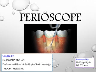
PERIOSCOPE
Presented By:
Dr.Deepali Jain
PG 2ND Year
Guided By:
Dr.RANJANA MOHAN
Professor and Head of the Dept of Periodontology
TMDCRC, Moradabad
WHAT IS PERIOSCOPE?
• Advancements in fiber optic technology now make it possible to have
access to plaque (bacteria) and calculus (tartar) under the gums.
• The perioscope is a tiny camera that lets the hygienist have a new vision
of your root surface and remove all the hard deposits in your deep
pocket. The root surface is magnified up to 45 times its actual size on a
flat screen TV.
• As the camera goes under the gum, the hygienist can see the shape of
the root. She can see where all the disease is hiding and remove it. Even
the smallest piece of infectious material can be seen thanks to the
camera and it’s remarkable magnification and resolution
INTRODUCTION
• Traditionally, anti-infective periodontal therapy has been performed by effective
plaque control and mechanical therapy by scaling and root planing.
• The efficacy of this treatment depends on different factors, like the anatomy of the
subgingival area and the presence of furcation defects, but also on the therapist’s
skills.
• Normally, the examination of the treated sites is accomplished by manual and
tactile exploration. However, the inability to detect some root deposits that have
not been eliminated, has been repeatedly demonstrated by different investigators
(brayer et al, 1989; rabbani et al, 1981; sherman et al, 1990).
• Visualization of the root surface during subgingival debridement may improve
the clinical results of the treated teeth.
• Recently, a non-invasive method for the examination of the hard and soft tissues
of the subgingival sulcus has been developed with the purpose to allow the
clinician a direct view of the subgingival area. This has been made possible, as a
result of the improvement in fiber-optic devices.
THE PERIOSCOPE
• This endoscope for dental purposes is manufactured by dentalview inc., Lake
forest, CA, USA .
• The endoscope has a flexible design that can be combined with other dental
instruments.
• The use of this technology has been previously described in a few case reports and
clinical studies (avradopoulos et al, 2004; stambaugh, 2002).
• The equipment contains a gradient index lens that is mounted on the end of a 2 m
long fused fiber-optic bundle containing 10,000 individual light guiding fibers
(pixels).
• Surrounding the fused bundle and lens are 15 large core plastic fiber-optic
strands for carrying illumination light from a remote lamp to the operative site.
• This assembly is encased in a flexible plastic tube resulting in a diameter of
0.85mm at the distal end. A spring- Activated connector is located 1 m from the
distal end to connect to a window sheath. This connector assures that the distal
lens remains in contact with the distal window of the sheath
• Endoscopic technology has been developed to facilitate real-
time visualization of the gingival sulcus during diagnostic and
therapeutic phases of periodontal care.
• The first generation of the periodontal endoscope, perioscope™
(perioscopy inc., Oakland, calif) was found to have technical
shortcomings and a steep learning curve.
• However, new technique changes and equipment modifications
have improved the reliability and a number of studies have
demonstrated improved efficacy for treatment of periodontal
disease
The Journal of Dental Hygiene Vol. 87 • No. 3 • June 2013
PERIOSCOPE form of endoscope used to explore and visualize the periodontal
pocket, producing an image of a diseased tooth's root
Gum surgery is the traditional means of removing the tartar from a root. The
perioscope is a revolutionary tool that lets the hygienist see under the gum without
cutting. The camera to have a perfect view of the root. Using specialized instruments
one can remove the disease causing tartar. The results of surgery and perioscopy are
the same in many circumstance
ENDOSCOPE SHEATH
• Sterilization is mandatory if the distal tip of the perioscope comes in direct
contact with the patient’s tissues.
• However, sterilization is time consuming and reduces the lifetime of the
endoscope (usually, the instrument must be replaced after 12 autoclave cycles).
• Thus, a sterile disposable sheath was developed, which provides a barrier against
pathogens and can be removed after use (fig. 2).
• The sheath is equipped with a sapphire window, allowing a clear view through
the endoscope. Furthermore, because subgingival bleeding may obscure the
vision through the endoscope, a separate water channel connected to a peristaltic
pump provides a water spray, which keeps the working field clear.
• Finally, a small plastic connector at the distal end of the sheath that fits on a
stainless steel receptacle built into each Instrument (curette, explorer and
ultrasonic adapter) allows a precise positioning of the endoscope while working
with the instrument.
• Visualization the endoscope is delivered with a medical grade CCD video
camera connected with a camera coupler.
• This coupler magnifies and focuses the transmitted image onto the CCD sensor.
The electrical signals from the sensor are digitized by the camera’s control unit
resulting in a standard svideo signal (Y/C) output to an attached monitor.
• The endoscope is delivered with a 12.1" diagonal active matrix backlit LCD
display (fig. 3), and the monitor has a resolution of 800 x 600 pixels. The
objective lens has a nominal 70° field view in air. Under water this field is
decreased due to the refraction index of water: 70°/1.33 =53°.
• The image of the root and sulcus projected on the monitor is magnified from
22x to 48x.
• The clinician can therefore indirectly observe the contents of the sulcus and
subgingival root surface with a highly magnified, illuminated view.
Total view of the DV2 PerioscopeTM.
(DentalView Inc., Lake Forest, CA, USA)
View of the endoscope and its disposable sheath.
(A) fiber-optic bundle, (B) sterile disposable sheath,
(C) spring tension connection,
(D) connector to the water supply (Luer Lock).
(Image: DentalView Inc., Lake Forest,
CA, USA)
Detail of the LCD Monitor.
Clinical application of the explorer.
• The perioscope consists of three units, in which the head reflects an
image of the area under examination through a magnifying lens, an
eyepiece by which we can see this image, and a lighting unit that
illuminates the area through optical fibers, the most important part is
the bead, which is the thinnest (1,2 mm in diameter) optical tube ever
developed, and is contained, with the lens and optical fibers, in a
stainless steel sheath, the instrument is described and shown in use in
figs 1 to 5
• These views were captured on film with a nikon 0M2- camera.
Which was connected to the camera lock pin of the eyepiece
The Perioscope.
Schematic illustration of the scope.
Quintessence International Volume 19, Number 7 1988
The tip and guide plate being inserted into the periodontal pocket.
Specifications of the Perioscope and its lighting unit
Visulization ol calcuii located 7 mm from
the gingival margin.
Residual calculi are seen after
one minute ot ultra-sonic
scaling.
Quintessence International Volume 19, Number 7 1988
Subgingival calculi at 8 mm.
Residual calculi after one minute of scaling
with a Columbia 4R/L curet.
Quintessence International Volume 19, Number 7 1988
Periodontal pocket tissue in an acute phase
Four days after root scaling: the healed tissue.
Direct observation of the root wall and pocket tissue .Quintessence International Volume 19, Number 7
Furcated bone which has been already replaced
by granulation tissue
Direct observation of the root wall and pocket tissue .Quintessence International Volume 19, Number 7
A tooth with advanced
periodontal disease
With the Perisocope we
can see the deeply buried
deposits of calculus that
before needed surgery to
discover
Because of the Perioscope we
can thoroughly clean the deeply
buried calculus deposits on the
root surface without the need of
surger
• Visualization and tactile perception are the most important
diagnostic tools for recognizing root wall calculus.
• Scaling and root planing have long been dependent on the
operator's ability. However, the perioscope can enable visualization
of the location, the amount, and the shape of subgingival calculus,
facilitating the reduction of residual calculus. Fit of restorations,
cement flow into the pocket, enameloma, root fracture, subgingival
caries, broken instruments in bone or in the pocket, and furcation
involvement can also be visualized
Direct observation of the root wall and pocket tissue .Quintessence International Volume 19, Number 7
• Diagnosing the disease activity properly may best be done by
observing color changes of the pocket tissue.
• observed distinct color changes in the acute stage but as yet have
been unable to associate it with disease activity related to bone
resorption.
• Presently, diagnosis of disease activity is conducted by inserting a
periodontal probe into the pocket and diagnosing the activity of the
disease by the blood that is produced.
• However, we are also able to visualize completely inflamed tissue
without calculus as well as locally inflamed areas containing
copious amounts of calculi.
• Directing efforts to finding the relationship between bleeding,
pocket appearance, and the active phase of bone resorption.
Direct observation of the root wall and pocket tissue .Quintessence International Volume 19, Number 7
• Visualizing subgingival accretions and the pocket wall is done as
follows:
1. rotate the guide plate against the gingiva, keeping the lens
proximal to the side of the tooth; and
2. insert the guide plate into the periodontal pocket and gently
enlarge the area under observation by depressing the gingiva.
• Calculus is easily visualized under the light radiating from around
the lens. This is done after the area is air-dried.
• Calculus is observed at x 4 magnification al a distance of 5 mm, so
it is generally unnecessary to situate the scope's tip subgingivally.
• After scaling, the scraped and widened gingival pocket allows
better visibility than was possible before scaling.
Direct observation of the root wall and pocket tissue .Quintessence International Volume 19, Number 7
• Exudate is easily flushed out, and water in the pocket does not
cause any visual problems.
• However, we recommend that the scope be used when the pocket
is deep and soft, or after the curet has been used. This is because it
is extremely hard to depress a healthy and shallow gingiva.
• The operator usually observes through the eyepiece, But the
instrument can be connected to a television camera and the view
projected on a sereen. Photographs can be taken from the screen or
by attaching a camera to the eyepiece.
Direct observation of the root wall and pocket tissue .Quintessence International Volume 19, Number 7
• An endoscope for visual inspection for calculus deposits has
been shown to be an aid to clinicians in removing
subgingival deposits from single-rooted teeth.
• However, recent research has shown that the endoscope as
an adjunct to removal of calculus in multirooted molar teeth
provided no significant improvement over traditional scaling
and root-planing procedures without an endoscope.''
• It has been introduced recently for use subgingivally in the
diagnosis and treatment of periodontal disease produced
by dental view, inc. And called the perioscopy system, it
consists of a 0.99 mm-diameter reusable fiberoptic
endoscope over which is fitted a disposable, sterile sheath
Perioscopy system: dental
endoscope(Courtesy
Perioscopy Incorporated,
Oakland, CA.)
• The fiberoptic endoscope fits onto periodontal probes and
ultrasonic instruments that have been designed to accept it
Viewing periodontal explorers (left/right/full viewing) for the Perios
(Courtesy Perioscopy Incorporated, Oakland, CA.)
• The sheath delivers water irrigation that flushes the pocket while
the endoscope is being used, keeping the field clear.
• The fiberoptic endoscope attaches to a medical-grade charged-
coupled device (CCD) video camera and light source that produces
an image on a flat-panel monitor for viewing during subgingival
exploration and instrumentation.
• This device allows clear visualization deeply into subgingival
pockets and furcations
• It permits operators to detect the presence and location of
subgingival deposits and guides them in the thorough removal
of these deposits.
• Magnification ranges from 24X to 48X, enabling visualization
of even minute deposits of plaque and calculus. Using this
device, operators can achieve levels of root
• Debridement and cleanliness that are much more difficult or
impossible to produce without it.
• The perioscopy system can also be used to evaluate subgingival
areas for caries, defective restorations, root fractures, and
resorption.
Pattison AM and Pattison GL. Scaling and root planning. Newman M, Takei H, Klokkevold P,
Carranza F. Carranza’s clinical periodontology. St Louis: Saunders. 2011; 10th Ed : 749-797
Contemporary Oral Hygiene May 2006
• Perioscope in the non-dominant hand (like the dental mirror) and an ultrasonic in
the dominant hand, viewing and instrumenting at the same time.
• Both piezo and magnetostrictive ultrasonics it was quickly apparent that
magnetostrictive instrumentation allows for quick and easy changing of
instruments.
• During a full mouth "scope" procedure we primarily us a 0.5mm diameter straight
tip, but on occasion we need to change to curved inserts and sometimes to diamond
coated tips.
• The diamond coated tips are for cutting overhanging restorations, residual bonded
cement, enamel pearls/projections, shallow caries or globular cementum. It is found
that ultrasonics were all that were necessary.
• Oraldent has launched a new dental endoscope, the DV2 perioscopy
system, specifically designed to assist in the diagnosis and treatment of
periodontal problems.
• The dv2 perioscope is an optical device which enables dentists and
hygienists to see minute details on their patients’ teeth and tooth roots,
offering views of subgingival root and tooth surfaces without resort to
invasive procedures.
• The instrument provides a direct, real-time, magnified visualisation of
the subgingival anatomy, allowing a more accurate and complete
diagnosis. Other benefits include the earlier identification of potential
clinical problems, reduced referrals an consequent loss of revenue with
more patients able to be treated within the practice, and enhanced
teaching/ learning opportunities.
BRITISH DENTAL JOURNAL VOLUME 199 NO. 5 SEPT 10 2005
BENEFITS OF THE PERIOSCOPE
• Because of the ability to see the diseased root surface, the perioscope usually allows
the clinician to treat periodontal disease without invasive surgical therapies.
• Additionally, the perioscope allows the clinician to see what could not be seen
before during periodontal surgery. Now, surgical therapies are far more effective
and reliable than in the past.
• Some of the breakthroughs that the periscope has made in the everyday practice of
periodontics:
 Increased effectiveness of non-surgical treatment methods, and thus a
reduction in the amount of surgical therapy required for the treatment of
periodontitis.
 Increased diagnostic accuracy; which leads to an increased appropriateness of
prescribe treatment methods.
 Increase effectiveness of surgical therapies which were limited by visibility
problems
• The perioscope has greatly increased the accuracy of periodontal
diagnostics and treatment prescription. Also, it has greatly increased the
effectiveness of non-surgical and surgical therapies.
• The perioscope has created a shift in the nature of periodontal care.
However, it does not diminish the power of, or importance of, periodontal
surgical therapies.
• Ultimately, periodontal therapy is about cleaning diseased tooth roots free
of bacterial contamination, and keeping them free of such accumulations.
Periodontal surgical therapy remains the most powerful of therapies to
achieve these goals.
• The perioscope contributes to the ability to achieve therapeutic results that
are similar to, and sometimes better than, those achieved by surgical
therapies — without the pain, disfigurement and cost of surgical therapies.
However, many periodontal disease situations cannot be adequately
resolved without surgical therapy
• The benefits of periodontal endoscopy are not just for people
suffering from the moderate to advanced stages of periodontal
disease.
• When used properly, the perioscope is a very powerful
diagnostic tool for the early detection and treatment of many
conditions.
• A routine examination and dental cleaning performed under
powerful magnification beneath the gums can reveal problems
frequently overlooked by traditional diagnostic methods, such
as decay, failing restorations and fractures.
• These problems left unchecked, including cavities and calculus
trapped beneath the gums, can lead to more serious conditions
DIAGNOSTIC PERIODONTAL ENDOSCOPY
CONCLUSION
• The perioscope improved calculus detection over the explorer at
each subject visit, indicating that a visual component is a positive
adjunct to tactile evaluation of subgingival calculus.
• Significantly more calculus was detected using the perioscope
than the explorer at each visit.
• Additionally, the perioscope facilitated calculus detection
between the reevaluation appointments, where the explorer did
not.
• Overall, the perioscope outperformed the explorer in residual
calculus detection
• Stambaugh RV: A clinician's 3-year experience with perioscopy. Compend contin
educ dent. 2002; 23(11a): 1061-1070.
• Stambaugh rv, myers g, stambaugh rv, et al: endoscopic visualization of the
submarginal gingiva dental sulcus and tooth root surfaces. J periodontol. 2002;
73(4): 374-382.
• Wilson tg, carnio j, schenk r, et al: absence of histologic signs of chronic
inflammation following closed subgingival scaling and root planning using the
dental endoscope: human biopsies—a pilot study. J periodontol. 2008; 79(11):
2036-2041.
• Wilson tg, harrel sk, nunn me, et al: the relationship between the presence of
tooth-borne subgingival deposits and inflammation found with a dental
endoscope. J periodontol. 2008; 79(11): 2029-2035.
• Pihlstrom bl, mchugh rb, oliphant th, et al: comparison of surgical and
nonsurgical treatment of periodontal disease: a review of current studies and
additional results after 6 years. J clin periodontol. 1983; 10:524
REFERENCES

More Related Content

What's hot

Bone loss and patterns of bone destruction
Bone loss and patterns of bone destructionBone loss and patterns of bone destruction
Bone loss and patterns of bone destructionvidushiKhanna1
 
Attached gingiva and its significance
Attached gingiva and its significanceAttached gingiva and its significance
Attached gingiva and its significanceMD Abdul Haleem
 
Alveolar bone in health and disease
Alveolar bone in health and disease Alveolar bone in health and disease
Alveolar bone in health and disease Chittoor Deals
 
Endodontic Periodontal Relationship, ENDO PERIO LESION
Endodontic Periodontal Relationship, ENDO PERIO LESIONEndodontic Periodontal Relationship, ENDO PERIO LESION
Endodontic Periodontal Relationship, ENDO PERIO LESIONDeepa jinan
 
Mechanism of bone loss and patterns of bone loss
Mechanism of bone loss and patterns of bone loss  Mechanism of bone loss and patterns of bone loss
Mechanism of bone loss and patterns of bone loss Kapil Arora
 
Dento gingival unit
Dento gingival unitDento gingival unit
Dento gingival unitsangeeta roy
 
Periodontal splinting
Periodontal splintingPeriodontal splinting
Periodontal splintingbibekjha
 
Resective osseous surgery
Resective osseous surgeryResective osseous surgery
Resective osseous surgeryShilpa Shiv
 
RADIOGRAPHIC AIDS IN THE DIAGNOSIS OF PERIODONTAL DISEASE
RADIOGRAPHIC AIDS IN THE DIAGNOSIS OF PERIODONTAL DISEASERADIOGRAPHIC AIDS IN THE DIAGNOSIS OF PERIODONTAL DISEASE
RADIOGRAPHIC AIDS IN THE DIAGNOSIS OF PERIODONTAL DISEASERupal Patle
 
Risk factors in Periodontal Disease
Risk factors in Periodontal DiseaseRisk factors in Periodontal Disease
Risk factors in Periodontal DiseaseNeil Pande
 
Root conditioning agents
Root conditioning agentsRoot conditioning agents
Root conditioning agentsmythreyeethakur
 
Bruxism and its effect on periodontium
Bruxism and its effect on periodontiumBruxism and its effect on periodontium
Bruxism and its effect on periodontiumRamya Ganesh
 
Advanced Periodontal diagnostic aids
Advanced Periodontal diagnostic aidsAdvanced Periodontal diagnostic aids
Advanced Periodontal diagnostic aidsJignesh Patel
 
Gingival recession classifications
Gingival recession classifications Gingival recession classifications
Gingival recession classifications Achi Joshi
 
Coronal advanced flap in combination with a connective tissue graft. Is the t...
Coronal advanced flap in combination with a connective tissue graft. Is the t...Coronal advanced flap in combination with a connective tissue graft. Is the t...
Coronal advanced flap in combination with a connective tissue graft. Is the t...MD Abdul Haleem
 
Sonic & ultrasonic instruments
Sonic & ultrasonic instruments Sonic & ultrasonic instruments
Sonic & ultrasonic instruments Shikha Arya
 

What's hot (20)

Aggressive periodontitis
Aggressive periodontitisAggressive periodontitis
Aggressive periodontitis
 
Bone loss and patterns of bone destruction
Bone loss and patterns of bone destructionBone loss and patterns of bone destruction
Bone loss and patterns of bone destruction
 
Attached gingiva and its significance
Attached gingiva and its significanceAttached gingiva and its significance
Attached gingiva and its significance
 
periodontal pocket
periodontal pocketperiodontal pocket
periodontal pocket
 
Alveolar bone in health and disease
Alveolar bone in health and disease Alveolar bone in health and disease
Alveolar bone in health and disease
 
Endodontic Periodontal Relationship, ENDO PERIO LESION
Endodontic Periodontal Relationship, ENDO PERIO LESIONEndodontic Periodontal Relationship, ENDO PERIO LESION
Endodontic Periodontal Relationship, ENDO PERIO LESION
 
Mechanism of bone loss and patterns of bone loss
Mechanism of bone loss and patterns of bone loss  Mechanism of bone loss and patterns of bone loss
Mechanism of bone loss and patterns of bone loss
 
Dento gingival unit
Dento gingival unitDento gingival unit
Dento gingival unit
 
Periodontal splinting
Periodontal splintingPeriodontal splinting
Periodontal splinting
 
Modified widman flap
Modified widman flapModified widman flap
Modified widman flap
 
Resective osseous surgery
Resective osseous surgeryResective osseous surgery
Resective osseous surgery
 
RADIOGRAPHIC AIDS IN THE DIAGNOSIS OF PERIODONTAL DISEASE
RADIOGRAPHIC AIDS IN THE DIAGNOSIS OF PERIODONTAL DISEASERADIOGRAPHIC AIDS IN THE DIAGNOSIS OF PERIODONTAL DISEASE
RADIOGRAPHIC AIDS IN THE DIAGNOSIS OF PERIODONTAL DISEASE
 
Risk factors in Periodontal Disease
Risk factors in Periodontal DiseaseRisk factors in Periodontal Disease
Risk factors in Periodontal Disease
 
Root conditioning agents
Root conditioning agentsRoot conditioning agents
Root conditioning agents
 
Dental plaque
Dental plaqueDental plaque
Dental plaque
 
Bruxism and its effect on periodontium
Bruxism and its effect on periodontiumBruxism and its effect on periodontium
Bruxism and its effect on periodontium
 
Advanced Periodontal diagnostic aids
Advanced Periodontal diagnostic aidsAdvanced Periodontal diagnostic aids
Advanced Periodontal diagnostic aids
 
Gingival recession classifications
Gingival recession classifications Gingival recession classifications
Gingival recession classifications
 
Coronal advanced flap in combination with a connective tissue graft. Is the t...
Coronal advanced flap in combination with a connective tissue graft. Is the t...Coronal advanced flap in combination with a connective tissue graft. Is the t...
Coronal advanced flap in combination with a connective tissue graft. Is the t...
 
Sonic & ultrasonic instruments
Sonic & ultrasonic instruments Sonic & ultrasonic instruments
Sonic & ultrasonic instruments
 

Viewers also liked

Boletín Informativo La Rioja Alta,SA Invierno 2011
Boletín Informativo La Rioja Alta,SA Invierno 2011Boletín Informativo La Rioja Alta,SA Invierno 2011
Boletín Informativo La Rioja Alta,SA Invierno 2011txamu
 
Eu somii newsletter 2014 web
Eu somii newsletter 2014 webEu somii newsletter 2014 web
Eu somii newsletter 2014 webPeter van Ooijen
 
Etica en el Yoga: Yamas y Niyamas, por Jaime Ocampo
Etica en el Yoga: Yamas y Niyamas, por Jaime OcampoEtica en el Yoga: Yamas y Niyamas, por Jaime Ocampo
Etica en el Yoga: Yamas y Niyamas, por Jaime OcampoAquarius en línea
 
Agenda de Actividades del Área de Cultura - Marzo 2015
Agenda de Actividades del Área de Cultura - Marzo 2015Agenda de Actividades del Área de Cultura - Marzo 2015
Agenda de Actividades del Área de Cultura - Marzo 2015Ayuntamiento de Málaga
 
Radio libre por Internet
Radio libre por InternetRadio libre por Internet
Radio libre por InternetPAT Extremadura
 
Actividad 3 (2)
Actividad 3 (2)Actividad 3 (2)
Actividad 3 (2)jmdsy
 
Listado de equipos OUTDOOR necesario antes de realizar travesias
Listado de equipos OUTDOOR necesario antes de realizar travesiasListado de equipos OUTDOOR necesario antes de realizar travesias
Listado de equipos OUTDOOR necesario antes de realizar travesiasKalluka North
 
MS 100 BA Balanced Armature Driver Earphones| Phiaton
MS 100 BA Balanced Armature Driver Earphones| PhiatonMS 100 BA Balanced Armature Driver Earphones| Phiaton
MS 100 BA Balanced Armature Driver Earphones| PhiatonPhiaton
 
Programa formativo en comunicación efectiva y gestión de equipos (en abiert...
Programa formativo en comunicación efectiva y gestión de equipos (en abiert...Programa formativo en comunicación efectiva y gestión de equipos (en abiert...
Programa formativo en comunicación efectiva y gestión de equipos (en abiert...Laura Pereiro
 
The Periodontal Instruments, dr anirudh singh chauhan
The Periodontal Instruments, dr anirudh singh chauhanThe Periodontal Instruments, dr anirudh singh chauhan
The Periodontal Instruments, dr anirudh singh chauhanAnirudh Singh Chauhan
 
Guía de orientacion para actores escolares - Gabinete Pedagógico Interdiscipl...
Guía de orientacion para actores escolares - Gabinete Pedagógico Interdiscipl...Guía de orientacion para actores escolares - Gabinete Pedagógico Interdiscipl...
Guía de orientacion para actores escolares - Gabinete Pedagógico Interdiscipl...Marcela Js
 
E-Business – Clase 04 – Implementación de una tienda ON LINE. PayPal. Ocho pi...
E-Business – Clase 04 – Implementación de una tienda ON LINE. PayPal. Ocho pi...E-Business – Clase 04 – Implementación de una tienda ON LINE. PayPal. Ocho pi...
E-Business – Clase 04 – Implementación de una tienda ON LINE. PayPal. Ocho pi...LEOnardo AMARaldo DELgado
 

Viewers also liked (20)

Boletín Informativo La Rioja Alta,SA Invierno 2011
Boletín Informativo La Rioja Alta,SA Invierno 2011Boletín Informativo La Rioja Alta,SA Invierno 2011
Boletín Informativo La Rioja Alta,SA Invierno 2011
 
Organismo y ambiente
Organismo y ambienteOrganismo y ambiente
Organismo y ambiente
 
Eu somii newsletter 2014 web
Eu somii newsletter 2014 webEu somii newsletter 2014 web
Eu somii newsletter 2014 web
 
Dansk Coaching Institut - Lean Coaching
Dansk Coaching Institut - Lean Coaching Dansk Coaching Institut - Lean Coaching
Dansk Coaching Institut - Lean Coaching
 
School Garden Materials and Resource Guide ~ Monterey County, California
School Garden Materials and Resource Guide ~ Monterey County, CaliforniaSchool Garden Materials and Resource Guide ~ Monterey County, California
School Garden Materials and Resource Guide ~ Monterey County, California
 
Etica en el Yoga: Yamas y Niyamas, por Jaime Ocampo
Etica en el Yoga: Yamas y Niyamas, por Jaime OcampoEtica en el Yoga: Yamas y Niyamas, por Jaime Ocampo
Etica en el Yoga: Yamas y Niyamas, por Jaime Ocampo
 
Agenda de Actividades del Área de Cultura - Marzo 2015
Agenda de Actividades del Área de Cultura - Marzo 2015Agenda de Actividades del Área de Cultura - Marzo 2015
Agenda de Actividades del Área de Cultura - Marzo 2015
 
Radio libre por Internet
Radio libre por InternetRadio libre por Internet
Radio libre por Internet
 
Actividad 3 (2)
Actividad 3 (2)Actividad 3 (2)
Actividad 3 (2)
 
07demaio oexpresso
07demaio oexpresso07demaio oexpresso
07demaio oexpresso
 
Listado de equipos OUTDOOR necesario antes de realizar travesias
Listado de equipos OUTDOOR necesario antes de realizar travesiasListado de equipos OUTDOOR necesario antes de realizar travesias
Listado de equipos OUTDOOR necesario antes de realizar travesias
 
MS 100 BA Balanced Armature Driver Earphones| Phiaton
MS 100 BA Balanced Armature Driver Earphones| PhiatonMS 100 BA Balanced Armature Driver Earphones| Phiaton
MS 100 BA Balanced Armature Driver Earphones| Phiaton
 
Programa formativo en comunicación efectiva y gestión de equipos (en abiert...
Programa formativo en comunicación efectiva y gestión de equipos (en abiert...Programa formativo en comunicación efectiva y gestión de equipos (en abiert...
Programa formativo en comunicación efectiva y gestión de equipos (en abiert...
 
The Periodontal Instruments, dr anirudh singh chauhan
The Periodontal Instruments, dr anirudh singh chauhanThe Periodontal Instruments, dr anirudh singh chauhan
The Periodontal Instruments, dr anirudh singh chauhan
 
Guía de orientacion para actores escolares - Gabinete Pedagógico Interdiscipl...
Guía de orientacion para actores escolares - Gabinete Pedagógico Interdiscipl...Guía de orientacion para actores escolares - Gabinete Pedagógico Interdiscipl...
Guía de orientacion para actores escolares - Gabinete Pedagógico Interdiscipl...
 
E-Business – Clase 04 – Implementación de una tienda ON LINE. PayPal. Ocho pi...
E-Business – Clase 04 – Implementación de una tienda ON LINE. PayPal. Ocho pi...E-Business – Clase 04 – Implementación de una tienda ON LINE. PayPal. Ocho pi...
E-Business – Clase 04 – Implementación de una tienda ON LINE. PayPal. Ocho pi...
 
Fuerza de ventas
Fuerza de ventasFuerza de ventas
Fuerza de ventas
 
Tesis tecniseg
Tesis tecnisegTesis tecniseg
Tesis tecniseg
 
Taller 1,taller2 y colaborativos
Taller 1,taller2 y colaborativosTaller 1,taller2 y colaborativos
Taller 1,taller2 y colaborativos
 
tics
ticstics
tics
 

Similar to Perioscope

Microscope 3/ orthodontic courses by indian dental academy
Microscope 3/ orthodontic courses by indian dental academyMicroscope 3/ orthodontic courses by indian dental academy
Microscope 3/ orthodontic courses by indian dental academyIndian dental academy
 
Microscope 3[1]/ orthodontic courses by indian dental academy
Microscope 3[1]/ orthodontic courses by indian dental academyMicroscope 3[1]/ orthodontic courses by indian dental academy
Microscope 3[1]/ orthodontic courses by indian dental academyIndian dental academy
 
Microscope 3 (2)/ orthodontic courses by indian dental academy
Microscope 3 (2)/ orthodontic courses by indian dental academyMicroscope 3 (2)/ orthodontic courses by indian dental academy
Microscope 3 (2)/ orthodontic courses by indian dental academyIndian dental academy
 
Keratoprsthesis
KeratoprsthesisKeratoprsthesis
KeratoprsthesisNikhil Rp
 
recent advances in prosthodontics/dental lab technology courses by Indian den...
recent advances in prosthodontics/dental lab technology courses by Indian den...recent advances in prosthodontics/dental lab technology courses by Indian den...
recent advances in prosthodontics/dental lab technology courses by Indian den...Indian dental academy
 
Recent advances in prosthodontics / crown & bridge courses by indian dental a...
Recent advances in prosthodontics / crown & bridge courses by indian dental a...Recent advances in prosthodontics / crown & bridge courses by indian dental a...
Recent advances in prosthodontics / crown & bridge courses by indian dental a...Indian dental academy
 
Apeceoctomy traditional and new concepts
Apeceoctomy traditional and new conceptsApeceoctomy traditional and new concepts
Apeceoctomy traditional and new conceptsAhmed Alrashedi
 
Recent advances in periodontal surgical technology
Recent advances in periodontal surgical technologyRecent advances in periodontal surgical technology
Recent advances in periodontal surgical technologyDr Aananyaa Khanna
 
Journal club on Magnification loupes
Journal club on Magnification loupesJournal club on Magnification loupes
Journal club on Magnification loupesDr Abhilasha
 
Keratoprosthesis
KeratoprosthesisKeratoprosthesis
Keratoprosthesispeterroy90
 
recent advances in implant dentistry
recent advances in implant dentistryrecent advances in implant dentistry
recent advances in implant dentistryDr.Pallavi Chavan
 
GINGIVAL SURGICAL TECHNIQUES IN PERIODONTOLOGY
GINGIVAL SURGICAL TECHNIQUES IN PERIODONTOLOGYGINGIVAL SURGICAL TECHNIQUES IN PERIODONTOLOGY
GINGIVAL SURGICAL TECHNIQUES IN PERIODONTOLOGYSupriya Bhat
 
Pterygium Excision with Free Conjunctival Limbal Autograft
Pterygium Excision with Free Conjunctival Limbal AutograftPterygium Excision with Free Conjunctival Limbal Autograft
Pterygium Excision with Free Conjunctival Limbal Autograftiosrjce
 

Similar to Perioscope (20)

Microscope 3/ orthodontic courses by indian dental academy
Microscope 3/ orthodontic courses by indian dental academyMicroscope 3/ orthodontic courses by indian dental academy
Microscope 3/ orthodontic courses by indian dental academy
 
Microscope 3[1]/ orthodontic courses by indian dental academy
Microscope 3[1]/ orthodontic courses by indian dental academyMicroscope 3[1]/ orthodontic courses by indian dental academy
Microscope 3[1]/ orthodontic courses by indian dental academy
 
Microscope 3 (2)/ orthodontic courses by indian dental academy
Microscope 3 (2)/ orthodontic courses by indian dental academyMicroscope 3 (2)/ orthodontic courses by indian dental academy
Microscope 3 (2)/ orthodontic courses by indian dental academy
 
Keratoprsthesis
KeratoprsthesisKeratoprsthesis
Keratoprsthesis
 
recent advances in prosthodontics/dental lab technology courses by Indian den...
recent advances in prosthodontics/dental lab technology courses by Indian den...recent advances in prosthodontics/dental lab technology courses by Indian den...
recent advances in prosthodontics/dental lab technology courses by Indian den...
 
Recent advances in prosthodontics / crown & bridge courses by indian dental a...
Recent advances in prosthodontics / crown & bridge courses by indian dental a...Recent advances in prosthodontics / crown & bridge courses by indian dental a...
Recent advances in prosthodontics / crown & bridge courses by indian dental a...
 
Apeceoctomy traditional and new concepts
Apeceoctomy traditional and new conceptsApeceoctomy traditional and new concepts
Apeceoctomy traditional and new concepts
 
Dental operating microscope
Dental operating microscope Dental operating microscope
Dental operating microscope
 
Kamra
KamraKamra
Kamra
 
Keratprosthesis
KeratprosthesisKeratprosthesis
Keratprosthesis
 
Periimplantitis
PeriimplantitisPeriimplantitis
Periimplantitis
 
Recent advances in periodontal surgical technology
Recent advances in periodontal surgical technologyRecent advances in periodontal surgical technology
Recent advances in periodontal surgical technology
 
Journal club on Magnification loupes
Journal club on Magnification loupesJournal club on Magnification loupes
Journal club on Magnification loupes
 
Keratoprosthesis
KeratoprosthesisKeratoprosthesis
Keratoprosthesis
 
recent advances in implant dentistry
recent advances in implant dentistryrecent advances in implant dentistry
recent advances in implant dentistry
 
GINGIVAL SURGICAL TECHNIQUES IN PERIODONTOLOGY
GINGIVAL SURGICAL TECHNIQUES IN PERIODONTOLOGYGINGIVAL SURGICAL TECHNIQUES IN PERIODONTOLOGY
GINGIVAL SURGICAL TECHNIQUES IN PERIODONTOLOGY
 
Pterygium Excision with Free Conjunctival Limbal Autograft
Pterygium Excision with Free Conjunctival Limbal AutograftPterygium Excision with Free Conjunctival Limbal Autograft
Pterygium Excision with Free Conjunctival Limbal Autograft
 
Ferrara Ring after DALK
Ferrara Ring after DALKFerrara Ring after DALK
Ferrara Ring after DALK
 
Iol master
Iol masterIol master
Iol master
 
Reconstructive periodontal therapy
Reconstructive periodontal therapyReconstructive periodontal therapy
Reconstructive periodontal therapy
 

Recently uploaded

Call Girls Service In Shyam Nagar Whatsapp 8445551418 Independent Escort Service
Call Girls Service In Shyam Nagar Whatsapp 8445551418 Independent Escort ServiceCall Girls Service In Shyam Nagar Whatsapp 8445551418 Independent Escort Service
Call Girls Service In Shyam Nagar Whatsapp 8445551418 Independent Escort Serviceparulsinha
 
Bangalore Call Girls Nelamangala Number 7001035870 Meetin With Bangalore Esc...
Bangalore Call Girls Nelamangala Number 7001035870  Meetin With Bangalore Esc...Bangalore Call Girls Nelamangala Number 7001035870  Meetin With Bangalore Esc...
Bangalore Call Girls Nelamangala Number 7001035870 Meetin With Bangalore Esc...narwatsonia7
 
(Rocky) Jaipur Call Girl - 9521753030 Escorts Service 50% Off with Cash ON De...
(Rocky) Jaipur Call Girl - 9521753030 Escorts Service 50% Off with Cash ON De...(Rocky) Jaipur Call Girl - 9521753030 Escorts Service 50% Off with Cash ON De...
(Rocky) Jaipur Call Girl - 9521753030 Escorts Service 50% Off with Cash ON De...indiancallgirl4rent
 
High Profile Call Girls Coimbatore Saanvi☎️ 8250192130 Independent Escort Se...
High Profile Call Girls Coimbatore Saanvi☎️  8250192130 Independent Escort Se...High Profile Call Girls Coimbatore Saanvi☎️  8250192130 Independent Escort Se...
High Profile Call Girls Coimbatore Saanvi☎️ 8250192130 Independent Escort Se...narwatsonia7
 
♛VVIP Hyderabad Call Girls Chintalkunta🖕7001035870🖕Riya Kappor Top Call Girl ...
♛VVIP Hyderabad Call Girls Chintalkunta🖕7001035870🖕Riya Kappor Top Call Girl ...♛VVIP Hyderabad Call Girls Chintalkunta🖕7001035870🖕Riya Kappor Top Call Girl ...
♛VVIP Hyderabad Call Girls Chintalkunta🖕7001035870🖕Riya Kappor Top Call Girl ...astropune
 
Call Girls Service Navi Mumbai Samaira 8617697112 Independent Escort Service ...
Call Girls Service Navi Mumbai Samaira 8617697112 Independent Escort Service ...Call Girls Service Navi Mumbai Samaira 8617697112 Independent Escort Service ...
Call Girls Service Navi Mumbai Samaira 8617697112 Independent Escort Service ...Call girls in Ahmedabad High profile
 
VIP Call Girls Indore Kirti 💚😋 9256729539 🚀 Indore Escorts
VIP Call Girls Indore Kirti 💚😋  9256729539 🚀 Indore EscortsVIP Call Girls Indore Kirti 💚😋  9256729539 🚀 Indore Escorts
VIP Call Girls Indore Kirti 💚😋 9256729539 🚀 Indore Escortsaditipandeya
 
Call Girls Service Pune Vaishnavi 9907093804 Short 1500 Night 6000 Best call ...
Call Girls Service Pune Vaishnavi 9907093804 Short 1500 Night 6000 Best call ...Call Girls Service Pune Vaishnavi 9907093804 Short 1500 Night 6000 Best call ...
Call Girls Service Pune Vaishnavi 9907093804 Short 1500 Night 6000 Best call ...Miss joya
 
Call Girl Coimbatore Prisha☎️ 8250192130 Independent Escort Service Coimbatore
Call Girl Coimbatore Prisha☎️  8250192130 Independent Escort Service CoimbatoreCall Girl Coimbatore Prisha☎️  8250192130 Independent Escort Service Coimbatore
Call Girl Coimbatore Prisha☎️ 8250192130 Independent Escort Service Coimbatorenarwatsonia7
 
CALL ON ➥9907093804 🔝 Call Girls Baramati ( Pune) Girls Service
CALL ON ➥9907093804 🔝 Call Girls Baramati ( Pune)  Girls ServiceCALL ON ➥9907093804 🔝 Call Girls Baramati ( Pune)  Girls Service
CALL ON ➥9907093804 🔝 Call Girls Baramati ( Pune) Girls ServiceMiss joya
 
Call Girls Service Jaipur Grishma WhatsApp ❤8445551418 VIP Call Girls Jaipur
Call Girls Service Jaipur Grishma WhatsApp ❤8445551418 VIP Call Girls JaipurCall Girls Service Jaipur Grishma WhatsApp ❤8445551418 VIP Call Girls Jaipur
Call Girls Service Jaipur Grishma WhatsApp ❤8445551418 VIP Call Girls Jaipurparulsinha
 
Best Rate (Hyderabad) Call Girls Jahanuma ⟟ 8250192130 ⟟ High Class Call Girl...
Best Rate (Hyderabad) Call Girls Jahanuma ⟟ 8250192130 ⟟ High Class Call Girl...Best Rate (Hyderabad) Call Girls Jahanuma ⟟ 8250192130 ⟟ High Class Call Girl...
Best Rate (Hyderabad) Call Girls Jahanuma ⟟ 8250192130 ⟟ High Class Call Girl...astropune
 
Call Girls Horamavu WhatsApp Number 7001035870 Meeting With Bangalore Escorts
Call Girls Horamavu WhatsApp Number 7001035870 Meeting With Bangalore EscortsCall Girls Horamavu WhatsApp Number 7001035870 Meeting With Bangalore Escorts
Call Girls Horamavu WhatsApp Number 7001035870 Meeting With Bangalore Escortsvidya singh
 
Bangalore Call Girls Marathahalli 📞 9907093804 High Profile Service 100% Safe
Bangalore Call Girls Marathahalli 📞 9907093804 High Profile Service 100% SafeBangalore Call Girls Marathahalli 📞 9907093804 High Profile Service 100% Safe
Bangalore Call Girls Marathahalli 📞 9907093804 High Profile Service 100% Safenarwatsonia7
 
Call Girl Number in Panvel Mumbai📲 9833363713 💞 Full Night Enjoy
Call Girl Number in Panvel Mumbai📲 9833363713 💞 Full Night EnjoyCall Girl Number in Panvel Mumbai📲 9833363713 💞 Full Night Enjoy
Call Girl Number in Panvel Mumbai📲 9833363713 💞 Full Night Enjoybabeytanya
 
VIP Call Girls Pune Vani 9907093804 Short 1500 Night 6000 Best call girls Ser...
VIP Call Girls Pune Vani 9907093804 Short 1500 Night 6000 Best call girls Ser...VIP Call Girls Pune Vani 9907093804 Short 1500 Night 6000 Best call girls Ser...
VIP Call Girls Pune Vani 9907093804 Short 1500 Night 6000 Best call girls Ser...Miss joya
 
Call Girl Number in Vashi Mumbai📲 9833363713 💞 Full Night Enjoy
Call Girl Number in Vashi Mumbai📲 9833363713 💞 Full Night EnjoyCall Girl Number in Vashi Mumbai📲 9833363713 💞 Full Night Enjoy
Call Girl Number in Vashi Mumbai📲 9833363713 💞 Full Night Enjoybabeytanya
 

Recently uploaded (20)

Call Girls Service In Shyam Nagar Whatsapp 8445551418 Independent Escort Service
Call Girls Service In Shyam Nagar Whatsapp 8445551418 Independent Escort ServiceCall Girls Service In Shyam Nagar Whatsapp 8445551418 Independent Escort Service
Call Girls Service In Shyam Nagar Whatsapp 8445551418 Independent Escort Service
 
Russian Call Girls in Delhi Tanvi ➡️ 9711199012 💋📞 Independent Escort Service...
Russian Call Girls in Delhi Tanvi ➡️ 9711199012 💋📞 Independent Escort Service...Russian Call Girls in Delhi Tanvi ➡️ 9711199012 💋📞 Independent Escort Service...
Russian Call Girls in Delhi Tanvi ➡️ 9711199012 💋📞 Independent Escort Service...
 
Bangalore Call Girls Nelamangala Number 7001035870 Meetin With Bangalore Esc...
Bangalore Call Girls Nelamangala Number 7001035870  Meetin With Bangalore Esc...Bangalore Call Girls Nelamangala Number 7001035870  Meetin With Bangalore Esc...
Bangalore Call Girls Nelamangala Number 7001035870 Meetin With Bangalore Esc...
 
(Rocky) Jaipur Call Girl - 9521753030 Escorts Service 50% Off with Cash ON De...
(Rocky) Jaipur Call Girl - 9521753030 Escorts Service 50% Off with Cash ON De...(Rocky) Jaipur Call Girl - 9521753030 Escorts Service 50% Off with Cash ON De...
(Rocky) Jaipur Call Girl - 9521753030 Escorts Service 50% Off with Cash ON De...
 
High Profile Call Girls Coimbatore Saanvi☎️ 8250192130 Independent Escort Se...
High Profile Call Girls Coimbatore Saanvi☎️  8250192130 Independent Escort Se...High Profile Call Girls Coimbatore Saanvi☎️  8250192130 Independent Escort Se...
High Profile Call Girls Coimbatore Saanvi☎️ 8250192130 Independent Escort Se...
 
♛VVIP Hyderabad Call Girls Chintalkunta🖕7001035870🖕Riya Kappor Top Call Girl ...
♛VVIP Hyderabad Call Girls Chintalkunta🖕7001035870🖕Riya Kappor Top Call Girl ...♛VVIP Hyderabad Call Girls Chintalkunta🖕7001035870🖕Riya Kappor Top Call Girl ...
♛VVIP Hyderabad Call Girls Chintalkunta🖕7001035870🖕Riya Kappor Top Call Girl ...
 
Call Girls Service Navi Mumbai Samaira 8617697112 Independent Escort Service ...
Call Girls Service Navi Mumbai Samaira 8617697112 Independent Escort Service ...Call Girls Service Navi Mumbai Samaira 8617697112 Independent Escort Service ...
Call Girls Service Navi Mumbai Samaira 8617697112 Independent Escort Service ...
 
sauth delhi call girls in Bhajanpura 🔝 9953056974 🔝 escort Service
sauth delhi call girls in Bhajanpura 🔝 9953056974 🔝 escort Servicesauth delhi call girls in Bhajanpura 🔝 9953056974 🔝 escort Service
sauth delhi call girls in Bhajanpura 🔝 9953056974 🔝 escort Service
 
VIP Call Girls Indore Kirti 💚😋 9256729539 🚀 Indore Escorts
VIP Call Girls Indore Kirti 💚😋  9256729539 🚀 Indore EscortsVIP Call Girls Indore Kirti 💚😋  9256729539 🚀 Indore Escorts
VIP Call Girls Indore Kirti 💚😋 9256729539 🚀 Indore Escorts
 
Call Girls Service Pune Vaishnavi 9907093804 Short 1500 Night 6000 Best call ...
Call Girls Service Pune Vaishnavi 9907093804 Short 1500 Night 6000 Best call ...Call Girls Service Pune Vaishnavi 9907093804 Short 1500 Night 6000 Best call ...
Call Girls Service Pune Vaishnavi 9907093804 Short 1500 Night 6000 Best call ...
 
Call Girl Coimbatore Prisha☎️ 8250192130 Independent Escort Service Coimbatore
Call Girl Coimbatore Prisha☎️  8250192130 Independent Escort Service CoimbatoreCall Girl Coimbatore Prisha☎️  8250192130 Independent Escort Service Coimbatore
Call Girl Coimbatore Prisha☎️ 8250192130 Independent Escort Service Coimbatore
 
CALL ON ➥9907093804 🔝 Call Girls Baramati ( Pune) Girls Service
CALL ON ➥9907093804 🔝 Call Girls Baramati ( Pune)  Girls ServiceCALL ON ➥9907093804 🔝 Call Girls Baramati ( Pune)  Girls Service
CALL ON ➥9907093804 🔝 Call Girls Baramati ( Pune) Girls Service
 
Call Girls Service Jaipur Grishma WhatsApp ❤8445551418 VIP Call Girls Jaipur
Call Girls Service Jaipur Grishma WhatsApp ❤8445551418 VIP Call Girls JaipurCall Girls Service Jaipur Grishma WhatsApp ❤8445551418 VIP Call Girls Jaipur
Call Girls Service Jaipur Grishma WhatsApp ❤8445551418 VIP Call Girls Jaipur
 
Best Rate (Hyderabad) Call Girls Jahanuma ⟟ 8250192130 ⟟ High Class Call Girl...
Best Rate (Hyderabad) Call Girls Jahanuma ⟟ 8250192130 ⟟ High Class Call Girl...Best Rate (Hyderabad) Call Girls Jahanuma ⟟ 8250192130 ⟟ High Class Call Girl...
Best Rate (Hyderabad) Call Girls Jahanuma ⟟ 8250192130 ⟟ High Class Call Girl...
 
Call Girls Horamavu WhatsApp Number 7001035870 Meeting With Bangalore Escorts
Call Girls Horamavu WhatsApp Number 7001035870 Meeting With Bangalore EscortsCall Girls Horamavu WhatsApp Number 7001035870 Meeting With Bangalore Escorts
Call Girls Horamavu WhatsApp Number 7001035870 Meeting With Bangalore Escorts
 
Bangalore Call Girls Marathahalli 📞 9907093804 High Profile Service 100% Safe
Bangalore Call Girls Marathahalli 📞 9907093804 High Profile Service 100% SafeBangalore Call Girls Marathahalli 📞 9907093804 High Profile Service 100% Safe
Bangalore Call Girls Marathahalli 📞 9907093804 High Profile Service 100% Safe
 
Escort Service Call Girls In Sarita Vihar,, 99530°56974 Delhi NCR
Escort Service Call Girls In Sarita Vihar,, 99530°56974 Delhi NCREscort Service Call Girls In Sarita Vihar,, 99530°56974 Delhi NCR
Escort Service Call Girls In Sarita Vihar,, 99530°56974 Delhi NCR
 
Call Girl Number in Panvel Mumbai📲 9833363713 💞 Full Night Enjoy
Call Girl Number in Panvel Mumbai📲 9833363713 💞 Full Night EnjoyCall Girl Number in Panvel Mumbai📲 9833363713 💞 Full Night Enjoy
Call Girl Number in Panvel Mumbai📲 9833363713 💞 Full Night Enjoy
 
VIP Call Girls Pune Vani 9907093804 Short 1500 Night 6000 Best call girls Ser...
VIP Call Girls Pune Vani 9907093804 Short 1500 Night 6000 Best call girls Ser...VIP Call Girls Pune Vani 9907093804 Short 1500 Night 6000 Best call girls Ser...
VIP Call Girls Pune Vani 9907093804 Short 1500 Night 6000 Best call girls Ser...
 
Call Girl Number in Vashi Mumbai📲 9833363713 💞 Full Night Enjoy
Call Girl Number in Vashi Mumbai📲 9833363713 💞 Full Night EnjoyCall Girl Number in Vashi Mumbai📲 9833363713 💞 Full Night Enjoy
Call Girl Number in Vashi Mumbai📲 9833363713 💞 Full Night Enjoy
 

Perioscope

  • 1. PERIOSCOPE Presented By: Dr.Deepali Jain PG 2ND Year Guided By: Dr.RANJANA MOHAN Professor and Head of the Dept of Periodontology TMDCRC, Moradabad
  • 2. WHAT IS PERIOSCOPE? • Advancements in fiber optic technology now make it possible to have access to plaque (bacteria) and calculus (tartar) under the gums. • The perioscope is a tiny camera that lets the hygienist have a new vision of your root surface and remove all the hard deposits in your deep pocket. The root surface is magnified up to 45 times its actual size on a flat screen TV. • As the camera goes under the gum, the hygienist can see the shape of the root. She can see where all the disease is hiding and remove it. Even the smallest piece of infectious material can be seen thanks to the camera and it’s remarkable magnification and resolution
  • 3. INTRODUCTION • Traditionally, anti-infective periodontal therapy has been performed by effective plaque control and mechanical therapy by scaling and root planing. • The efficacy of this treatment depends on different factors, like the anatomy of the subgingival area and the presence of furcation defects, but also on the therapist’s skills. • Normally, the examination of the treated sites is accomplished by manual and tactile exploration. However, the inability to detect some root deposits that have not been eliminated, has been repeatedly demonstrated by different investigators (brayer et al, 1989; rabbani et al, 1981; sherman et al, 1990). • Visualization of the root surface during subgingival debridement may improve the clinical results of the treated teeth. • Recently, a non-invasive method for the examination of the hard and soft tissues of the subgingival sulcus has been developed with the purpose to allow the clinician a direct view of the subgingival area. This has been made possible, as a result of the improvement in fiber-optic devices.
  • 4. THE PERIOSCOPE • This endoscope for dental purposes is manufactured by dentalview inc., Lake forest, CA, USA . • The endoscope has a flexible design that can be combined with other dental instruments. • The use of this technology has been previously described in a few case reports and clinical studies (avradopoulos et al, 2004; stambaugh, 2002). • The equipment contains a gradient index lens that is mounted on the end of a 2 m long fused fiber-optic bundle containing 10,000 individual light guiding fibers (pixels). • Surrounding the fused bundle and lens are 15 large core plastic fiber-optic strands for carrying illumination light from a remote lamp to the operative site. • This assembly is encased in a flexible plastic tube resulting in a diameter of 0.85mm at the distal end. A spring- Activated connector is located 1 m from the distal end to connect to a window sheath. This connector assures that the distal lens remains in contact with the distal window of the sheath
  • 5. • Endoscopic technology has been developed to facilitate real- time visualization of the gingival sulcus during diagnostic and therapeutic phases of periodontal care. • The first generation of the periodontal endoscope, perioscope™ (perioscopy inc., Oakland, calif) was found to have technical shortcomings and a steep learning curve. • However, new technique changes and equipment modifications have improved the reliability and a number of studies have demonstrated improved efficacy for treatment of periodontal disease The Journal of Dental Hygiene Vol. 87 • No. 3 • June 2013
  • 6. PERIOSCOPE form of endoscope used to explore and visualize the periodontal pocket, producing an image of a diseased tooth's root
  • 7.
  • 8. Gum surgery is the traditional means of removing the tartar from a root. The perioscope is a revolutionary tool that lets the hygienist see under the gum without cutting. The camera to have a perfect view of the root. Using specialized instruments one can remove the disease causing tartar. The results of surgery and perioscopy are the same in many circumstance
  • 9.
  • 10.
  • 11. ENDOSCOPE SHEATH • Sterilization is mandatory if the distal tip of the perioscope comes in direct contact with the patient’s tissues. • However, sterilization is time consuming and reduces the lifetime of the endoscope (usually, the instrument must be replaced after 12 autoclave cycles). • Thus, a sterile disposable sheath was developed, which provides a barrier against pathogens and can be removed after use (fig. 2). • The sheath is equipped with a sapphire window, allowing a clear view through the endoscope. Furthermore, because subgingival bleeding may obscure the vision through the endoscope, a separate water channel connected to a peristaltic pump provides a water spray, which keeps the working field clear. • Finally, a small plastic connector at the distal end of the sheath that fits on a stainless steel receptacle built into each Instrument (curette, explorer and ultrasonic adapter) allows a precise positioning of the endoscope while working with the instrument.
  • 12. • Visualization the endoscope is delivered with a medical grade CCD video camera connected with a camera coupler. • This coupler magnifies and focuses the transmitted image onto the CCD sensor. The electrical signals from the sensor are digitized by the camera’s control unit resulting in a standard svideo signal (Y/C) output to an attached monitor. • The endoscope is delivered with a 12.1" diagonal active matrix backlit LCD display (fig. 3), and the monitor has a resolution of 800 x 600 pixels. The objective lens has a nominal 70° field view in air. Under water this field is decreased due to the refraction index of water: 70°/1.33 =53°. • The image of the root and sulcus projected on the monitor is magnified from 22x to 48x. • The clinician can therefore indirectly observe the contents of the sulcus and subgingival root surface with a highly magnified, illuminated view.
  • 13. Total view of the DV2 PerioscopeTM. (DentalView Inc., Lake Forest, CA, USA) View of the endoscope and its disposable sheath. (A) fiber-optic bundle, (B) sterile disposable sheath, (C) spring tension connection, (D) connector to the water supply (Luer Lock). (Image: DentalView Inc., Lake Forest, CA, USA)
  • 14. Detail of the LCD Monitor. Clinical application of the explorer.
  • 15.
  • 16. • The perioscope consists of three units, in which the head reflects an image of the area under examination through a magnifying lens, an eyepiece by which we can see this image, and a lighting unit that illuminates the area through optical fibers, the most important part is the bead, which is the thinnest (1,2 mm in diameter) optical tube ever developed, and is contained, with the lens and optical fibers, in a stainless steel sheath, the instrument is described and shown in use in figs 1 to 5 • These views were captured on film with a nikon 0M2- camera. Which was connected to the camera lock pin of the eyepiece
  • 18. Schematic illustration of the scope. Quintessence International Volume 19, Number 7 1988
  • 19. The tip and guide plate being inserted into the periodontal pocket.
  • 20. Specifications of the Perioscope and its lighting unit
  • 21. Visulization ol calcuii located 7 mm from the gingival margin. Residual calculi are seen after one minute ot ultra-sonic scaling. Quintessence International Volume 19, Number 7 1988
  • 22. Subgingival calculi at 8 mm. Residual calculi after one minute of scaling with a Columbia 4R/L curet. Quintessence International Volume 19, Number 7 1988
  • 23. Periodontal pocket tissue in an acute phase Four days after root scaling: the healed tissue. Direct observation of the root wall and pocket tissue .Quintessence International Volume 19, Number 7
  • 24. Furcated bone which has been already replaced by granulation tissue Direct observation of the root wall and pocket tissue .Quintessence International Volume 19, Number 7
  • 25. A tooth with advanced periodontal disease With the Perisocope we can see the deeply buried deposits of calculus that before needed surgery to discover Because of the Perioscope we can thoroughly clean the deeply buried calculus deposits on the root surface without the need of surger
  • 26. • Visualization and tactile perception are the most important diagnostic tools for recognizing root wall calculus. • Scaling and root planing have long been dependent on the operator's ability. However, the perioscope can enable visualization of the location, the amount, and the shape of subgingival calculus, facilitating the reduction of residual calculus. Fit of restorations, cement flow into the pocket, enameloma, root fracture, subgingival caries, broken instruments in bone or in the pocket, and furcation involvement can also be visualized Direct observation of the root wall and pocket tissue .Quintessence International Volume 19, Number 7
  • 27. • Diagnosing the disease activity properly may best be done by observing color changes of the pocket tissue. • observed distinct color changes in the acute stage but as yet have been unable to associate it with disease activity related to bone resorption. • Presently, diagnosis of disease activity is conducted by inserting a periodontal probe into the pocket and diagnosing the activity of the disease by the blood that is produced. • However, we are also able to visualize completely inflamed tissue without calculus as well as locally inflamed areas containing copious amounts of calculi. • Directing efforts to finding the relationship between bleeding, pocket appearance, and the active phase of bone resorption. Direct observation of the root wall and pocket tissue .Quintessence International Volume 19, Number 7
  • 28. • Visualizing subgingival accretions and the pocket wall is done as follows: 1. rotate the guide plate against the gingiva, keeping the lens proximal to the side of the tooth; and 2. insert the guide plate into the periodontal pocket and gently enlarge the area under observation by depressing the gingiva. • Calculus is easily visualized under the light radiating from around the lens. This is done after the area is air-dried. • Calculus is observed at x 4 magnification al a distance of 5 mm, so it is generally unnecessary to situate the scope's tip subgingivally. • After scaling, the scraped and widened gingival pocket allows better visibility than was possible before scaling. Direct observation of the root wall and pocket tissue .Quintessence International Volume 19, Number 7
  • 29. • Exudate is easily flushed out, and water in the pocket does not cause any visual problems. • However, we recommend that the scope be used when the pocket is deep and soft, or after the curet has been used. This is because it is extremely hard to depress a healthy and shallow gingiva. • The operator usually observes through the eyepiece, But the instrument can be connected to a television camera and the view projected on a sereen. Photographs can be taken from the screen or by attaching a camera to the eyepiece. Direct observation of the root wall and pocket tissue .Quintessence International Volume 19, Number 7
  • 30. • An endoscope for visual inspection for calculus deposits has been shown to be an aid to clinicians in removing subgingival deposits from single-rooted teeth. • However, recent research has shown that the endoscope as an adjunct to removal of calculus in multirooted molar teeth provided no significant improvement over traditional scaling and root-planing procedures without an endoscope.''
  • 31. • It has been introduced recently for use subgingivally in the diagnosis and treatment of periodontal disease produced by dental view, inc. And called the perioscopy system, it consists of a 0.99 mm-diameter reusable fiberoptic endoscope over which is fitted a disposable, sterile sheath Perioscopy system: dental endoscope(Courtesy Perioscopy Incorporated, Oakland, CA.)
  • 32. • The fiberoptic endoscope fits onto periodontal probes and ultrasonic instruments that have been designed to accept it Viewing periodontal explorers (left/right/full viewing) for the Perios (Courtesy Perioscopy Incorporated, Oakland, CA.)
  • 33. • The sheath delivers water irrigation that flushes the pocket while the endoscope is being used, keeping the field clear. • The fiberoptic endoscope attaches to a medical-grade charged- coupled device (CCD) video camera and light source that produces an image on a flat-panel monitor for viewing during subgingival exploration and instrumentation. • This device allows clear visualization deeply into subgingival pockets and furcations
  • 34. • It permits operators to detect the presence and location of subgingival deposits and guides them in the thorough removal of these deposits. • Magnification ranges from 24X to 48X, enabling visualization of even minute deposits of plaque and calculus. Using this device, operators can achieve levels of root • Debridement and cleanliness that are much more difficult or impossible to produce without it. • The perioscopy system can also be used to evaluate subgingival areas for caries, defective restorations, root fractures, and resorption.
  • 35. Pattison AM and Pattison GL. Scaling and root planning. Newman M, Takei H, Klokkevold P, Carranza F. Carranza’s clinical periodontology. St Louis: Saunders. 2011; 10th Ed : 749-797
  • 36.
  • 39. • Perioscope in the non-dominant hand (like the dental mirror) and an ultrasonic in the dominant hand, viewing and instrumenting at the same time. • Both piezo and magnetostrictive ultrasonics it was quickly apparent that magnetostrictive instrumentation allows for quick and easy changing of instruments. • During a full mouth "scope" procedure we primarily us a 0.5mm diameter straight tip, but on occasion we need to change to curved inserts and sometimes to diamond coated tips. • The diamond coated tips are for cutting overhanging restorations, residual bonded cement, enamel pearls/projections, shallow caries or globular cementum. It is found that ultrasonics were all that were necessary.
  • 40. • Oraldent has launched a new dental endoscope, the DV2 perioscopy system, specifically designed to assist in the diagnosis and treatment of periodontal problems. • The dv2 perioscope is an optical device which enables dentists and hygienists to see minute details on their patients’ teeth and tooth roots, offering views of subgingival root and tooth surfaces without resort to invasive procedures. • The instrument provides a direct, real-time, magnified visualisation of the subgingival anatomy, allowing a more accurate and complete diagnosis. Other benefits include the earlier identification of potential clinical problems, reduced referrals an consequent loss of revenue with more patients able to be treated within the practice, and enhanced teaching/ learning opportunities. BRITISH DENTAL JOURNAL VOLUME 199 NO. 5 SEPT 10 2005
  • 41. BRITISH DENTAL JOURNAL VOLUME 199 NO. 5 SEPT 10 2005
  • 42. BENEFITS OF THE PERIOSCOPE • Because of the ability to see the diseased root surface, the perioscope usually allows the clinician to treat periodontal disease without invasive surgical therapies. • Additionally, the perioscope allows the clinician to see what could not be seen before during periodontal surgery. Now, surgical therapies are far more effective and reliable than in the past. • Some of the breakthroughs that the periscope has made in the everyday practice of periodontics:  Increased effectiveness of non-surgical treatment methods, and thus a reduction in the amount of surgical therapy required for the treatment of periodontitis.  Increased diagnostic accuracy; which leads to an increased appropriateness of prescribe treatment methods.  Increase effectiveness of surgical therapies which were limited by visibility problems
  • 43. • The perioscope has greatly increased the accuracy of periodontal diagnostics and treatment prescription. Also, it has greatly increased the effectiveness of non-surgical and surgical therapies. • The perioscope has created a shift in the nature of periodontal care. However, it does not diminish the power of, or importance of, periodontal surgical therapies. • Ultimately, periodontal therapy is about cleaning diseased tooth roots free of bacterial contamination, and keeping them free of such accumulations. Periodontal surgical therapy remains the most powerful of therapies to achieve these goals. • The perioscope contributes to the ability to achieve therapeutic results that are similar to, and sometimes better than, those achieved by surgical therapies — without the pain, disfigurement and cost of surgical therapies. However, many periodontal disease situations cannot be adequately resolved without surgical therapy
  • 44. • The benefits of periodontal endoscopy are not just for people suffering from the moderate to advanced stages of periodontal disease. • When used properly, the perioscope is a very powerful diagnostic tool for the early detection and treatment of many conditions. • A routine examination and dental cleaning performed under powerful magnification beneath the gums can reveal problems frequently overlooked by traditional diagnostic methods, such as decay, failing restorations and fractures. • These problems left unchecked, including cavities and calculus trapped beneath the gums, can lead to more serious conditions DIAGNOSTIC PERIODONTAL ENDOSCOPY
  • 45. CONCLUSION • The perioscope improved calculus detection over the explorer at each subject visit, indicating that a visual component is a positive adjunct to tactile evaluation of subgingival calculus. • Significantly more calculus was detected using the perioscope than the explorer at each visit. • Additionally, the perioscope facilitated calculus detection between the reevaluation appointments, where the explorer did not. • Overall, the perioscope outperformed the explorer in residual calculus detection
  • 46. • Stambaugh RV: A clinician's 3-year experience with perioscopy. Compend contin educ dent. 2002; 23(11a): 1061-1070. • Stambaugh rv, myers g, stambaugh rv, et al: endoscopic visualization of the submarginal gingiva dental sulcus and tooth root surfaces. J periodontol. 2002; 73(4): 374-382. • Wilson tg, carnio j, schenk r, et al: absence of histologic signs of chronic inflammation following closed subgingival scaling and root planning using the dental endoscope: human biopsies—a pilot study. J periodontol. 2008; 79(11): 2036-2041. • Wilson tg, harrel sk, nunn me, et al: the relationship between the presence of tooth-borne subgingival deposits and inflammation found with a dental endoscope. J periodontol. 2008; 79(11): 2029-2035. • Pihlstrom bl, mchugh rb, oliphant th, et al: comparison of surgical and nonsurgical treatment of periodontal disease: a review of current studies and additional results after 6 years. J clin periodontol. 1983; 10:524 REFERENCES