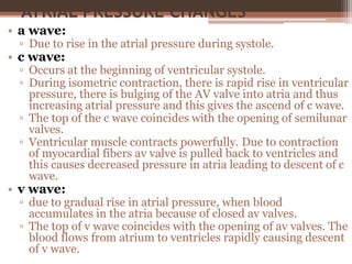
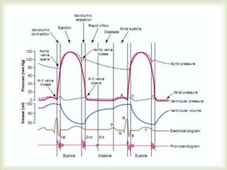
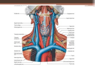
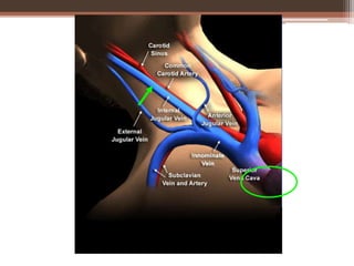
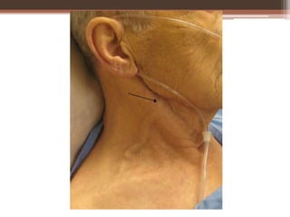
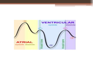
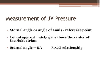
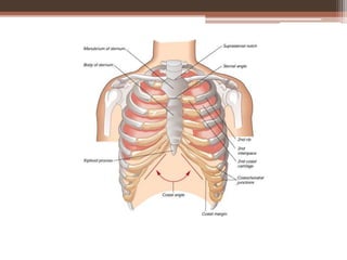
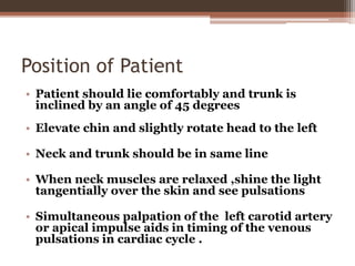
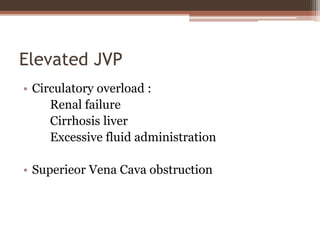
Atrial Pressure Changes
ATRIAL PRESSURE CHANGES
• Atria also undergoes systole and diastole.
• Duration of atrial systole is 0.11 sec where as atrial
diastole is 0.7 sec.
• Much prolonged atrial diastole which helps in filling.
• During each cardiac cycle, three waves are recorded
from atria i.e. a wave, c wave and v wave.
• a wave
• c wave
• v wave
ATRIAL PRESSURE CHANGES
• a wave:
▫ Due to rise in the atrial pressure during systole.
• c wave:
▫ Occurs at the beginning of ventricular systole.
▫ During isometric contraction, there is rapid rise in ventricular
pressure, there is bulging of the AV valve into atria and thus
increasing atrial pressure and this gives the ascend of c wave.
▫ The top of the c wave coincides with the opening of semilunar
valves.
▫ Ventricular muscle contracts powerfully. Due to contraction
of myocardial fibers av valve is pulled back to ventricles and
this causes decreased pressure in atria leading to descent of c
wave.
• v wave:
▫ due to gradual rise in atrial pressure, when blood
accumulates in the atria because of closed av valves.
▫ The top of v wave coincides with the opening of av valves. The
blood flows from atrium to ventricles rapidly causing descent
of v wave.
ATRIAL PRESSURE CHANGES
• In the right atrium, the pressure may rise upto 4 -5
mm of hg. And in left atrium the pressure may rise
upto 6-8 m of hg.
• Pulsations recorded from Right jugular vein is called
JVP.
• JVP is due to backward transmission of pressure
changes of right atrium to neck veins and so in JVP we
see same waves.
Jugular Venous Pulse (JVP)
Jugular venous pulse
These pressure changes can be recorded from
internal jugular vein which is directly connected to
atria.
Such a record is known as jugular venous pulse
(JVP)
Becomes apparent in
• Right Heart Failure
• Congestive cardiac failure
Causes of these waves:
‘a’ wave: RA contraction.
‘c’ wave: Bulging of TV into RA during
isovolumetric contraction phase.
 ‘x’ descent: Downward displacement of TV
during rapid ejection phase.
‘v’ wave:  RA press due to filling of atrium
with blood, (venous return.)
‘y’ descent: Rapid blood flow from RA to RV.
Measurement of JV Pressure
• Sternal angle or angle of Louis - reference point
• Found approximately 5 cm above the center of
the right atrium
• Sternal angle – RA Fixed relationship
Position of Patient
• Patient should lie comfortably and trunk is
inclined by an angle of 45 degrees
• Elevate chin and slightly rotate head to the left
• Neck and trunk should be in same line
• When neck muscles are relaxed ,shine the light
tangentially over the skin and see pulsations
• Simultaneous palpation of the left carotid artery
or apical impulse aids in timing of the venous
pulsations in cardiac cycle .
Measurement of JVP
• Two scale method is commonly used
• Normally JV pressure does not exceed 1-2 cm
above the sternal angle
• Elevated JVP : JVP of >4 cm above sternal angle .
Elevated JVP
• Increased RVP :
Pulmonary stenosis
Pulmonary hypertension
Right ventricular failure
RV infarction
• RV inflow impedance:
Tricuspid stenosis / atresia
RA myxoma
Constrictive pericarditis
Elevated JVP
• Circulatory overload :
Renal failure
Cirrhosis liver
Excessive fluid administration
• Superieor Vena Cava obstruction

Atrial pressure changes and Jugular Venous pressure

  • 1.
  • 3.
    ATRIAL PRESSURE CHANGES •Atria also undergoes systole and diastole. • Duration of atrial systole is 0.11 sec where as atrial diastole is 0.7 sec. • Much prolonged atrial diastole which helps in filling. • During each cardiac cycle, three waves are recorded from atria i.e. a wave, c wave and v wave. • a wave • c wave • v wave
  • 4.
    ATRIAL PRESSURE CHANGES •a wave: ▫ Due to rise in the atrial pressure during systole. • c wave: ▫ Occurs at the beginning of ventricular systole. ▫ During isometric contraction, there is rapid rise in ventricular pressure, there is bulging of the AV valve into atria and thus increasing atrial pressure and this gives the ascend of c wave. ▫ The top of the c wave coincides with the opening of semilunar valves. ▫ Ventricular muscle contracts powerfully. Due to contraction of myocardial fibers av valve is pulled back to ventricles and this causes decreased pressure in atria leading to descent of c wave. • v wave: ▫ due to gradual rise in atrial pressure, when blood accumulates in the atria because of closed av valves. ▫ The top of v wave coincides with the opening of av valves. The blood flows from atrium to ventricles rapidly causing descent of v wave.
  • 6.
    ATRIAL PRESSURE CHANGES •In the right atrium, the pressure may rise upto 4 -5 mm of hg. And in left atrium the pressure may rise upto 6-8 m of hg. • Pulsations recorded from Right jugular vein is called JVP. • JVP is due to backward transmission of pressure changes of right atrium to neck veins and so in JVP we see same waves.
  • 7.
  • 8.
    Jugular venous pulse Thesepressure changes can be recorded from internal jugular vein which is directly connected to atria. Such a record is known as jugular venous pulse (JVP) Becomes apparent in • Right Heart Failure • Congestive cardiac failure
  • 13.
    Causes of thesewaves: ‘a’ wave: RA contraction. ‘c’ wave: Bulging of TV into RA during isovolumetric contraction phase.  ‘x’ descent: Downward displacement of TV during rapid ejection phase. ‘v’ wave:  RA press due to filling of atrium with blood, (venous return.) ‘y’ descent: Rapid blood flow from RA to RV.
  • 14.
    Measurement of JVPressure • Sternal angle or angle of Louis - reference point • Found approximately 5 cm above the center of the right atrium • Sternal angle – RA Fixed relationship
  • 17.
    Position of Patient •Patient should lie comfortably and trunk is inclined by an angle of 45 degrees • Elevate chin and slightly rotate head to the left • Neck and trunk should be in same line • When neck muscles are relaxed ,shine the light tangentially over the skin and see pulsations • Simultaneous palpation of the left carotid artery or apical impulse aids in timing of the venous pulsations in cardiac cycle .
  • 19.
    Measurement of JVP •Two scale method is commonly used • Normally JV pressure does not exceed 1-2 cm above the sternal angle • Elevated JVP : JVP of >4 cm above sternal angle .
  • 22.
    Elevated JVP • IncreasedRVP : Pulmonary stenosis Pulmonary hypertension Right ventricular failure RV infarction • RV inflow impedance: Tricuspid stenosis / atresia RA myxoma Constrictive pericarditis
  • 23.
    Elevated JVP • Circulatoryoverload : Renal failure Cirrhosis liver Excessive fluid administration • Superieor Vena Cava obstruction