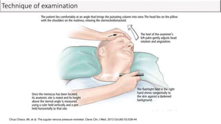
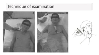
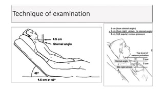
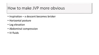
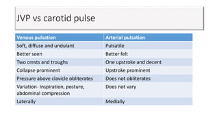
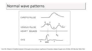
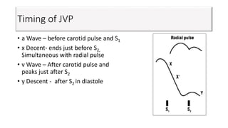
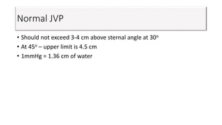
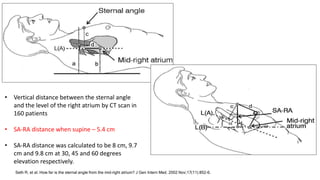
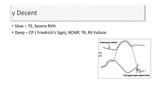
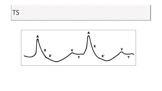
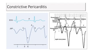
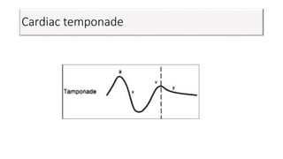
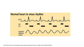
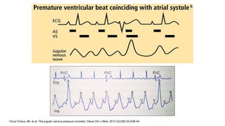
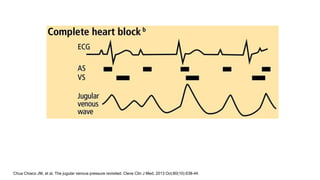
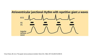
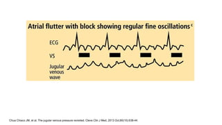
Jugular Venous Pressure
Dr. Lokesh Khandelwal
DM Cardiology
GB Pant Hospital, Delhi
JVP
• James Mackenzie first described JVP waves in 1870.
• Thomas Lewis described technique of bedside JVP assessment in
1930.*
*Montinari MR et al. The first 200 years of cardiac auscultation and future perspectives. J Multidiscip Healthc. 2019;12;183-9
Definition
• Oscillating top of column of blood in IJV.
• Reflects RA pressure at all stages of cardiac cycle.
• Reasonable estimate of CVP*
• Imparts prognostic information in patients with HF*
*Drazner MH, et al. Prognostic importance of elevated jugular venous pressure and a third heart sound in patients with heart
failure. N Engl J Med 2001; 345:574.
IJV or EJV
• IJV - In direct line with SVC and RA
• EJV has valves
• EJV passes through facial planes
• EJV not visible in increased sympathetic activity eg. Shock
Right IJV vs Left IJV
• Right IJV in direct line
• Compression of left innominate vein
• Left SVC draing into coronary sinus
Right IJV vs Left IJV
Technique of examination
Chua Chiaco JM, et al. The jugular venous pressure revisited. Cleve Clin J Med. 2013 Oct;80(10):638-44.
Technique of examination
How to make JVP more obvious
• Inspiration – x descent becomes brisker
• Horizontal posture
• Leg elevation
• Abdominal compression
• IV fluids
Effect of respiration
JVP vs carotid pulse
Venous pulsation Arterial pulsation
Soft, diffuse and undulant Pulsatile
Better seen Better felt
Two crests and troughs One upstroke and decent
Collapse prominent Upstroke prominent
Pressure above clavicle obliterates Does not obliterates
Variation- Inspiration, posture,
abdominal compression
Does not vary
Laterally Medially
JVP vs carotid pulse
Normal wave patterns
Conn RD, O'Keefe JH. Simplified evaluation of the jugular venous pressure: significance of inspiratory collapse of jugular veins. Mo Med. 2012 Mar-Apr;109(2):150-2.
Timing of JVP
• a Wave – before carotid pulse and S1
• x Decent- ends just before S2,
Simultaneous with radial pulse
• v Wave – After carotid pulse and
peaks just after S2
• y Descent - after S2 in diastole
Normal JVP
• Should not exceed 3-4 cm above sternal angle at 30o
• At 45o – upper limit is 4.5 cm
• 1mmHg = 1.36 cm of water
Abrams J. Prim Cardiol. 1982
Seth R, et al. How far is the sternal angle from the mid-right atrium? J Gen Intern Med. 2002 Nov;17(11):852-6.
• Vertical distance between the sternal angle
and the level of the right atrium by CT scan in
160 patients
• SA-RA distance when supine – 5.4 cm
• SA-RA distance was calculated to be 8 cm, 9.7
cm and 9.8 cm at 30, 45 and 60 degrees
elevation respectively.
Seth R, et al. How far is the sternal angle from the mid-right atrium? J Gen Intern Med. 2002 Nov;17(11):852-6.
Variation with body habitus
• 52 consecutive patients underwent CT.
• The Angle of Louis and mid RA distance was accessed.
• There was a positive correlation between patients' weight or BMI and
the distance of the Angle of Louis to right atrium.
• Using the traditional 5 cm in an obese patient usually will result in an
underestimation of RA pressure.
Ramana RK, Sanagala T, Lichtenberg R. A new angle on the Angle of Louis. Congest Heart Fail. 2006 Jul-Aug;12(4):196-9.
Constant J. The X prime descent in jugular contour nomenclature and recognition. Am Heart J. 1974 Sep;88(3):372-9.
Carotid artifact
Constant J. The X prime descent in jugular contour nomenclature and recognition. Am Heart J. 1974 Sep;88(3):372-9.
h Wave
Abdominojugular reflux
• Hepatojugular reflux – first described by
Pasteur in TR.*
• Reflux not reflex
• Duration of pressure- 10 to 15 sec
• Amount of pressure – 20 to 35 mmHg
• Rise of at least 3 cm for 15 seconds**
*Pasteur W. Note on a new physical sigh of tricuspid regurgitation. Lancet. 1885;2:524
**Am J Medicine. 2000:109:59
Abdominojugular reflux
Am J Medicine. 2000;109:59
Abdominojugular reflux
• Constriction
• Restriction
• RVMI
• Incipient RHF
• PCWP more than 15mmHg
• False positive
• COPD
• Increased blood volume
Wiese J. The abdominojugular reflux sign. Am J Med. 2000 Jul;109(1):59-61.
Alterations of ‘a’ wave
Giant a wave
Severe pulmonary hypertension
Severe PS
Bernheim’s effect
Tricuspid stenosis
Alterations of ‘a’ wave
Regular cannon ‘a’ waves Irregular cannon ‘a’ waves
Junctional rhythm CHB
PSVT APC
Extremely prolonged PR
interval
VPC
VT
Alterations of ‘a’ wave
• Absent a waves – AF, Ebstein’s anomaly.
x Decent
x Decent
• Absent – AF, severe TR
• Small – RA myxoma, RA hypertrophy secondary to TS, absent
pericardium
• Prominent - Large a waves present
Increased RV contraction – PS, PAH, ASD, CP and cardiac
temponade.
v Wave
• Prominent – TR (Lancisi’s sign), ASD, CP, post RA surgery, PAPVC,
TAPVC
y Decent
• Slow – TS, Severe RVH
• Deep – CP ( Friedrich’s Sign), RCMP, TR, RV Failure
Kussmaul’s sign
• Failure to fall or increase in JVP during inspiration.
• Causes- CP, RVMI, RCMP, Massive pulmonary embolism, TS, RA or RV
tumors
ASD
• Wide a wave
• Deep x decent
• Tall v wave ≥ a wave
• Deep y decent
TR
• Large v wave
• Obliteration of x decent
• Ventricularization of RA pressure in severe TR
TS
Constrictive Pericarditis
Cardiac temponade
Chua Chiaco JM, et al. The jugular venous pressure revisited. Cleve Clin J Med. 2013 Oct;80(10):638-44.
THANK YOU

Jugular venous pressure

  • 1.
    Jugular Venous Pressure Dr.Lokesh Khandelwal DM Cardiology GB Pant Hospital, Delhi
  • 2.
    JVP • James Mackenziefirst described JVP waves in 1870. • Thomas Lewis described technique of bedside JVP assessment in 1930.* *Montinari MR et al. The first 200 years of cardiac auscultation and future perspectives. J Multidiscip Healthc. 2019;12;183-9
  • 3.
    Definition • Oscillating topof column of blood in IJV. • Reflects RA pressure at all stages of cardiac cycle. • Reasonable estimate of CVP* • Imparts prognostic information in patients with HF* *Drazner MH, et al. Prognostic importance of elevated jugular venous pressure and a third heart sound in patients with heart failure. N Engl J Med 2001; 345:574.
  • 4.
    IJV or EJV •IJV - In direct line with SVC and RA • EJV has valves • EJV passes through facial planes • EJV not visible in increased sympathetic activity eg. Shock
  • 5.
    Right IJV vsLeft IJV • Right IJV in direct line • Compression of left innominate vein • Left SVC draing into coronary sinus
  • 6.
    Right IJV vsLeft IJV
  • 7.
    Technique of examination ChuaChiaco JM, et al. The jugular venous pressure revisited. Cleve Clin J Med. 2013 Oct;80(10):638-44.
  • 8.
  • 9.
  • 10.
    How to makeJVP more obvious • Inspiration – x descent becomes brisker • Horizontal posture • Leg elevation • Abdominal compression • IV fluids
  • 11.
  • 12.
    JVP vs carotidpulse Venous pulsation Arterial pulsation Soft, diffuse and undulant Pulsatile Better seen Better felt Two crests and troughs One upstroke and decent Collapse prominent Upstroke prominent Pressure above clavicle obliterates Does not obliterates Variation- Inspiration, posture, abdominal compression Does not vary Laterally Medially
  • 13.
  • 14.
    Normal wave patterns ConnRD, O'Keefe JH. Simplified evaluation of the jugular venous pressure: significance of inspiratory collapse of jugular veins. Mo Med. 2012 Mar-Apr;109(2):150-2.
  • 15.
    Timing of JVP •a Wave – before carotid pulse and S1 • x Decent- ends just before S2, Simultaneous with radial pulse • v Wave – After carotid pulse and peaks just after S2 • y Descent - after S2 in diastole
  • 16.
    Normal JVP • Shouldnot exceed 3-4 cm above sternal angle at 30o • At 45o – upper limit is 4.5 cm • 1mmHg = 1.36 cm of water
  • 17.
    Abrams J. PrimCardiol. 1982
  • 18.
    Seth R, etal. How far is the sternal angle from the mid-right atrium? J Gen Intern Med. 2002 Nov;17(11):852-6. • Vertical distance between the sternal angle and the level of the right atrium by CT scan in 160 patients • SA-RA distance when supine – 5.4 cm • SA-RA distance was calculated to be 8 cm, 9.7 cm and 9.8 cm at 30, 45 and 60 degrees elevation respectively.
  • 19.
    Seth R, etal. How far is the sternal angle from the mid-right atrium? J Gen Intern Med. 2002 Nov;17(11):852-6.
  • 20.
    Variation with bodyhabitus • 52 consecutive patients underwent CT. • The Angle of Louis and mid RA distance was accessed. • There was a positive correlation between patients' weight or BMI and the distance of the Angle of Louis to right atrium. • Using the traditional 5 cm in an obese patient usually will result in an underestimation of RA pressure. Ramana RK, Sanagala T, Lichtenberg R. A new angle on the Angle of Louis. Congest Heart Fail. 2006 Jul-Aug;12(4):196-9.
  • 21.
    Constant J. TheX prime descent in jugular contour nomenclature and recognition. Am Heart J. 1974 Sep;88(3):372-9.
  • 22.
    Carotid artifact Constant J.The X prime descent in jugular contour nomenclature and recognition. Am Heart J. 1974 Sep;88(3):372-9.
  • 23.
  • 24.
    Abdominojugular reflux • Hepatojugularreflux – first described by Pasteur in TR.* • Reflux not reflex • Duration of pressure- 10 to 15 sec • Amount of pressure – 20 to 35 mmHg • Rise of at least 3 cm for 15 seconds** *Pasteur W. Note on a new physical sigh of tricuspid regurgitation. Lancet. 1885;2:524 **Am J Medicine. 2000:109:59
  • 25.
    Abdominojugular reflux Am JMedicine. 2000;109:59
  • 26.
    Abdominojugular reflux • Constriction •Restriction • RVMI • Incipient RHF • PCWP more than 15mmHg • False positive • COPD • Increased blood volume Wiese J. The abdominojugular reflux sign. Am J Med. 2000 Jul;109(1):59-61.
  • 27.
    Alterations of ‘a’wave Giant a wave Severe pulmonary hypertension Severe PS Bernheim’s effect Tricuspid stenosis
  • 28.
    Alterations of ‘a’wave Regular cannon ‘a’ waves Irregular cannon ‘a’ waves Junctional rhythm CHB PSVT APC Extremely prolonged PR interval VPC VT
  • 29.
    Alterations of ‘a’wave • Absent a waves – AF, Ebstein’s anomaly.
  • 30.
  • 31.
    x Decent • Absent– AF, severe TR • Small – RA myxoma, RA hypertrophy secondary to TS, absent pericardium • Prominent - Large a waves present Increased RV contraction – PS, PAH, ASD, CP and cardiac temponade.
  • 32.
    v Wave • Prominent– TR (Lancisi’s sign), ASD, CP, post RA surgery, PAPVC, TAPVC
  • 33.
    y Decent • Slow– TS, Severe RVH • Deep – CP ( Friedrich’s Sign), RCMP, TR, RV Failure
  • 34.
    Kussmaul’s sign • Failureto fall or increase in JVP during inspiration. • Causes- CP, RVMI, RCMP, Massive pulmonary embolism, TS, RA or RV tumors
  • 35.
    ASD • Wide awave • Deep x decent • Tall v wave ≥ a wave • Deep y decent
  • 36.
    TR • Large vwave • Obliteration of x decent • Ventricularization of RA pressure in severe TR
  • 37.
  • 38.
  • 39.
  • 40.
    Chua Chiaco JM,et al. The jugular venous pressure revisited. Cleve Clin J Med. 2013 Oct;80(10):638-44.
  • 41.
    Chua Chiaco JM,et al. The jugular venous pressure revisited. Cleve Clin J Med. 2013 Oct;80(10):638-44.
  • 42.
    Chua Chiaco JM,et al. The jugular venous pressure revisited. Cleve Clin J Med. 2013 Oct;80(10):638-44.
  • 43.
    Chua Chiaco JM,et al. The jugular venous pressure revisited. Cleve Clin J Med. 2013 Oct;80(10):638-44.
  • 44.
    Chua Chiaco JM,et al. The jugular venous pressure revisited. Cleve Clin J Med. 2013 Oct;80(10):638-44.
  • 45.