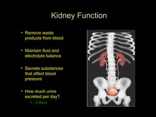
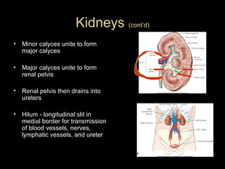
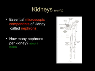
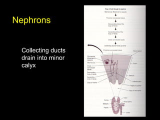
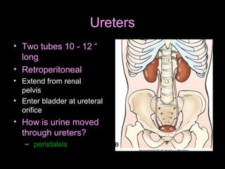
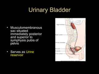
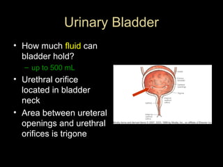
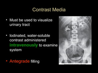
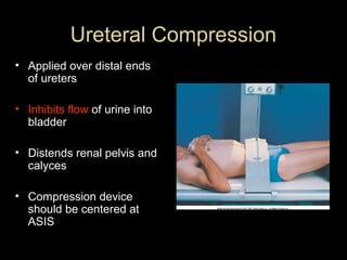
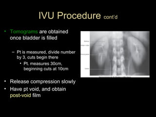
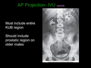
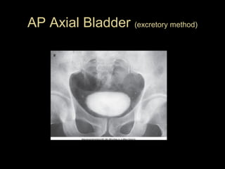
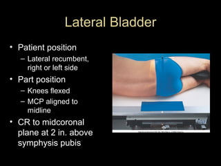
Urinary System
• Often called the excretory system
•
•
•
•

Two kidneys
Two ureters
One urinary bladder
One urethra
2 bean shaped bodies situated
behind peritoneum
Asymmetrical - left is slightly
longer and narrower than right
Why Rt kidney slightly lower
than Lt kidney?
Liver Lie in an oblique plane
Normally extend from T-12 to L3

Kidneys
Kidney Function
• Remove waste
products from blood
• Maintain fluid and
electrolyte balance
• Secrete substances
that affect blood
pressure
• How much urine
excreted per day?
1 - 2 liters
Kidneys (cont’d)
•

Minor calyces unite to form
major calyces

•

Major calyces unite to form
renal pelvis

•

Renal pelvis then drains into
ureters

•

Hilum - longitudinal slit in
medial border for transmission
of blood vessels, nerves,
lymphatic vessels, and ureter
Kidneys (cont’d)
• Essential microscopic
components of kidney
called nephrons
• How many nephrons
per kidney? about 1
million
Nephrons
Collecting ducts
drain into minor
calyx
Adrenal Glands
Cannot be seen on plain

radiographs

Not part of urinary system
Chiefly responsible for
regulating stress
response through
adrenaline etc
Ureters
• Two tubes 10 - 12 “
long
• Retroperitoneal
• Extend from renal
pelvis
• Enter bladder at ureteral
orifice

• How is urine moved
through ureters?
– peristalsis
Urinary Bladder
• Musculomembranous
sac situated
immediately posterior
and superior to
symphysis pubis of
pelvis
• Serves as Urine
reservoir
Urinary Bladder
• How much fluid can
bladder hold?
– up to 500 mL

• Urethral orifice
located in bladder
neck
• Area between ureteral
openings and urethral
orifices is trigone
Urethra
•
•
•
•
•
•
•
•

Carries urine from bladder to?
exterior of body
How long is it in females?
About 1.5′′
In males?
About 7′′ to 8′′
Sphincter at neck of bladder
controls flow
Male urethra contains following
parts:
– Prostate
– Membranous area
– Spongy area
Prostate
• Gland surrounding
proximal part of male
urethra
• Considered part of male
reproductive system, but
due to location, often
described with urinary
system
• Prostate secretes fluid
that mixes with seminal
fluid to create ejaculate
Radiography of Urinary System

aka

Urography
Radiographic investigation of renal drainage or
collecting system
IVU- Intravenous Urogram !
Formerly known as IVP-Intravenous
pyelogram!
– pyelo refers to renal pelvis and calyces only
– study also shows ureters, bladder, and
sometimes urethra
Indications For Urography
• Demonstrate physiologic function and structure
of urinary system
• Evaluate abd. Masses, renal cysts and tumors
• Stones
• Pyelonephritis (Inflammation of kidney)
• Hydronephrosis (distension of renal pelvis and calyces with urine)
• Effects of trauma
• Pre-op evaluation
• Renal hypertension
Contraindications
• Inability to filter contrast medium from
blood
• Allergy to contrast
• Abnormal BUN and Creatinine levels
Preparation Of Pt
• Pt should follow low residue diet for 1-2 days
prior to exam
• laxative taken day before
• NPO after midnight
• Pts with multiple myeloma, high uric acid levels,
or diabetes should be well hydrated before IVP
exam
– Dehydration leads to increased risk of renal
failure
Patient preparation:
•

Bowel is purged with strong laxative
and gas-absorbent tabs.
• Patient should take nothing by mouth
after midnight on the day of
examination.
Contrast Media
• Must be used to visualize
urinary tract
• Iodinated, water-soluble
contrast administered
intravenously to examine
system

• Antegrade filling
Contrast Media
• Excretory urography (IVU) generally uses a 50 to
70% iodine solution
• Lower concentrations for bladder studies due to
large amount required to fill bladder (30%)
• Non-ionic contrast is generally used
– More expensive, but– Patients less likely to have reactions with nonionic
Contrast Media and Adverse Reactions
• Crucial not to leave pt alone for first 5 minutes after
injection!

• Mild reactions
– warmth
– flushing
– hives, Nausea/Vomiting, respiratory edema
(accumulation of fluid in lungs)

• Severe reactions
– Anaphylactic shock

(sudden allergic response associated with a

sudden drop in blood pressure and difficulty breathing). Can lead to death in a
matter of minutes)
Injection Procedure
•
•
•
•

Obtain allergy history
Explain exam to pt
Prepare contrast and supplies (sterile tech.)
Assist radiologist as necessary
– or

• Perform injection if IVcertified
Injection Supplies (cont.d)
•
•
•
•
•
•
•

Tourniquet
IV arm board
Towels
Emergency kit
Alcohol wipes, swabs
Contrast
19-22 G needle, butterfly or angiocath for
infusion
• Extension tubing
• Tape or clear-type dressing
IVU Procedure
• Scout – KUB
• Contrast is injected
• Timed sequence of films obtained until bladder
begins to fill– Immediate image of kidneys
– 5 minute image of abd. or kidneys
– Compression applied
Ureteral Compression
• Applied over distal ends
of ureters
• Inhibits flow of urine into
bladder
• Distends renal pelvis and
calyces
• Compression device
should be centered at
ASIS
Ureteral Compression
• As much compression as
pt can tolerate!
• Should not be applied
when:
– stones, abd. mass or
aneurysm, colostomy,
suprapubic catheter, recent
abd. surgery or trauma
•

(Because of improvement of contrast
agents, compression no longer
generally used)

(cont’d)
IVU Procedure cont’d
• Tomograms are obtained
once bladder is filled
– Pt is measured, divide number
by 3, cuts begin there
• Pt. measures 30cm,
beginning cuts at 10cm

• Release compression slowly
• Have pt void, and obtain
post-void film
Radiation Protection
• Radiographer is responsible!
• Gonadal shield - if it does not interfere
with examination objective
• Close collimation
• Avoid repeat exposures
• Shield males for all urinary studies, except
when urethra is of primary interest
Radiation Protection
• Shield females when IR centered over
kidneys
• Rule out chance of pregnancy before
examination
(Emergency cases may not allow time)
Radiographic Positions IVU
AP Projection-IVU
• KUB
• (All exposures at end
of expiration for any
urinary system study)
AP Projection- IVU (cont’d)

Must include entire
KUB region
Should include
prostatic region on
older males
AP Projection Variations
• Trendelenberg
– Lower head 15 - 20 degrees
– Helps demonstrate lower ureters

• Upright
– Center lower - organs change position

• Prone
– Demonstrates ureteropelvic region
– Fills obstructed ureter in cases of hydronephrosis
(distension of renal pelvis and calyces with urine)
AP Oblique Projections - RPO/LPO
• Patient is supine
• Patient rotated to
30 degrees
• CR to iliac crest, 2
in. lateral to
midline
– Center to side up
AP Oblique Projections - RPO/LPO
• Elevated
kidney will be
parallel to
cassette
• Kidney
closest to
cassette will
be
perpendicular
• Entire KUB
region must
be included
AP Axial Bladder
• CR( similar to coccyx projection)
– Angled 10 to 15
degrees caudad to
center of IR
– Enters 2′′ above
upper border of
pubic symphysis
AP Axial Bladder (excretory method)
AP Oblique Bladder
• Pt position
– 40- to 60-degree
– RPO or LPO
depending on
physician
preference
AP Oblique Bladder
CR
– Perpendicular to center of
IR
– CR 2′′ above upper border
of pubic symphysis and 2′′
medial to upper ASIS
– If bladder neck and
proximal urethra is of
interest, 10-degree caudal
angle of CR will project
pubic bones below them
Lateral Bladder
• Patient position
– Lateral recumbent,
right or left side

• Part position
– Knees flexed
– MCP aligned to
midline

• CR to midcoronal
plane at 2 in. above
symphysis pubis
Lateral Bladder
– Demonstrates
anterior/posterior
bladder walls
– Base of bladder
– Any vesicovaginal
or vesicorectal
fistulae

Ivu

  • 1.
    Urinary System • Oftencalled the excretory system • • • • Two kidneys Two ureters One urinary bladder One urethra
  • 2.
    2 bean shapedbodies situated behind peritoneum Asymmetrical - left is slightly longer and narrower than right Why Rt kidney slightly lower than Lt kidney? Liver Lie in an oblique plane Normally extend from T-12 to L3 Kidneys
  • 3.
    Kidney Function • Removewaste products from blood • Maintain fluid and electrolyte balance • Secrete substances that affect blood pressure • How much urine excreted per day? 1 - 2 liters
  • 4.
    Kidneys (cont’d) • Minor calycesunite to form major calyces • Major calyces unite to form renal pelvis • Renal pelvis then drains into ureters • Hilum - longitudinal slit in medial border for transmission of blood vessels, nerves, lymphatic vessels, and ureter
  • 5.
    Kidneys (cont’d) • Essentialmicroscopic components of kidney called nephrons • How many nephrons per kidney? about 1 million
  • 6.
  • 7.
    Adrenal Glands Cannot beseen on plain radiographs Not part of urinary system Chiefly responsible for regulating stress response through adrenaline etc
  • 8.
    Ureters • Two tubes10 - 12 “ long • Retroperitoneal • Extend from renal pelvis • Enter bladder at ureteral orifice • How is urine moved through ureters? – peristalsis
  • 9.
    Urinary Bladder • Musculomembranous sacsituated immediately posterior and superior to symphysis pubis of pelvis • Serves as Urine reservoir
  • 10.
    Urinary Bladder • Howmuch fluid can bladder hold? – up to 500 mL • Urethral orifice located in bladder neck • Area between ureteral openings and urethral orifices is trigone
  • 11.
    Urethra • • • • • • • • Carries urine frombladder to? exterior of body How long is it in females? About 1.5′′ In males? About 7′′ to 8′′ Sphincter at neck of bladder controls flow Male urethra contains following parts: – Prostate – Membranous area – Spongy area
  • 12.
    Prostate • Gland surrounding proximalpart of male urethra • Considered part of male reproductive system, but due to location, often described with urinary system • Prostate secretes fluid that mixes with seminal fluid to create ejaculate
  • 13.
    Radiography of UrinarySystem aka Urography Radiographic investigation of renal drainage or collecting system
  • 14.
    IVU- Intravenous Urogram! Formerly known as IVP-Intravenous pyelogram! – pyelo refers to renal pelvis and calyces only – study also shows ureters, bladder, and sometimes urethra
  • 15.
    Indications For Urography •Demonstrate physiologic function and structure of urinary system • Evaluate abd. Masses, renal cysts and tumors • Stones • Pyelonephritis (Inflammation of kidney) • Hydronephrosis (distension of renal pelvis and calyces with urine) • Effects of trauma • Pre-op evaluation • Renal hypertension
  • 16.
    Contraindications • Inability tofilter contrast medium from blood • Allergy to contrast • Abnormal BUN and Creatinine levels
  • 17.
    Preparation Of Pt •Pt should follow low residue diet for 1-2 days prior to exam • laxative taken day before • NPO after midnight • Pts with multiple myeloma, high uric acid levels, or diabetes should be well hydrated before IVP exam – Dehydration leads to increased risk of renal failure
  • 18.
    Patient preparation: • Bowel ispurged with strong laxative and gas-absorbent tabs. • Patient should take nothing by mouth after midnight on the day of examination.
  • 19.
    Contrast Media • Mustbe used to visualize urinary tract • Iodinated, water-soluble contrast administered intravenously to examine system • Antegrade filling
  • 20.
    Contrast Media • Excretoryurography (IVU) generally uses a 50 to 70% iodine solution • Lower concentrations for bladder studies due to large amount required to fill bladder (30%) • Non-ionic contrast is generally used – More expensive, but– Patients less likely to have reactions with nonionic
  • 21.
    Contrast Media andAdverse Reactions • Crucial not to leave pt alone for first 5 minutes after injection! • Mild reactions – warmth – flushing – hives, Nausea/Vomiting, respiratory edema (accumulation of fluid in lungs) • Severe reactions – Anaphylactic shock (sudden allergic response associated with a sudden drop in blood pressure and difficulty breathing). Can lead to death in a matter of minutes)
  • 22.
    Injection Procedure • • • • Obtain allergyhistory Explain exam to pt Prepare contrast and supplies (sterile tech.) Assist radiologist as necessary – or • Perform injection if IVcertified
  • 23.
    Injection Supplies (cont.d) • • • • • • • Tourniquet IVarm board Towels Emergency kit Alcohol wipes, swabs Contrast 19-22 G needle, butterfly or angiocath for infusion • Extension tubing • Tape or clear-type dressing
  • 24.
    IVU Procedure • Scout– KUB • Contrast is injected • Timed sequence of films obtained until bladder begins to fill– Immediate image of kidneys – 5 minute image of abd. or kidneys – Compression applied
  • 25.
    Ureteral Compression • Appliedover distal ends of ureters • Inhibits flow of urine into bladder • Distends renal pelvis and calyces • Compression device should be centered at ASIS
  • 26.
    Ureteral Compression • Asmuch compression as pt can tolerate! • Should not be applied when: – stones, abd. mass or aneurysm, colostomy, suprapubic catheter, recent abd. surgery or trauma • (Because of improvement of contrast agents, compression no longer generally used) (cont’d)
  • 27.
    IVU Procedure cont’d •Tomograms are obtained once bladder is filled – Pt is measured, divide number by 3, cuts begin there • Pt. measures 30cm, beginning cuts at 10cm • Release compression slowly • Have pt void, and obtain post-void film
  • 28.
    Radiation Protection • Radiographeris responsible! • Gonadal shield - if it does not interfere with examination objective • Close collimation • Avoid repeat exposures • Shield males for all urinary studies, except when urethra is of primary interest
  • 29.
    Radiation Protection • Shieldfemales when IR centered over kidneys • Rule out chance of pregnancy before examination (Emergency cases may not allow time)
  • 30.
  • 31.
    AP Projection-IVU • KUB •(All exposures at end of expiration for any urinary system study)
  • 32.
    AP Projection- IVU(cont’d) Must include entire KUB region Should include prostatic region on older males
  • 33.
    AP Projection Variations •Trendelenberg – Lower head 15 - 20 degrees – Helps demonstrate lower ureters • Upright – Center lower - organs change position • Prone – Demonstrates ureteropelvic region – Fills obstructed ureter in cases of hydronephrosis (distension of renal pelvis and calyces with urine)
  • 34.
    AP Oblique Projections- RPO/LPO • Patient is supine • Patient rotated to 30 degrees • CR to iliac crest, 2 in. lateral to midline – Center to side up
  • 35.
    AP Oblique Projections- RPO/LPO • Elevated kidney will be parallel to cassette • Kidney closest to cassette will be perpendicular • Entire KUB region must be included
  • 36.
    AP Axial Bladder •CR( similar to coccyx projection) – Angled 10 to 15 degrees caudad to center of IR – Enters 2′′ above upper border of pubic symphysis
  • 37.
    AP Axial Bladder(excretory method)
  • 38.
    AP Oblique Bladder •Pt position – 40- to 60-degree – RPO or LPO depending on physician preference
  • 39.
    AP Oblique Bladder CR –Perpendicular to center of IR – CR 2′′ above upper border of pubic symphysis and 2′′ medial to upper ASIS – If bladder neck and proximal urethra is of interest, 10-degree caudal angle of CR will project pubic bones below them
  • 40.
    Lateral Bladder • Patientposition – Lateral recumbent, right or left side • Part position – Knees flexed – MCP aligned to midline • CR to midcoronal plane at 2 in. above symphysis pubis
  • 41.
    Lateral Bladder – Demonstrates anterior/posterior bladderwalls – Base of bladder – Any vesicovaginal or vesicorectal fistulae