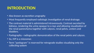
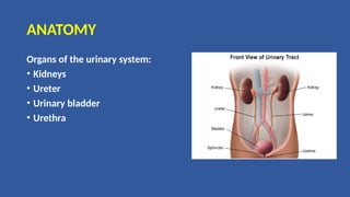
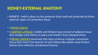
RADIOGRAPHIC PROCEDURE :IVU
PRESENTED BY:PRATIVA KHANAL
BSC.MIT 2ND
YEAR
NMCTH
INTRODUCTION
• Also known as excretion urography
• Most frequently employed radiologic investigation of renal drainage.
• The contrast material is administered intravenously. Contrast excreted by
kidneys, rendering the urine opaque to x-rays and allowing visualization of
the renal parenchyma together with calyces, renal pelvis, ureters and
bladders.
• Pyelography - radiographic demonstration of the renal pelvis and calyces.
• So, IVP is a misnomer.
• Term “pyelogram” is reserved for retrograde studies visualizing only the
collecting system
ANATOMY
Organs of the urinary system:
• Kidneys
• Ureter
• Urinary bladder
• Urethra
KIDNEY-EXTERNAL ANATOMY
• KIDNEYS –held in place on the posterior body wall and protected by three
external layers of connective tissue
• 1.RENAL FASCIA
• 2.ADIPOSE CAPSULE: middle and thickest layer,consist of adipose tissue
that wedges each kidney in place and shield it from physical shock
• 3.RENAL CAPSULE:extremely thin layer of dense irregular connective
tissue that covers the exterior of each kidney like plastic wrap,Protect
kidney from infection and physical trauma
Kidney: Internal structure
• The parenchyma of kidney is divided into two major parts: superficial
is the renal cortex and deep is the renal medulla
• Grossly, these structures take the shape of 8 to 18 cone shaped renal
lobes, each containing renal cortex surrounding a portion of medulla
called the renal pyramid(of Malpighi)
• Between the renal pyramids are the projections of cortex called renal
cortex of Bertini
• Nephrons, the urine producing functional structures of the kidney,
span the cortex and medulla
Kidney: Internal structure
• The tip, or papilla, of each pyramid
empties urine into minor calyx
• Minor calyces empty into major
calyces, and major calyces empty into
the renal pelvis, which becomes the
ureter
• Minor calyces:8-10 in number. Multiple
papillae drain into single calyx
• Major calyces:2-4 or more connects
minor calyces to renal pelvis
Position of kidney
• Located in lumbar fossa
• Left kidney is slightly higher than the right
• Upper pole of left kidney is at the level of T11-T12
• Upper pole of right kidney is at the level of T12-L1
• Lower limit of kidney is way above iliac crest, at the
level of L3 or L3-l4 IV disc.
• Medial border is parallel to psoas margin
• The psoas muscles cause the longitudinal plane of the
kidney to form a vertical angle of about 20 degree with
the mid saggital plane
ureter
• A pair of thick wall cylindrical tubes
• Convey urine from corresponding kidneys to U.B
• 25-30 cm long and has three parts:
• Abdominal ureter: from the renal pelvis to the
pelvic brim
• Pelvic ureter: from the pelvic brim to the
bladder
• Intravesical or intramural ureter: within the
bladder wall
Course
• The ureter begins its descent to the bladder by
running along the medial aspect of the psoas
muscle. Here, the ureter lies anteriorly and slightly
medial to the tips of the L2-L5 transverse processes
• It enters the pelvis anteriorly to the sacroiliac joint
at the bifurcation of the common iliac vessels (at
the pelvic brim) and then courses anteriorly to
the internal iliac artery down the lateral pelvic
sidewall
• At the level of the ischial spine it turns forward and
medially to enter the posterolateral wall of the
bladder, where it runs an oblique 1-2 cm course,
before opening into the bladder at the internal
ureteric orifice
NORMAL CONSTRICTIONS
Along their course the ureters are constricted
• At the ureteropelvic junction ,just inferior to the
kidney
• Point where the ureters cross the common iliac
vessels at the pelvic brim
• Crossing of ureter by ductus deferens in male
and broad ligament of uterus in female
• Where ureters enter the wall of the
bladder(narrow one)
• Opening at the lateral angle of trigone of bladder
URINARY BLADDER
• Muscular sac
• Has ability to expand and collapse
• Stores and expels urine
POSITION:
• Full bladder-spherical(Expands into the abdominal cavity)
• Empty bladder-lies entirely within the pelvis
SHAPE:
• Tetrahedral-when empty
• Ovoid-when distended
CAPACITY
• Average-120 to 320ml urine
• Loss of voluntary control over
micturition-at 800ml urine
PARTS
• Body:major part,collects urine
• Neck:funnel shaped extension of
the body connecting with urethra
PHYSIOLOGY
• For the delineation and differentiation of cysts and tumor masses within the
kidney , the renal parenchyma is opacified by the iv introduction of contrast
media.
• Once the contrast media enters the blood stream,it is conveyed to the renal
glomeruli and is discharged into the capsules with glomerular filtrate,which
is excreted as urine.
• With the reabsorption of water the contrast media becomes sufficiently
concentrated to render the urinary canals opaque
• In recent years, there has been a decline in the intravenous urogram
because of:
• Development of newer imaging modalities like CT Scan, USG, MRI
• Adverse effects of contrast media.
• Cost containment.
INDICATIONS
In Adults
• Suspected urinary tract pathology
• Investigation of persistent or frank hematuria
• Renal /ureteric calculi (prior to endourological procedure)
• complex urinary tract infection (including Renal TB)
• Ureteric fistulas and strictures
• Suspected transitional cell carcinoma
• Obstructive uropathy
In Children:
• Evaluation of VATER anomalies- 90% has Renal anomalies
• Malformation of genitalia –hypospadiasis
• Enuresis
• Constant or intermittent dampness in girls to rule out ectopically
inserted ureter
• Anorectal anomalies
CONTRAINDICATIONS
Absolute CIs:
• Past h/o severe adverse reaction to contrast media.
• HOCM carries 20% risk and LOCM decrease risk to 5% , and in those cases
radioisotope scan , USG, CT,
• MRI provide alternative means of investigations proven hypersensitivity
to iodine.
Relative CIs: (@ABCD MS)
• Asthma /significant allergic history
• B-blockers
• Chronic Renal insufficiency
• Cardiac disease –Cardiac failure /arrhythmias may be precipitated and in
these cases lower risk with LOCM
• Diabetes Mellitus
• Dehydration
• Multiple Myeloma
• Metformin therapy: Co-administration of metformin (glucophage)+ iv
contrast to diabetics may lead to acute alteration of renal function and lactic
acidosis, therefore metformin is withheld
• Sickle cell anemia
• Thyrotoxicosis
• Pregnancy
• A contrast material is excreted by a similar mechanism to creatinine, a
serum creatinine level above 200micromol/l would indicate a patient
who would unlikely to excrete contrast satisfactorily.
• So, cautions in diabetics and patients with severe disturbances of
liver and kidneys
Procedure: Equipments
• 600 mA fluoroscopy guided x-ray unit
• Abdominal compression equipment
• Medium/Regular film screen combination in variety of sizes.
• Pads and immobilization aids
• Intravenous administration equipment:50ml syringe, filling needle, skin
prep, sticky tape
• Selection of needles, venflon 19 gauge
• Torniquet or blood pressure cuff
• Emergency drugs and equipments
Contrast medium and injection data
• Contrast medium and injection data
• Ionic and non-ionic are available, both of which are excreted by
different mechanisms. The ionic group is excreted mainly by
glomerular filtration causing a peak concentration of iodine in the
renal cortex faster compared to nonionic which is mainly excreted by
proximal tubules
• The timing for first radiograph to demonstrate parenchymal phase
best will thus differ
• HOCM or LOCM 370 are acceptable but the following “high risk”
group should receive LOCM.
• Infants/small children/elderly
• Poorly hydrated patients
• Those with renal /cardiac failure
• Patients with diabetes, myelomatosis, sickle cell disease
• Patients with previous contrast medium reactions/ strong allergic
history
Adults Children
Non ionic contrast media( Iohexol-
omniopque)
300 mg I/ml-40-80 ml or
350 mg I/ml 40-80ml
240 mg I/ml 300mg I/ml
< 7 kg :4 ml/kg 3 ml/kg
> 7 Kg :3 ml/kg 2 ml/kg
Ionic contrast media 300 to 600 mg Iodine
equivalent/kg body weight.
Maximum of 40 gm of Iodine.
Meglumine iothalamate or
diatrizoate 60% containing
equivalent of 280 mg I/ I
ml of iodine. Dose is 1-2 ml /kg
body weight
Below 6 months: 10ml
6 months to 2 years:20ml
2-10 years:20-40ml
PATENT PREPARATION
• Bowel preparation is important as abdomen should ideally be free of
radio-opaque fecal matter and gas
• NPO (No food for 4-6 hr prior to examination)
• Should have plenty of oral fluids
• Laxatives- Dulcolax 2-4 tabs at bed time for 2 days prior to procedure
• Bowel preparation is now generally regarded as unhelpful and it is
unpleasant to the patient
IS FLUID DEPRIVATION INDICATEED ?
• Traditionally fluid was restricted prior to IVU in order to improve
opacification of collecting system
• However, dehydration increase risk of nephrotoxicity which may be
permanent in patients with DM, Multiple Myeloma, Hyperuricemia,
Sickle Cell Disease and pre-existing renal disease
• Risk of irreversible renal damage to renal function in previously
healthy kidney due to contrast agent is very low
• Also, with the advent of modern non-ionic contrast agents which do
not provoke an osmotic diuresis, degree of opacification is unlikely to
be significantly altered by dehydration.
• So, fluid restriction should be avoided and if there is a risk that the
patient is dehydrated before the IVU, this should be corrected first.
SIGNING INFORMED CONSENT FORM
• Venipuncture is an invasive procedure that carries risks for
complications, especially when contrast media is injected. Before
beginning the procedure, the technologist must ensure that the
patient is fully aware of these potential risks and has signed an
informed consent form.
• If a child is undergoing venipuncture, the procedure should be
explained to both the child and the guardian, and the guardian should
sign the informed consent form.
RADIATION PROTECTION
• “Pregnancy rule” should be applied
• If whole of renal tract is to be visualized, no gonad shielding
is possible for the females, but for males the testis can be
protected by placing a lead rubber sheet over upper thighs
below lower edge of symphysis pubis
• When bladder and lower ureters are not included then
female can also be given gonad protection
•Most adverse reactions are likely to occur within few
minutes after injection. So, Emergency drugs (eg.
Adrenaline), Oxygen and Resuscitation equipments
should also be readily available
•Doctor (radiologist) should be available in the
department
Mode of injection
• Contrast media is usually given as a IV bolus injection within 30-
60
seconds
• The density of the nephrogram is directly proportional to
the plasma concentration of contrast media
• More iodine increases the density of the nephrogram
• Large doses of contrast media increase diuresis which distends
the collecting system thus increasing the diagnostic information
from the urogram
PROCEDURE
• Patient should be placed in supine position with pelvis at cathode side of the
tube
• A support should be placed under patient's knees to reduce lordotic curvature
of lumbosacral spine and provide comfort
• A scout film should be taken including the kidneys, ureters, bladder and urethral
regions on a large size film
• Contrast media should be injected intravenously into a prominent vein in the
arm
• Test injection of 1ml of contrast should be given and patient should
observed for 1 min to look for any contrast reactions
• Then the rest of the contrast should rapidly injected within 30-60 seconds
Classic series of plain flims
• Preliminary/Control film
• Immediate film- Nephrogram
• 5-min film
• 15-min compression film
• 15-min release film
• Post-micturition film
Stereotypical appearance of normal IVU
• Takes 15-20 seconds for contrast to reach renal arteries following iv
injection
• At this stage, its concentration is maximum in the vascular
compartment
• This falls rapidly as contrast medium begins to escape into the
extracellular compartment and undergoes rapid glomerular filtration
and enters renal tubules.
• In first minute of IVU, healthy kidneys (assuming a normal
cardiovascular system) shows diffuse enhancement-Nephrogram
• During this phase renal size and outline are seen.
• In roughly first half minute-contrast in the vascular compartment
dominated and cortex is much enhanced than the medulla.
• The differentiation is sometimes visible in the immediate film of IVU
seris( but regularly visible on the CT performed at this stage)
• In second half minute- contrast in the tubule increases and
enhancement of the kidneys in more diffuse.
• At 1 minute: Contrast begins to appear in the calyces
• After 1 minute: Contrast in the normal calyces begins to drain
immediately into the pelvis and ureter- Pyelogram
• On the release of the compression, there is transient increase in flow
down the ureter and release film offers the best chance of
demonstrating the ureters.
• Normal ureters exhibit peristalsis and on single film it is uncommon to
demonstrate the entire length of both( or even either) ureter.
Preliminary/Control film
• Plain film to demonstrate the urinary
tract prior to the administration of
contrast media
• kVp=70-80(low kvp), mAs= 60-70
• Supine full length AP view of the
abdomen on inspiration
• It provides valuable information and
sometimes indicates probable
diagnosis.
NEED OF CONTROL FILM
• To check exposure factor and centering
• To check state of bowel preparation
• Obvious pre-existing pathology-urinary tract calculi/calcification
IMMEDIATE FILM/NEPHROGRAM
• Immediate film shows nephrogram
• Exposed 10-14 seconds after contrast
injection
• Renal parenchymal is opacified by contrast
media in renal tubules
• Aim is to see renal outlines
5- minute film
• 5 minute film shows nephrogram, renal pelvis, upper part of ureter
• Compression band is now applied on patient's abdomen and the
balloon is positioned on anterior superior iliac spine where ureters
cross pelvic brim. This is to produce better pelvicalyceal distension
• Compression inhibits ureteric drainage and promotes distension of
pelvicalyceal system, optimizing their visualization
5-min film
BEFORE COMPRESSION AFTER COMPRESSION
Compression is contraindicated in
• Renal trauma
• Large abdominal mass
• Abdominal aneurysm
• After abdominal surgery
• If 5 minute film already shows dilated calyces or if calyces and pelvis
are not adequately opacified, obstruction exists and compression
band should not be applied.
• If compression is applied, a film is taken after 5 minutes of
compression i.e. 10 minute film, centred on kidneys to demonstrate
distended collecting system and proximal ureters
15-min Compression film
• AP view of renal areas
• Adequate distension of pelvicalyceal system
with opaque urine
• Compression is removed when satisfactory
demonstration of pelvicalyceal system has
been achieved
15-min release film
• Supine AP film
• To show whole urinary system
Post Micturition film
• Based on the clinical findings and
radiological findings of earlier films , either a
full length abdominal film or a coned view
of the bladder with tube angled 15 degree
caudad and centered 5 cm above pubic
symphysis
• It is used to assess for residual urine,
bladder mucosal lesions, diverticula,
bladder tumor, outlet obstruction and
vesiculoureteric reflux
Main aim of films is to
• Assess bladder emptying
• To demonstrate return of dilated upper tracts with relief of bladder
pressure
• Aid diagnosis of VUJ calculi
• Diagnosis of bladder tumors
• Demonstrate urethral diverticulum
Special views in IVU
• Oblique view: To project the ureter away from spine and to separate
overlying radio opaque shadows mimicking calculi. Oblique
views are also used for visualisation of posterolateral aspects of
bladder; differentiation of extrinsic or intrinsic renal, ureteral or
bladder masses and for doubtful urethral masses.
• Erect film is used to
• Provoke emptying of urinary tract;
• Demonstrate layering of calculi in cysts and abscesses;
• Detect urinary tract gas not seen in other films;
• Have optimum demonstration of renal ptosis, bladder hernia,
cystocele and areas of obstruction in ureter
• Prone film is used for
• Viewing of ureteral areas not seen in supine films,
• Demonstration of renal ptosis and bladder hernia
• Delayed films in IVU are taken 1-24 hours after injection. Patients should
always be instructed to void prior to delayed films so that a calculus in the
distal ureter is seen well.
Usual sequence of delayed films is after 1 hr, 3hrs, 6 hrs, 12 hrs and 24 hrs
Delayed films are used in
• Cases of obstruction where early nephrogram is seen but collecting system is
not seen.
• Long standing hydronephrosis in which renal parenchyma is seen but
collecting system is not visualised until many hours later.
• Congenital lesions like non-visualised upper calyceal system with ectopic or
obstructed ureter
Modifications of Urogram
Diuretic urogram:
• It is useful when intermittent obstruction is suspected but
cannot be confirmed by standard urogram. Therefore the use
of diuretics shows an acutely developing hydronephrosis if true
intermittent hydronephrosis is present.
• I.V. frusemide is used to induce diuresis which distends the
renal pelvis. The dose of Lasix is 0.3-1 mg/kg in adults and
0.5 mg/kg in children.
• The film is taken 5-10 minutes after administering the diuretic.
Modifications of Urogram
• Tailored Urogram: It modifies the urogram to provide the information
needed to include or exclude the clinical problem and tailor the
urogram for that. The study is terminated as soon as the desired
information is available.
• Hypertensive Urogram:It is also called minute sequence urogram.
Films are taken 1, 2, 3, 5 minutes after injection of contrast media.
Although the findings are of value, IVU cannot be used for screening
of hypertensives as there are many false positive and false negative
results.
Modifications of Urogram
• Drip infusion urography: Contrast is given in 500 ml normal saline. Now,
this procedure is not widely used.
ADVANTAGES:
Nephrogram persists for longer time.
Enhanced diuresis from the additional contrast media and water
volume will distend the collecting system and ureters more fully.
 Collecting system is visualised for longer times.
No significant increase in contrast reactions.
Ureteral compression need not be used because excellent ureteral
visualization is usually obtained.
Administration is easy
DISADVANTAGES:
• Overloads the patient with more Iodine than necessary.
• Calyceal blunting may be produced, suggesting abnormal dilatation.
• May lead to pyelosinus extravasation and pain in patients with
partial obstruction.
• Increased diuresis may decrease visualization if there is low fixed
specific gravity.
• May cause CCF in patients with borderline cardiac complaints.
• An initial vascular nephrogram is not obtained.
Modifications of Urogram
Limited urography: The procedure is useful for follow up of earlier
pathology. Films taken :
• a.KUB
• b.15 min
• c.Post void.
Emergency urography: Done in the cases of ureteric colic. Films taken:
• a.KUB
• b.15 min
Complications
Due to contrast:
• Minor reactions (5%): Nausea, vomiting, mild rash, light headache,
mild dyspnoea.
• Intermediate reactions (1 %): Extensive urticaria, facial oedema,
bronchospasm, laryngeal oedema, dyspnoea, hypotension.
• Severe reactions (0.05%): Circulatory collapse, pulmonary oedema,
severe angina, myocardial infarction, convulsions, coma, cardiac or
respiratory arrest.
Complications
Due to technique:
• Upper arm or shoulder pain.
• Extravasation of contrast at the injection site.
AFTERCARE
• 1. Observation for 6 hours.
2. Watch for late contrast reactions.
3. Prevention of dehydration.
4. In high risk patients-renal function tests should be done to
watch for deterioration
ADVANTAGES
• The strength of urography are:
• Rapid overview of the entire urinary tract,
• Detailed anatomy of the collecting system
• Demonstration of calcifications
• It is sensitive for obstruction
• Low cost
DISADVANTAGES
• It depends on the kidney function
• Provides little assessment of the parenchymal structures
• The perinephric spaces is not demonstrated
• May miss small stones
• It provides no assessment of the glomerular filtration rate
Horseshoe Kidney: Flowervase Appearance
• The connection of
the horseshoe
kidneys in the
isthmus is usually
seen better on
nephrogram phase of
IVU
Ectopic Ureter: Drooping lily appearence
•The lower pole moiety is
displaced inferolaterally by an
upper pole hydronephrosis
Ureterocele:Cobra head appearance
•Seen as a non opacified
structure surrounded by
urine in the bladder
Medullary sponge kidney:
•Ectasia(fusiform or cystic) of the
collecting ducts within the renal
pyramids give paint brush
appearance
Polycystic Kidney Disease
• The calyces have a classical
stretched pattern due to
presence of multiple cysts
Crescent/Rim sign
• Contrast may be seen with
a curvilinear configuration
just peripheral to the
calyces
• This appearance has been
termed `crescents' and is
thought to represent
contrast stasis in collecting
ducts displaced around
distended calyces
REFLUX NEPHROPATHY
• Small Kidney
• Widespread cortical loss (esp. at upper
pole)
• Clubbing of calyces
CALICEAL DIVERTICULUM
COLLAPSED BLADDER
TRANSITIONAL CELL CARCINOMA
REFERENCES
• Textbook of Radiology, David Sutton, 7th edition
• Fundamentals of Radiology, Brynts and Helms
• Radiological procedure,Bushan N Lakhar
•THANK YOU

RADIOGRAPHIC PROCEDURE IVU.pptx.........

  • 1.
    RADIOGRAPHIC PROCEDURE :IVU PRESENTEDBY:PRATIVA KHANAL BSC.MIT 2ND YEAR NMCTH
  • 2.
    INTRODUCTION • Also knownas excretion urography • Most frequently employed radiologic investigation of renal drainage. • The contrast material is administered intravenously. Contrast excreted by kidneys, rendering the urine opaque to x-rays and allowing visualization of the renal parenchyma together with calyces, renal pelvis, ureters and bladders. • Pyelography - radiographic demonstration of the renal pelvis and calyces. • So, IVP is a misnomer. • Term “pyelogram” is reserved for retrograde studies visualizing only the collecting system
  • 3.
    ANATOMY Organs of theurinary system: • Kidneys • Ureter • Urinary bladder • Urethra
  • 4.
    KIDNEY-EXTERNAL ANATOMY • KIDNEYS–held in place on the posterior body wall and protected by three external layers of connective tissue • 1.RENAL FASCIA • 2.ADIPOSE CAPSULE: middle and thickest layer,consist of adipose tissue that wedges each kidney in place and shield it from physical shock • 3.RENAL CAPSULE:extremely thin layer of dense irregular connective tissue that covers the exterior of each kidney like plastic wrap,Protect kidney from infection and physical trauma
  • 5.
    Kidney: Internal structure •The parenchyma of kidney is divided into two major parts: superficial is the renal cortex and deep is the renal medulla • Grossly, these structures take the shape of 8 to 18 cone shaped renal lobes, each containing renal cortex surrounding a portion of medulla called the renal pyramid(of Malpighi) • Between the renal pyramids are the projections of cortex called renal cortex of Bertini • Nephrons, the urine producing functional structures of the kidney, span the cortex and medulla
  • 6.
    Kidney: Internal structure •The tip, or papilla, of each pyramid empties urine into minor calyx • Minor calyces empty into major calyces, and major calyces empty into the renal pelvis, which becomes the ureter • Minor calyces:8-10 in number. Multiple papillae drain into single calyx • Major calyces:2-4 or more connects minor calyces to renal pelvis
  • 7.
    Position of kidney •Located in lumbar fossa • Left kidney is slightly higher than the right • Upper pole of left kidney is at the level of T11-T12 • Upper pole of right kidney is at the level of T12-L1 • Lower limit of kidney is way above iliac crest, at the level of L3 or L3-l4 IV disc. • Medial border is parallel to psoas margin • The psoas muscles cause the longitudinal plane of the kidney to form a vertical angle of about 20 degree with the mid saggital plane
  • 8.
    ureter • A pairof thick wall cylindrical tubes • Convey urine from corresponding kidneys to U.B • 25-30 cm long and has three parts: • Abdominal ureter: from the renal pelvis to the pelvic brim • Pelvic ureter: from the pelvic brim to the bladder • Intravesical or intramural ureter: within the bladder wall
  • 9.
    Course • The ureterbegins its descent to the bladder by running along the medial aspect of the psoas muscle. Here, the ureter lies anteriorly and slightly medial to the tips of the L2-L5 transverse processes • It enters the pelvis anteriorly to the sacroiliac joint at the bifurcation of the common iliac vessels (at the pelvic brim) and then courses anteriorly to the internal iliac artery down the lateral pelvic sidewall • At the level of the ischial spine it turns forward and medially to enter the posterolateral wall of the bladder, where it runs an oblique 1-2 cm course, before opening into the bladder at the internal ureteric orifice
  • 10.
    NORMAL CONSTRICTIONS Along theircourse the ureters are constricted • At the ureteropelvic junction ,just inferior to the kidney • Point where the ureters cross the common iliac vessels at the pelvic brim • Crossing of ureter by ductus deferens in male and broad ligament of uterus in female • Where ureters enter the wall of the bladder(narrow one) • Opening at the lateral angle of trigone of bladder
  • 11.
    URINARY BLADDER • Muscularsac • Has ability to expand and collapse • Stores and expels urine POSITION: • Full bladder-spherical(Expands into the abdominal cavity) • Empty bladder-lies entirely within the pelvis
  • 12.
    SHAPE: • Tetrahedral-when empty •Ovoid-when distended CAPACITY • Average-120 to 320ml urine • Loss of voluntary control over micturition-at 800ml urine PARTS • Body:major part,collects urine • Neck:funnel shaped extension of the body connecting with urethra
  • 14.
    PHYSIOLOGY • For thedelineation and differentiation of cysts and tumor masses within the kidney , the renal parenchyma is opacified by the iv introduction of contrast media. • Once the contrast media enters the blood stream,it is conveyed to the renal glomeruli and is discharged into the capsules with glomerular filtrate,which is excreted as urine. • With the reabsorption of water the contrast media becomes sufficiently concentrated to render the urinary canals opaque
  • 15.
    • In recentyears, there has been a decline in the intravenous urogram because of: • Development of newer imaging modalities like CT Scan, USG, MRI • Adverse effects of contrast media. • Cost containment.
  • 16.
    INDICATIONS In Adults • Suspectedurinary tract pathology • Investigation of persistent or frank hematuria • Renal /ureteric calculi (prior to endourological procedure) • complex urinary tract infection (including Renal TB) • Ureteric fistulas and strictures • Suspected transitional cell carcinoma • Obstructive uropathy
  • 17.
    In Children: • Evaluationof VATER anomalies- 90% has Renal anomalies • Malformation of genitalia –hypospadiasis • Enuresis • Constant or intermittent dampness in girls to rule out ectopically inserted ureter • Anorectal anomalies
  • 18.
    CONTRAINDICATIONS Absolute CIs: • Pasth/o severe adverse reaction to contrast media. • HOCM carries 20% risk and LOCM decrease risk to 5% , and in those cases radioisotope scan , USG, CT, • MRI provide alternative means of investigations proven hypersensitivity to iodine. Relative CIs: (@ABCD MS) • Asthma /significant allergic history • B-blockers
  • 19.
    • Chronic Renalinsufficiency • Cardiac disease –Cardiac failure /arrhythmias may be precipitated and in these cases lower risk with LOCM • Diabetes Mellitus • Dehydration • Multiple Myeloma • Metformin therapy: Co-administration of metformin (glucophage)+ iv contrast to diabetics may lead to acute alteration of renal function and lactic acidosis, therefore metformin is withheld • Sickle cell anemia
  • 20.
    • Thyrotoxicosis • Pregnancy •A contrast material is excreted by a similar mechanism to creatinine, a serum creatinine level above 200micromol/l would indicate a patient who would unlikely to excrete contrast satisfactorily. • So, cautions in diabetics and patients with severe disturbances of liver and kidneys
  • 21.
    Procedure: Equipments • 600mA fluoroscopy guided x-ray unit • Abdominal compression equipment • Medium/Regular film screen combination in variety of sizes. • Pads and immobilization aids • Intravenous administration equipment:50ml syringe, filling needle, skin prep, sticky tape • Selection of needles, venflon 19 gauge • Torniquet or blood pressure cuff • Emergency drugs and equipments
  • 22.
    Contrast medium andinjection data • Contrast medium and injection data • Ionic and non-ionic are available, both of which are excreted by different mechanisms. The ionic group is excreted mainly by glomerular filtration causing a peak concentration of iodine in the renal cortex faster compared to nonionic which is mainly excreted by proximal tubules • The timing for first radiograph to demonstrate parenchymal phase best will thus differ
  • 23.
    • HOCM orLOCM 370 are acceptable but the following “high risk” group should receive LOCM. • Infants/small children/elderly • Poorly hydrated patients • Those with renal /cardiac failure • Patients with diabetes, myelomatosis, sickle cell disease • Patients with previous contrast medium reactions/ strong allergic history
  • 24.
    Adults Children Non ioniccontrast media( Iohexol- omniopque) 300 mg I/ml-40-80 ml or 350 mg I/ml 40-80ml 240 mg I/ml 300mg I/ml < 7 kg :4 ml/kg 3 ml/kg > 7 Kg :3 ml/kg 2 ml/kg Ionic contrast media 300 to 600 mg Iodine equivalent/kg body weight. Maximum of 40 gm of Iodine. Meglumine iothalamate or diatrizoate 60% containing equivalent of 280 mg I/ I ml of iodine. Dose is 1-2 ml /kg body weight Below 6 months: 10ml 6 months to 2 years:20ml 2-10 years:20-40ml
  • 25.
    PATENT PREPARATION • Bowelpreparation is important as abdomen should ideally be free of radio-opaque fecal matter and gas • NPO (No food for 4-6 hr prior to examination) • Should have plenty of oral fluids • Laxatives- Dulcolax 2-4 tabs at bed time for 2 days prior to procedure • Bowel preparation is now generally regarded as unhelpful and it is unpleasant to the patient
  • 26.
    IS FLUID DEPRIVATIONINDICATEED ? • Traditionally fluid was restricted prior to IVU in order to improve opacification of collecting system • However, dehydration increase risk of nephrotoxicity which may be permanent in patients with DM, Multiple Myeloma, Hyperuricemia, Sickle Cell Disease and pre-existing renal disease
  • 27.
    • Risk ofirreversible renal damage to renal function in previously healthy kidney due to contrast agent is very low • Also, with the advent of modern non-ionic contrast agents which do not provoke an osmotic diuresis, degree of opacification is unlikely to be significantly altered by dehydration. • So, fluid restriction should be avoided and if there is a risk that the patient is dehydrated before the IVU, this should be corrected first.
  • 28.
    SIGNING INFORMED CONSENTFORM • Venipuncture is an invasive procedure that carries risks for complications, especially when contrast media is injected. Before beginning the procedure, the technologist must ensure that the patient is fully aware of these potential risks and has signed an informed consent form. • If a child is undergoing venipuncture, the procedure should be explained to both the child and the guardian, and the guardian should sign the informed consent form.
  • 29.
    RADIATION PROTECTION • “Pregnancyrule” should be applied • If whole of renal tract is to be visualized, no gonad shielding is possible for the females, but for males the testis can be protected by placing a lead rubber sheet over upper thighs below lower edge of symphysis pubis • When bladder and lower ureters are not included then female can also be given gonad protection
  • 30.
    •Most adverse reactionsare likely to occur within few minutes after injection. So, Emergency drugs (eg. Adrenaline), Oxygen and Resuscitation equipments should also be readily available •Doctor (radiologist) should be available in the department
  • 31.
    Mode of injection •Contrast media is usually given as a IV bolus injection within 30- 60 seconds • The density of the nephrogram is directly proportional to the plasma concentration of contrast media • More iodine increases the density of the nephrogram • Large doses of contrast media increase diuresis which distends the collecting system thus increasing the diagnostic information from the urogram
  • 32.
    PROCEDURE • Patient shouldbe placed in supine position with pelvis at cathode side of the tube • A support should be placed under patient's knees to reduce lordotic curvature of lumbosacral spine and provide comfort • A scout film should be taken including the kidneys, ureters, bladder and urethral regions on a large size film • Contrast media should be injected intravenously into a prominent vein in the arm • Test injection of 1ml of contrast should be given and patient should observed for 1 min to look for any contrast reactions • Then the rest of the contrast should rapidly injected within 30-60 seconds
  • 33.
    Classic series ofplain flims • Preliminary/Control film • Immediate film- Nephrogram • 5-min film • 15-min compression film • 15-min release film • Post-micturition film
  • 34.
    Stereotypical appearance ofnormal IVU • Takes 15-20 seconds for contrast to reach renal arteries following iv injection • At this stage, its concentration is maximum in the vascular compartment • This falls rapidly as contrast medium begins to escape into the extracellular compartment and undergoes rapid glomerular filtration and enters renal tubules. • In first minute of IVU, healthy kidneys (assuming a normal cardiovascular system) shows diffuse enhancement-Nephrogram • During this phase renal size and outline are seen.
  • 35.
    • In roughlyfirst half minute-contrast in the vascular compartment dominated and cortex is much enhanced than the medulla. • The differentiation is sometimes visible in the immediate film of IVU seris( but regularly visible on the CT performed at this stage) • In second half minute- contrast in the tubule increases and enhancement of the kidneys in more diffuse.
  • 36.
    • At 1minute: Contrast begins to appear in the calyces • After 1 minute: Contrast in the normal calyces begins to drain immediately into the pelvis and ureter- Pyelogram • On the release of the compression, there is transient increase in flow down the ureter and release film offers the best chance of demonstrating the ureters. • Normal ureters exhibit peristalsis and on single film it is uncommon to demonstrate the entire length of both( or even either) ureter.
  • 37.
    Preliminary/Control film • Plainfilm to demonstrate the urinary tract prior to the administration of contrast media • kVp=70-80(low kvp), mAs= 60-70 • Supine full length AP view of the abdomen on inspiration • It provides valuable information and sometimes indicates probable diagnosis.
  • 38.
    NEED OF CONTROLFILM • To check exposure factor and centering • To check state of bowel preparation • Obvious pre-existing pathology-urinary tract calculi/calcification
  • 39.
    IMMEDIATE FILM/NEPHROGRAM • Immediatefilm shows nephrogram • Exposed 10-14 seconds after contrast injection • Renal parenchymal is opacified by contrast media in renal tubules • Aim is to see renal outlines
  • 40.
    5- minute film •5 minute film shows nephrogram, renal pelvis, upper part of ureter • Compression band is now applied on patient's abdomen and the balloon is positioned on anterior superior iliac spine where ureters cross pelvic brim. This is to produce better pelvicalyceal distension • Compression inhibits ureteric drainage and promotes distension of pelvicalyceal system, optimizing their visualization
  • 42.
  • 43.
    Compression is contraindicatedin • Renal trauma • Large abdominal mass • Abdominal aneurysm • After abdominal surgery • If 5 minute film already shows dilated calyces or if calyces and pelvis are not adequately opacified, obstruction exists and compression band should not be applied. • If compression is applied, a film is taken after 5 minutes of compression i.e. 10 minute film, centred on kidneys to demonstrate distended collecting system and proximal ureters
  • 44.
    15-min Compression film •AP view of renal areas • Adequate distension of pelvicalyceal system with opaque urine • Compression is removed when satisfactory demonstration of pelvicalyceal system has been achieved
  • 45.
    15-min release film •Supine AP film • To show whole urinary system
  • 46.
    Post Micturition film •Based on the clinical findings and radiological findings of earlier films , either a full length abdominal film or a coned view of the bladder with tube angled 15 degree caudad and centered 5 cm above pubic symphysis • It is used to assess for residual urine, bladder mucosal lesions, diverticula, bladder tumor, outlet obstruction and vesiculoureteric reflux
  • 47.
    Main aim offilms is to • Assess bladder emptying • To demonstrate return of dilated upper tracts with relief of bladder pressure • Aid diagnosis of VUJ calculi • Diagnosis of bladder tumors • Demonstrate urethral diverticulum
  • 48.
    Special views inIVU • Oblique view: To project the ureter away from spine and to separate overlying radio opaque shadows mimicking calculi. Oblique views are also used for visualisation of posterolateral aspects of bladder; differentiation of extrinsic or intrinsic renal, ureteral or bladder masses and for doubtful urethral masses. • Erect film is used to • Provoke emptying of urinary tract; • Demonstrate layering of calculi in cysts and abscesses; • Detect urinary tract gas not seen in other films; • Have optimum demonstration of renal ptosis, bladder hernia, cystocele and areas of obstruction in ureter
  • 49.
    • Prone filmis used for • Viewing of ureteral areas not seen in supine films, • Demonstration of renal ptosis and bladder hernia • Delayed films in IVU are taken 1-24 hours after injection. Patients should always be instructed to void prior to delayed films so that a calculus in the distal ureter is seen well. Usual sequence of delayed films is after 1 hr, 3hrs, 6 hrs, 12 hrs and 24 hrs Delayed films are used in • Cases of obstruction where early nephrogram is seen but collecting system is not seen. • Long standing hydronephrosis in which renal parenchyma is seen but collecting system is not visualised until many hours later. • Congenital lesions like non-visualised upper calyceal system with ectopic or obstructed ureter
  • 50.
    Modifications of Urogram Diureticurogram: • It is useful when intermittent obstruction is suspected but cannot be confirmed by standard urogram. Therefore the use of diuretics shows an acutely developing hydronephrosis if true intermittent hydronephrosis is present. • I.V. frusemide is used to induce diuresis which distends the renal pelvis. The dose of Lasix is 0.3-1 mg/kg in adults and 0.5 mg/kg in children. • The film is taken 5-10 minutes after administering the diuretic.
  • 51.
    Modifications of Urogram •Tailored Urogram: It modifies the urogram to provide the information needed to include or exclude the clinical problem and tailor the urogram for that. The study is terminated as soon as the desired information is available. • Hypertensive Urogram:It is also called minute sequence urogram. Films are taken 1, 2, 3, 5 minutes after injection of contrast media. Although the findings are of value, IVU cannot be used for screening of hypertensives as there are many false positive and false negative results.
  • 52.
    Modifications of Urogram •Drip infusion urography: Contrast is given in 500 ml normal saline. Now, this procedure is not widely used. ADVANTAGES: Nephrogram persists for longer time. Enhanced diuresis from the additional contrast media and water volume will distend the collecting system and ureters more fully.  Collecting system is visualised for longer times. No significant increase in contrast reactions. Ureteral compression need not be used because excellent ureteral visualization is usually obtained. Administration is easy
  • 53.
    DISADVANTAGES: • Overloads thepatient with more Iodine than necessary. • Calyceal blunting may be produced, suggesting abnormal dilatation. • May lead to pyelosinus extravasation and pain in patients with partial obstruction. • Increased diuresis may decrease visualization if there is low fixed specific gravity. • May cause CCF in patients with borderline cardiac complaints. • An initial vascular nephrogram is not obtained.
  • 54.
    Modifications of Urogram Limitedurography: The procedure is useful for follow up of earlier pathology. Films taken : • a.KUB • b.15 min • c.Post void. Emergency urography: Done in the cases of ureteric colic. Films taken: • a.KUB • b.15 min
  • 55.
    Complications Due to contrast: •Minor reactions (5%): Nausea, vomiting, mild rash, light headache, mild dyspnoea. • Intermediate reactions (1 %): Extensive urticaria, facial oedema, bronchospasm, laryngeal oedema, dyspnoea, hypotension. • Severe reactions (0.05%): Circulatory collapse, pulmonary oedema, severe angina, myocardial infarction, convulsions, coma, cardiac or respiratory arrest.
  • 56.
    Complications Due to technique: •Upper arm or shoulder pain. • Extravasation of contrast at the injection site.
  • 57.
    AFTERCARE • 1. Observationfor 6 hours. 2. Watch for late contrast reactions. 3. Prevention of dehydration. 4. In high risk patients-renal function tests should be done to watch for deterioration
  • 58.
    ADVANTAGES • The strengthof urography are: • Rapid overview of the entire urinary tract, • Detailed anatomy of the collecting system • Demonstration of calcifications • It is sensitive for obstruction • Low cost
  • 59.
    DISADVANTAGES • It dependson the kidney function • Provides little assessment of the parenchymal structures • The perinephric spaces is not demonstrated • May miss small stones • It provides no assessment of the glomerular filtration rate
  • 60.
    Horseshoe Kidney: FlowervaseAppearance • The connection of the horseshoe kidneys in the isthmus is usually seen better on nephrogram phase of IVU
  • 61.
    Ectopic Ureter: Droopinglily appearence •The lower pole moiety is displaced inferolaterally by an upper pole hydronephrosis
  • 62.
    Ureterocele:Cobra head appearance •Seenas a non opacified structure surrounded by urine in the bladder
  • 63.
    Medullary sponge kidney: •Ectasia(fusiformor cystic) of the collecting ducts within the renal pyramids give paint brush appearance
  • 64.
    Polycystic Kidney Disease •The calyces have a classical stretched pattern due to presence of multiple cysts
  • 65.
    Crescent/Rim sign • Contrastmay be seen with a curvilinear configuration just peripheral to the calyces • This appearance has been termed `crescents' and is thought to represent contrast stasis in collecting ducts displaced around distended calyces
  • 66.
    REFLUX NEPHROPATHY • SmallKidney • Widespread cortical loss (esp. at upper pole) • Clubbing of calyces
  • 67.
  • 68.
  • 69.
  • 70.
    REFERENCES • Textbook ofRadiology, David Sutton, 7th edition • Fundamentals of Radiology, Brynts and Helms • Radiological procedure,Bushan N Lakhar
  • 71.

Editor's Notes

  • #2 Intravenous pyelogram is a misinomer as it implies visualization of the pelvis and calyces without parenchyma..the term pyelogram is reserved for retrograde studies visaualizating only the collecting system..
  • #4 Kidneys are reddish brown in colour
  • #7 Because of large size of right lobe of liver
  • #12 The wall of the bladder is comprised of smooth muscle fibers oriented in multiple different directions. These smooth muscle fibers are collectively known as the detrusor muscle. This interwoven orientation provides the bladder with the ability to stretch in response to the presence of urine.
  • #17 Veretbral,anal,trachea oesophageal and renal aneomalies
  • #32 Venous access via the median antecubital vein is the preferred injection site because flow is retarded in the cephalic vein as it pierces the clavipectoral fascia
  • #39 Film is
  • #42 a..collecting system is bilaterally underfilled and poorly demonstrated ……. After comprssion was applied ,distension of the collecting system is significantly improved
  • #46 Visualized residual urine
  • #47 vesicoureteric junction
  • #61 This usually occurs due to obstruction of the upper pole moiety ureter at its orifice associated with ectopic insertion or a ureterocele
  • #63 Paint brush appearance
  • #64 Spider Leg Appearance
  • #67 Caliceal diverticulum. (a) Preliminary image demonstrates clustered calcifications in theupper pole of the right kidney. (b) Urographic pyelogram shows slowed temporal filling of a large cavity in communication with, but peripheral to, the uppermost calix, a finding that is consistent with a caliceal diverticulum.
  • #68 Collapsed bladder. (a) On a radiograph obtained during bladder filling, the contrast material is smoothly defined and the bladder wall has become less evident. A normal uterine impression on the superior margin is noted. (b) Postvoid radiograph shows a normal collapsed bladder with mucosal fold
  • #69 demonstrates a large papillary filling defect (arrow) with irregularity of the contrast material in the renal pelvis and proximal ureteral lumen. This proved to be a transitional cell carcinoma Urographic image shows multiple filling defects in the left renal pelvis and ureter.