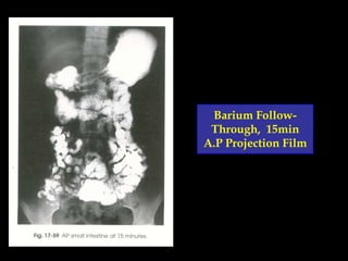
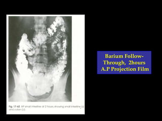
Dr. Mustafa Zuhair Mahmoud
B.Sc; SUST {Khartoum, Sudan}
M.Sc; AAU {Khartoum, Sudan} & JUREI {Philadelphia, USA}
Ph.D, Ludes {Lugano, Swiss}
Ph.D, SUST {Khartoum, Sudan}
Digestive
System
Radiography
Quick Anatomical Review:
Digestive System
 The

Digestive System consist of two parts:
Alimentary Canal.
Accessory Glands.
Quick Anatomical Review:


The Alimentary Canal include:
 Mouth, pharynx & esophagus.
 Stomach.
 Small intestine.
 Large intestine.
Quick Anatomical Review:


The Accessory Glands include:
 Salivary glands.
 Liver & gallbladder.
 Pancreas.
Mouth, Pharynx & Esophagus:
Stomach:
Duodenum:
Jejunum & Ileum:
Large Intestine:
Large Intestine:
What Is Digestive System
Radiography??


Gastrointestinal tract radiography, is an x-ray
examination of the elementary canal that uses
a special form of x-ray called fluoroscopy and
an orally ingested contrast media called
barium.



Fluoroscopy makes it possible to see internal
organs in motion. When the GI tract is coated
with barium, the radiologist is able to view and
assess the anatomy and function of the
digestive system.
Contrast Media:


The contrast media used is Barium Sulphate.
Indications For Imaging (Upper GIT):


An upper GI examination helps evaluate
digestive function and to detect:
Ulcers.
 Tumours.
 Inflammation of the esophagus, stomach and
duodenum.
 Hiatal hernias.
 Blockages.
 Abnormalities of the muscular wall of GI tissues.
 Difficulty swallowing.
 Blood in the stool (indicating internal GI bleeding).

Indications For Imaging (Lower GIT):


The lower GI examination helps evaluate
digestive function and to detect:
Tumors.
 Causes of other intestinal illnesses.
 Chronic diarrhea.
 Blood in stools.
 Constipation.
 IBS (irritable bowel syndrome).
 Unexplained weight loss.

Patients Preparation:


For upper GI tract:


To ensure the best possible image quality, stomach
must be empty of food.



Women should always inform their physician or xray technologist if there is any possibility that they
are pregnant.
Patients Preparation:


For lower GI tract:


On the day before the procedure you will likely be
asked not to eat, and to drink only clear liquids.



Women should always inform their physician or xray technologist if there is any possibility that they
are pregnant.
Esophagus
Radiograph
y
Especial Radiographic Examination of the
Esophagus Name as Barium Swallow.
Barium Swallow, A.P Projection
Barium Swallow, Lat. Projection
Barium Swallow, Rt. Anterior Oblique Position
Stomach
Radiograph
y
Especial Radiographic Examination of the Stomach
Name as Barium Meal.
Barium Meal, Rt. Anterior Oblique Position
Barium Meal, Lt. Posterior Oblique Position
Barium Meal, Lat. Projection
Small
Intestine
Radiograph
y
Especial Radiographic Examination of the SmallIntestine Name as Barium Follow-Through.
Barium FollowThrough, 15min
A.P Projection Film
Barium FollowThrough, 30min
A.P Projection Film
Barium FollowThrough, 1hours
A.P Projection Film
Barium FollowThrough, 2hours
A.P Projection Film
Barium FollowThrough, Lt.
Posterior Oblique
Position
Large
Intestine
Radiograph
y
Especial Radiographic Examination of the LargeIntestine Name as Barium Enema.
Filling Colon With Barium:
Lat. Projection, Barium Enema
A.P Projection, Barium Enema
Lt. Posterior Oblique, Barium Enema
Rt. Posterior Oblique, Barium Enema
Digestive system radiography

Digestive system radiography