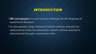
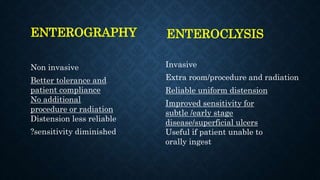
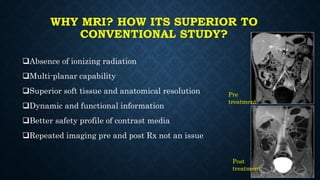
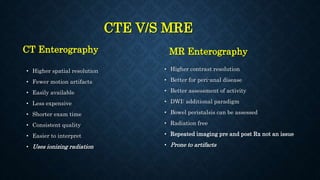
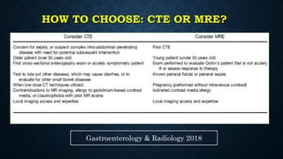
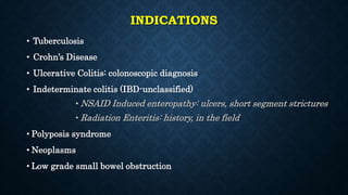
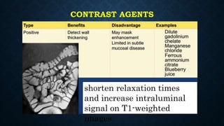
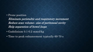
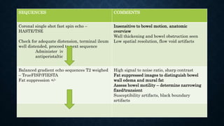
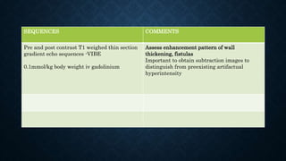
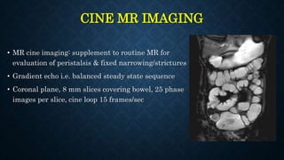
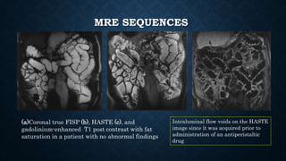
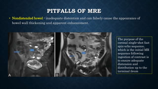
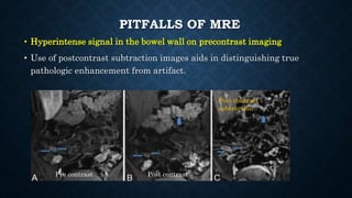
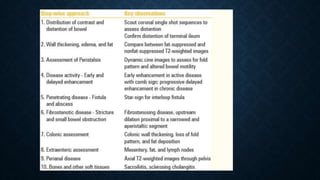
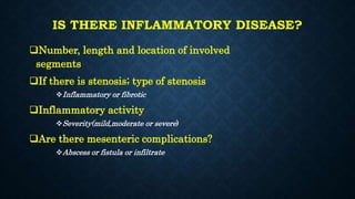
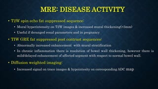
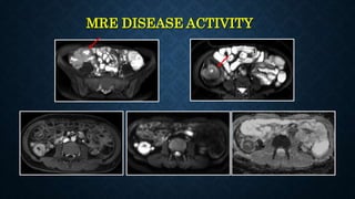
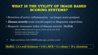
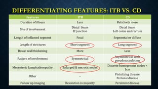
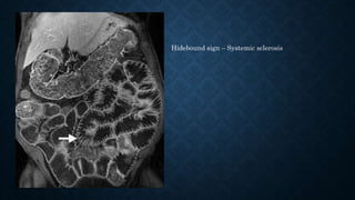
MR ENTEROGRAPHY/ENTEROCLYSIS
Moderator- Dr Anupama
PG - Dr Nihal Ahmed
Department of Radiology
SRIHER
INTRODUCTION
• MR enterography is a non-invasive technique for the diagnosis of
small bowel disorders.
• In enterography, large volumes of enteric contrast material are
administered orally. In enteroclysis, enteric contrast material is
administered through a nasoenteric tube.
ENTEROGRAPHY
Non invasive
Better tolerance and
patient compliance
No additional
procedure or radiation
Distension less reliable
?sensitivity diminished
ENTEROCLYSIS
Invasive
Extra room/procedure and radiation
Reliable uniform distension
Improved sensitivity for
subtle /early stage
disease/superficial ulcers
Useful if patient unable to
orally ingest
WHY MRI? HOW ITS SUPERIOR TO
CONVENTIONAL STUDY?
Absence of ionizing radiation
Multi-planar capability
Superior soft tissue and anatomical resolution
Dynamic and functional information
Better safety profile of contrast media
Repeated imaging pre and post Rx not an issue
Pre
treatment
Post
treatment
CTE V/S MRE
CT Enterography
• Higher spatial resolution
• Fewer motion artifacts
• Easily available
• Less expensive
• Shorter exam time
• Consistent quality
• Easier to interpret
• Uses ionizing radiation
MR Enterography
• Higher contrast resolution
• Better for peri-anal disease
• Better assessment of activity
• DWI: additional paradigm
• Bowel peristalsis can be assessed
• Radiation free
• Repeated imaging pre and post Rx not an issue
• Prone to artifacts
HOW TO CHOOSE: CTE OR MRE?
Gastroenterology & Radiology 2018
INDICATIONS
• Tuberculosis
• Crohn’s Disease
• Ulcerative Colitis: colonoscopic diagnosis
• Indeterminate colitis (IBD-unclassified)
• NSAID Induced enteropathy: ulcers, short segment strictures
• Radiation Enteritis: history, in the field
• Polyposis syndrome
• Neoplasms
• Low grade small bowel obstruction
CONTRAST AGENTS
shorten relaxation times
and increase intraluminal
signal on T1-weighted
images
Induce local field
inhomogeneity and
marked T2* shortening
Biphasic contrast agents are all water-based, appearing dark on
T1-weighted and bright on T2-weighted images.
Biphasic
1. Water
2. Mannitol
3. Polyethylene
glycol(PEG)
4. Methylcellulose
MRE TECHNIQUE & PROTOCOL
Preparati
on
6 hours fasting
Oral
contrast
Water + Mannitol (300 ml of 20% mannitol in 1500 ml water)
Technique
500/500/250/250/250 ml over duration of 1 hour
500 ml in the first 15 min, followed by
500 ml in the next 15 min
250 ml in the next 15 min
250 ml in the next 15 min and finally
250 ml of plain water on the table just before image
acquisition
MRE TECHNIQUE & PROTOCOL
• The contrast medium used at MR enteroclysis was administered using
nasojejunal catheter in two phases.
• 1st phase
flow rate of 80–150 mL/min
contrast medium had reached the terminal ileum.
• 2nd phase,
Increase in the flow rate to 200 mL/min
Reflex atony
• Reflex atony and administration of antiperistaltic drugs (I.V glucagon)
essential to acquire images free of motion artifacts.
• Prone position
Eliminate peristaltic and respiratory movement
Reduce scan volume– size of peritoneal cavity
Help separation of bowel loops
• Gadolinium 0.1-0.2 mmol/kg
• Time to peak enhancement typically 60-70 s
SEQUENCES COMMENTS
Coronal single shot fast spin echo –
HASTE/TSE
Check for adequate distension, terminal ileum
well distended, proceed to next sequence
Administer iv
antiperistaltic
Insensitive to bowel motion, anatomic
overview
Wall thickening and bowel obstruction seen
Low spatial resolution, flow void artifacts
Balanced gradient echo sequences T2 weighed
– TrueFISP/FIESTA
Fat suppression +/-
High signal to noise ratio, sharp contrast
Fat suppressed images to distinguish bowel
wall edema and mural fat
Assess bowel motility – determine narrowing
fixed/transient
Susceptibility artifacts, black boundary
artifacts
SEQUENCES COMMENTS
Pre and post contrast T1 weighed thin section
gradient echo sequences -VIBE
0.1mmol/kg body weight iv gadolinium
Assess enhancement pattern of wall
thickening, fistulas
Important to obtain subtraction images to
distinguish from preexisting artifactual
hyperintensity
CINE MR IMAGING
• MR cine imaging: supplement to routine MR for
evaluation of peristalsis & fixed narrowing/strictures
• Gradient echo i.e. balanced steady state sequence
• Coronal plane, 8 mm slices covering bowel, 25 phase
images per slice, cine loop 15 frames/sec
MRE SEQUENCES
(a)Coronal true FISP (b), HASTE (c), and
gadolinium-enhanced T1 post contrast with fat
saturation in a patient with no abnormal findings
Intraluminal flow voids on the HASTE
image since it was acquired prior to
administration of an antiperistaltic
drug
susceptibility artifact
Black
boundary
artifact
PITFALLS OF MRE
• Nondistended bowel - inadequate distention and can falsely cause the appearance of
bowel wall thickening and apparent enhancement.
The purpose of the
coronal single-shot fast
spin-echo sequence,
which is the initial MR
sequence following
ingestion of contrast is
to ensure adequate
distension and
distribution up to the
terminal ileum
PITFALLS OF MRE
• Hyperintense signal in the bowel wall on precontrast imaging
• Use of postcontrast subtraction images aids in distinguishing true
pathologic enhancement from artifact.
Pre contrast Post contrast
Post contrast
subtraction
IS THERE INFLAMMATORY DISEASE?
Number, length and location of involved
segments
If there is stenosis; type of stenosis
Inflammatory or fibrotic
Inflammatory activity
Severity(mild,moderate or severe)
Are there mesenteric complications?
Abscess or fistula or infiltrate
MRE: DISEASE ACTIVITY
• T2W spin echo fat suppressed sequence:
• Mural hyperintensity on T2W images & increased mural thickening(>3mm)
• Useful if deranged renal parameters and in pregnancy
• TIW GRE fat suppressed post contrast sequences:
• Abnormally increased enhancement with mural stratification
• In chronic inflammation there is resolution of bowel wall thickening, however there is
mild/delayed enhancement of affected segment with respect to normal bowel wall.
• Diffusion weighted imaging:
• Increased signal on trace images & hypointesity on corresponding ADC map
MRE DISEASE ACTIVITY
PATTERN OF ENHANCEMENT
Enhancement of the bowel wall can be categorized
in one of the following patterns:
1.Homogeneous
2.Mucosal
3.Layered
The latter two enhancement patterns can only be
appreciated when the wall is thickened.
A layered pattern is regarded to depict more severe
disease activity compared to the mucosal pattern,
which in turn is more severe than a homogeneous
pattern
WHAT IS THE UTILITY OF IMAGE BASED
SCORING SYSTEMS?
• Detection of active inflammation - no longer main goalpost
• Disease severity most crucial aspect in diagnostic algorithms
• Magnetic resonance index of disease activity -MaRIA
o Objective MRI based score to assess activity and severity of CD
o Excludes nodal enlargement (low
prevalence and high variability)
 High correlation with CDEIS(endoscopic severity score)
MaRIA: 1.5 x wall thickness + 0.02 x RCE + 5 x edema + 10 x ulceration
DIFFERENTIATING FEATURES: ITB VS. CD
Features ITB CD
Duration of illness Less Relatively more
Site of involvement
Distal ileum
IC junction
Distal ileum
Left colon and rectum
Length of inflamed segment Focal Segmental or diffuse
Length of strictures Short segment Long segment
Bowel wall thickening More Less
Pattern of involvement Symmetrical
Asymmetrical with
pseudosacculation
Mesenteric Lymphadenopathy Enlarged & necrotic nodes
Discrete homogenous nodes <
1cm
Other -
Fistulizing disease
Perianal disease
Follow up imaging Resolution in majority Persistent disease
• Disconnect between biologic inflammation & the signs,
symptoms
• Penetrating & stricturing complications may be present in
asymptomatic patients
Enterography is important as 50% patients with small bowel CD
may have active inflammation when endoscopy is normal
TYPICAL IMAGING FEATURES: ITB
• ITB may involve any part of gastro-intestinal tract
• Ileocecal region with adjacent terminal ileum & cecum: most common site
90% cases. Other common site - jejunum
Fleischner sign in ITB
refers to a widely gaping,
thickened, patulous
ileocecal valve & a
narrowed, ulcerated
terminal ileum
ILEOCECAL TB
• Cecum is usually involved(>ileum): conical, shrunken & retracted out of right iliac fossa due
to contraction of mesocolon
• Loss of normal ileocecal angle & ileum may appear suspended from shrunken cecum (Goose
neck deformity)
T1W FS CE coronal images
showing “goose neck
deformity”
Thickening of IC valve with
contracted pulled up cecum
Necrotic
mesenteric &
RP lymph
nodes
CROHN’S DISEASE
• CD: any portion of GIT, mainly distal ileum like ITB (about 50%), left colon and rectum
• Granulomatous disease: transmural inflammation, discontinuous involvement (skip lesions),
asymmetric inflammation - more along mesenteric border
• Mesenteric changes – Edema, fibrofatty proliferation, increased vascularity, venous thrombosis
Mural edema vs
mural fat
Best evaluated by
comparing the bowel
wall between fat-
suppressed and
nonfat-suppressed
T2-weighted images
PHENOTYPES
• Stricturing disease (upstream dilatation > 3cm)
• Majority strictures have inflammatory & fibrotic component
• If fixed narrowing but no dilatation: “probable stricture”
• Penetrating disease: sinus, fistula, abscess, perforation
• Perianal disease added to any of these phenotypes
Dynamic disease which can wax & wane but is often
progressive
It is actually a continuum & combination of different stages may
be seen
STRICTURE: IS IT INFLAMMATORY OR
FIBROTIC?
INFLAMMATORY
• Narrowing with upstream dilatation >3cm
• Wall thickening: T2 hyperintensity
• Stratified enhancement
• Engorged vasa recta: Comb sign
• Mesenteric fat stranding
• Restricted diffusion
• Respond to immunosuppression
FIBROTIC
• Narrowing with upstream dilatation >3cm
• Wall thickening: T2 dark
• No/ homogenous/delayed enhancement
• No comb sign
• Fibrofatty proliferation
• Restricted diffusion
• Responds to surgery, dilation
MRE images of 19 year male with CD who presented with
fecaluria
Penetrating or Fistulizing Stage: Indication for biologicals
ASSESSMENT OF PENETRATING DISEASE:
FISTULA AND ABSCESS FORMATION
• Two types of fistulas - intraabdominal (interloop, enterovesical,
enterovaginal,enterocutaneous) and perianal fistula
• Sensitivity of MRE for the detection of fistulizing/penetrating disease
range from 83.3–84.4% with a specificity of 100%
• Best seen on postcontrast and fat-suppressed T2-weighted images.
• Not all penetrating diseases result in the formation of fluid-filled sinus
tracts or fistulae - Desmoplastic reaction can result in band-like areas of
fibrosis.
ABSCESS
• Abscess - intramural or inter loop or mesenteric.
• On T2-weighted images, the detection of interloop abscess
may be limited. Use of negative contrast agents that provide
hypointense T2 signal - better delineate interloop abscesses.
PERIANAL DISEASE
St. James University classification
system
(1) Simple linear inter sphincteric fistula
(2) Intersphincteric fistula with inter
sphincteric abscess
(3) Transsphincteric fistula
(4) Transsphincteric fistula with abscess
or secondary track within the ischioanal
or ischiorectal fossa
(5) Supralevator disease
(6) Extrasphincteric disease
• Detect sphincter involvement, secondary tracts and abscesses
• Active fistulous tracts appear hypointense on T1 and hyperintense on
T2-weighted images, DWI +, peripheral rim enhancement on
postcontrast T1-weighted images
• Inactive or chronic fistula lack the T2 hyperintense signal due to
presence of scarring and fibrous tissue.
COMB SIGN
TARGET SIGN
Mural stratification
with
hyperenhancement of
the inner mucosa and
outer serosa (arrows)
and nonenhancing
intervening edematous
submucosa
(arrowheads)
STAR SIGN
Enterocutaneous fistula
(arrows) between the anterior
abdominal wall and multiple
tethered small bowel loops,
consisting of a stellate pattern
of mesenteric enhancement
extending between multiple
small bowel loops.
CREEPING FAT SIGN
Creeping fat/fibrofatty
proliferation/fat wrapping-
hypertrophy of the
subserosal fat.
It is a common finding in
longstanding Crohn's
disease.
ROLE OF MRI IN SBO?
The most common cause of small-bowel obstructions is postoperative
adhesions.
• Abrupt change in bowel caliber without evidence of another cause of
obstruction in the vicinity of the transition point.
• Low-signal-intensity soft-tissue bands may be seen coursing through high-
signal-intensity mesenteric fat on T2-weighted images.
• Clustering or deformation of bowel loops also may be seen.
High grade obstruction – CT is preferred modality
Coronal FISP image - ileal loop dilatation (curved
arrow), a transition point (straight arrow), and
normal distal caliber (arrowhead). No mass, bowel
wall thickening, stricture, or other specific cause
of obstruction was identified
58-year-old
woman with
GI tract
bleeding and
recurrent low-
grade SBO
Dx- Brunner
gland
hamartoma
ADENOCARCINOMA
annular soft-tissue mass
homogeneous wall enhancement
lymphadenopathy
CARCINOID TUMORS
• Avidly enhancing, , multifocal enhancing submucosal polypoid lesions in a
segmental distribution
• Hypervascular metastases may be seen in the liver
Mass (arrow) with a mural location(eccentric)
Artifacts are more
centrally located
GIST
• Most common mesenchymal
neoplasm of the GI tract
• Originate from Muscularis propria
• Exoenteric heterogenous mass
• Satellite adenopathy is lacking
exophytic duodenal lesion
Peutz Jeghers syndrome Lymphoma
Metastases
Celiac disease Diverticulosis
Hidebound sign – Systemic sclerosis
TAKE HOME POINTS
• HOW TO CHOOSE CTE/MRE
• INDICATIONS
• MR SEQUENCES AND ARTIFACTS– Fast spin echo /Balanced
gradient/T1 Pre and Post contrast
• ILEOCAECAL TB VS CROHNS DISEASE
• ACTIVE VS CHRONIC DISEASE
• INFLAMMATORY VS FIBROTIC STRICTURES
• ASSESS FISTULA
• LOW GRADE SMALL BOWEL OBSTRUCTION
REFERENCES
Thank You For Your Attention

MR ENTEROGRAPHY

  • 1.
    MR ENTEROGRAPHY/ENTEROCLYSIS Moderator- DrAnupama PG - Dr Nihal Ahmed Department of Radiology SRIHER
  • 2.
    INTRODUCTION • MR enterographyis a non-invasive technique for the diagnosis of small bowel disorders. • In enterography, large volumes of enteric contrast material are administered orally. In enteroclysis, enteric contrast material is administered through a nasoenteric tube.
  • 3.
    ENTEROGRAPHY Non invasive Better toleranceand patient compliance No additional procedure or radiation Distension less reliable ?sensitivity diminished ENTEROCLYSIS Invasive Extra room/procedure and radiation Reliable uniform distension Improved sensitivity for subtle /early stage disease/superficial ulcers Useful if patient unable to orally ingest
  • 4.
    WHY MRI? HOWITS SUPERIOR TO CONVENTIONAL STUDY? Absence of ionizing radiation Multi-planar capability Superior soft tissue and anatomical resolution Dynamic and functional information Better safety profile of contrast media Repeated imaging pre and post Rx not an issue Pre treatment Post treatment
  • 5.
    CTE V/S MRE CTEnterography • Higher spatial resolution • Fewer motion artifacts • Easily available • Less expensive • Shorter exam time • Consistent quality • Easier to interpret • Uses ionizing radiation MR Enterography • Higher contrast resolution • Better for peri-anal disease • Better assessment of activity • DWI: additional paradigm • Bowel peristalsis can be assessed • Radiation free • Repeated imaging pre and post Rx not an issue • Prone to artifacts
  • 6.
    HOW TO CHOOSE:CTE OR MRE? Gastroenterology & Radiology 2018
  • 7.
    INDICATIONS • Tuberculosis • Crohn’sDisease • Ulcerative Colitis: colonoscopic diagnosis • Indeterminate colitis (IBD-unclassified) • NSAID Induced enteropathy: ulcers, short segment strictures • Radiation Enteritis: history, in the field • Polyposis syndrome • Neoplasms • Low grade small bowel obstruction
  • 8.
    CONTRAST AGENTS shorten relaxationtimes and increase intraluminal signal on T1-weighted images
  • 9.
    Induce local field inhomogeneityand marked T2* shortening
  • 10.
    Biphasic contrast agentsare all water-based, appearing dark on T1-weighted and bright on T2-weighted images. Biphasic 1. Water 2. Mannitol 3. Polyethylene glycol(PEG) 4. Methylcellulose
  • 11.
    MRE TECHNIQUE &PROTOCOL Preparati on 6 hours fasting Oral contrast Water + Mannitol (300 ml of 20% mannitol in 1500 ml water) Technique 500/500/250/250/250 ml over duration of 1 hour 500 ml in the first 15 min, followed by 500 ml in the next 15 min 250 ml in the next 15 min 250 ml in the next 15 min and finally 250 ml of plain water on the table just before image acquisition
  • 12.
    MRE TECHNIQUE &PROTOCOL • The contrast medium used at MR enteroclysis was administered using nasojejunal catheter in two phases. • 1st phase flow rate of 80–150 mL/min contrast medium had reached the terminal ileum. • 2nd phase, Increase in the flow rate to 200 mL/min Reflex atony • Reflex atony and administration of antiperistaltic drugs (I.V glucagon) essential to acquire images free of motion artifacts.
  • 13.
    • Prone position Eliminateperistaltic and respiratory movement Reduce scan volume– size of peritoneal cavity Help separation of bowel loops • Gadolinium 0.1-0.2 mmol/kg • Time to peak enhancement typically 60-70 s
  • 14.
    SEQUENCES COMMENTS Coronal singleshot fast spin echo – HASTE/TSE Check for adequate distension, terminal ileum well distended, proceed to next sequence Administer iv antiperistaltic Insensitive to bowel motion, anatomic overview Wall thickening and bowel obstruction seen Low spatial resolution, flow void artifacts Balanced gradient echo sequences T2 weighed – TrueFISP/FIESTA Fat suppression +/- High signal to noise ratio, sharp contrast Fat suppressed images to distinguish bowel wall edema and mural fat Assess bowel motility – determine narrowing fixed/transient Susceptibility artifacts, black boundary artifacts
  • 15.
    SEQUENCES COMMENTS Pre andpost contrast T1 weighed thin section gradient echo sequences -VIBE 0.1mmol/kg body weight iv gadolinium Assess enhancement pattern of wall thickening, fistulas Important to obtain subtraction images to distinguish from preexisting artifactual hyperintensity
  • 16.
    CINE MR IMAGING •MR cine imaging: supplement to routine MR for evaluation of peristalsis & fixed narrowing/strictures • Gradient echo i.e. balanced steady state sequence • Coronal plane, 8 mm slices covering bowel, 25 phase images per slice, cine loop 15 frames/sec
  • 17.
    MRE SEQUENCES (a)Coronal trueFISP (b), HASTE (c), and gadolinium-enhanced T1 post contrast with fat saturation in a patient with no abnormal findings Intraluminal flow voids on the HASTE image since it was acquired prior to administration of an antiperistaltic drug
  • 18.
  • 19.
    PITFALLS OF MRE •Nondistended bowel - inadequate distention and can falsely cause the appearance of bowel wall thickening and apparent enhancement. The purpose of the coronal single-shot fast spin-echo sequence, which is the initial MR sequence following ingestion of contrast is to ensure adequate distension and distribution up to the terminal ileum
  • 20.
    PITFALLS OF MRE •Hyperintense signal in the bowel wall on precontrast imaging • Use of postcontrast subtraction images aids in distinguishing true pathologic enhancement from artifact. Pre contrast Post contrast Post contrast subtraction
  • 22.
    IS THERE INFLAMMATORYDISEASE? Number, length and location of involved segments If there is stenosis; type of stenosis Inflammatory or fibrotic Inflammatory activity Severity(mild,moderate or severe) Are there mesenteric complications? Abscess or fistula or infiltrate
  • 23.
    MRE: DISEASE ACTIVITY •T2W spin echo fat suppressed sequence: • Mural hyperintensity on T2W images & increased mural thickening(>3mm) • Useful if deranged renal parameters and in pregnancy • TIW GRE fat suppressed post contrast sequences: • Abnormally increased enhancement with mural stratification • In chronic inflammation there is resolution of bowel wall thickening, however there is mild/delayed enhancement of affected segment with respect to normal bowel wall. • Diffusion weighted imaging: • Increased signal on trace images & hypointesity on corresponding ADC map
  • 24.
  • 25.
    PATTERN OF ENHANCEMENT Enhancementof the bowel wall can be categorized in one of the following patterns: 1.Homogeneous 2.Mucosal 3.Layered The latter two enhancement patterns can only be appreciated when the wall is thickened. A layered pattern is regarded to depict more severe disease activity compared to the mucosal pattern, which in turn is more severe than a homogeneous pattern
  • 26.
    WHAT IS THEUTILITY OF IMAGE BASED SCORING SYSTEMS? • Detection of active inflammation - no longer main goalpost • Disease severity most crucial aspect in diagnostic algorithms • Magnetic resonance index of disease activity -MaRIA o Objective MRI based score to assess activity and severity of CD o Excludes nodal enlargement (low prevalence and high variability)  High correlation with CDEIS(endoscopic severity score) MaRIA: 1.5 x wall thickness + 0.02 x RCE + 5 x edema + 10 x ulceration
  • 27.
    DIFFERENTIATING FEATURES: ITBVS. CD Features ITB CD Duration of illness Less Relatively more Site of involvement Distal ileum IC junction Distal ileum Left colon and rectum Length of inflamed segment Focal Segmental or diffuse Length of strictures Short segment Long segment Bowel wall thickening More Less Pattern of involvement Symmetrical Asymmetrical with pseudosacculation Mesenteric Lymphadenopathy Enlarged & necrotic nodes Discrete homogenous nodes < 1cm Other - Fistulizing disease Perianal disease Follow up imaging Resolution in majority Persistent disease
  • 28.
    • Disconnect betweenbiologic inflammation & the signs, symptoms • Penetrating & stricturing complications may be present in asymptomatic patients Enterography is important as 50% patients with small bowel CD may have active inflammation when endoscopy is normal
  • 29.
    TYPICAL IMAGING FEATURES:ITB • ITB may involve any part of gastro-intestinal tract • Ileocecal region with adjacent terminal ileum & cecum: most common site 90% cases. Other common site - jejunum Fleischner sign in ITB refers to a widely gaping, thickened, patulous ileocecal valve & a narrowed, ulcerated terminal ileum
  • 30.
    ILEOCECAL TB • Cecumis usually involved(>ileum): conical, shrunken & retracted out of right iliac fossa due to contraction of mesocolon • Loss of normal ileocecal angle & ileum may appear suspended from shrunken cecum (Goose neck deformity) T1W FS CE coronal images showing “goose neck deformity” Thickening of IC valve with contracted pulled up cecum Necrotic mesenteric & RP lymph nodes
  • 31.
    CROHN’S DISEASE • CD:any portion of GIT, mainly distal ileum like ITB (about 50%), left colon and rectum • Granulomatous disease: transmural inflammation, discontinuous involvement (skip lesions), asymmetric inflammation - more along mesenteric border • Mesenteric changes – Edema, fibrofatty proliferation, increased vascularity, venous thrombosis
  • 32.
    Mural edema vs muralfat Best evaluated by comparing the bowel wall between fat- suppressed and nonfat-suppressed T2-weighted images
  • 33.
    PHENOTYPES • Stricturing disease(upstream dilatation > 3cm) • Majority strictures have inflammatory & fibrotic component • If fixed narrowing but no dilatation: “probable stricture” • Penetrating disease: sinus, fistula, abscess, perforation • Perianal disease added to any of these phenotypes Dynamic disease which can wax & wane but is often progressive It is actually a continuum & combination of different stages may be seen
  • 34.
    STRICTURE: IS ITINFLAMMATORY OR FIBROTIC? INFLAMMATORY • Narrowing with upstream dilatation >3cm • Wall thickening: T2 hyperintensity • Stratified enhancement • Engorged vasa recta: Comb sign • Mesenteric fat stranding • Restricted diffusion • Respond to immunosuppression FIBROTIC • Narrowing with upstream dilatation >3cm • Wall thickening: T2 dark • No/ homogenous/delayed enhancement • No comb sign • Fibrofatty proliferation • Restricted diffusion • Responds to surgery, dilation
  • 35.
    MRE images of19 year male with CD who presented with fecaluria Penetrating or Fistulizing Stage: Indication for biologicals
  • 36.
    ASSESSMENT OF PENETRATINGDISEASE: FISTULA AND ABSCESS FORMATION • Two types of fistulas - intraabdominal (interloop, enterovesical, enterovaginal,enterocutaneous) and perianal fistula • Sensitivity of MRE for the detection of fistulizing/penetrating disease range from 83.3–84.4% with a specificity of 100% • Best seen on postcontrast and fat-suppressed T2-weighted images. • Not all penetrating diseases result in the formation of fluid-filled sinus tracts or fistulae - Desmoplastic reaction can result in band-like areas of fibrosis.
  • 37.
    ABSCESS • Abscess -intramural or inter loop or mesenteric. • On T2-weighted images, the detection of interloop abscess may be limited. Use of negative contrast agents that provide hypointense T2 signal - better delineate interloop abscesses.
  • 38.
    PERIANAL DISEASE St. JamesUniversity classification system (1) Simple linear inter sphincteric fistula (2) Intersphincteric fistula with inter sphincteric abscess (3) Transsphincteric fistula (4) Transsphincteric fistula with abscess or secondary track within the ischioanal or ischiorectal fossa (5) Supralevator disease (6) Extrasphincteric disease
  • 39.
    • Detect sphincterinvolvement, secondary tracts and abscesses • Active fistulous tracts appear hypointense on T1 and hyperintense on T2-weighted images, DWI +, peripheral rim enhancement on postcontrast T1-weighted images • Inactive or chronic fistula lack the T2 hyperintense signal due to presence of scarring and fibrous tissue.
  • 40.
  • 41.
    TARGET SIGN Mural stratification with hyperenhancementof the inner mucosa and outer serosa (arrows) and nonenhancing intervening edematous submucosa (arrowheads)
  • 42.
    STAR SIGN Enterocutaneous fistula (arrows)between the anterior abdominal wall and multiple tethered small bowel loops, consisting of a stellate pattern of mesenteric enhancement extending between multiple small bowel loops.
  • 43.
    CREEPING FAT SIGN Creepingfat/fibrofatty proliferation/fat wrapping- hypertrophy of the subserosal fat. It is a common finding in longstanding Crohn's disease.
  • 44.
    ROLE OF MRIIN SBO? The most common cause of small-bowel obstructions is postoperative adhesions. • Abrupt change in bowel caliber without evidence of another cause of obstruction in the vicinity of the transition point. • Low-signal-intensity soft-tissue bands may be seen coursing through high- signal-intensity mesenteric fat on T2-weighted images. • Clustering or deformation of bowel loops also may be seen. High grade obstruction – CT is preferred modality
  • 45.
    Coronal FISP image- ileal loop dilatation (curved arrow), a transition point (straight arrow), and normal distal caliber (arrowhead). No mass, bowel wall thickening, stricture, or other specific cause of obstruction was identified 58-year-old woman with GI tract bleeding and recurrent low- grade SBO Dx- Brunner gland hamartoma
  • 46.
  • 47.
    CARCINOID TUMORS • Avidlyenhancing, , multifocal enhancing submucosal polypoid lesions in a segmental distribution • Hypervascular metastases may be seen in the liver Mass (arrow) with a mural location(eccentric) Artifacts are more centrally located
  • 48.
    GIST • Most commonmesenchymal neoplasm of the GI tract • Originate from Muscularis propria • Exoenteric heterogenous mass • Satellite adenopathy is lacking exophytic duodenal lesion
  • 49.
  • 50.
  • 51.
    Hidebound sign –Systemic sclerosis
  • 52.
    TAKE HOME POINTS •HOW TO CHOOSE CTE/MRE • INDICATIONS • MR SEQUENCES AND ARTIFACTS– Fast spin echo /Balanced gradient/T1 Pre and Post contrast • ILEOCAECAL TB VS CROHNS DISEASE • ACTIVE VS CHRONIC DISEASE • INFLAMMATORY VS FIBROTIC STRICTURES • ASSESS FISTULA • LOW GRADE SMALL BOWEL OBSTRUCTION
  • 53.
  • 56.
    Thank You ForYour Attention