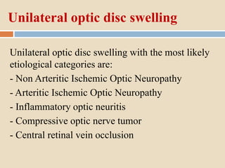
SlideShare a Scribd company logo
PAPILLOEDEMA
Dr/ Reyad Abo Yossif (MRCOphth)
Ophthalmology specialist
Shebin Elkom ophthalmology hospital
Definitions
- Optic disc oedema.
- Unilateral optic disc swelling.
- Bilateral optic disc swelling.
- Papilloedema.
- Pseudo- Papilloedema.
Optic disc oedema
Optic disc edema refers to the ophthalmoscopic swelling
of the optic disc with a concurrent increase in fluid within
or surrounding the axons.
Unilateral optic disc swelling
Unilateral optic disc swelling with the most likely
etiological categories are:
- Non Arteritic Ischemic Optic Neuropathy
- Arteritic Ischemic Optic Neuropathy
- Inflammatory optic neuritis
- Compressive optic nerve tumor
- Central retinal vein occlusion
Unilateral optic disc swelling
Optic nerve functions are affected:
- Visual Acuity diminution
- Pupillary affection (+ve RAPD)
- Color vision affection
Bilateral optic disc swelling
Bilateral optic disc swelling with the most likely
etiological categories are:
- Increased intracranial pressure (ICP) (Papilloedema)
- Infectious/inflammatory
- Demyelinating
- Toxic-metabolic/nutritional,
and Hypertensive emergency (ie, malignant HTN).
Bilateral optic disc swelling
Optic nerve functions are normal
- Normal Visual Acuity
- Normal Pupillary response
- No Color vision problem
(except late if optic atrophy occurred)
Papilloedema
Bilateral optic disc swelling due to increased ICP can be
caused by many disease processes including :
- intracranial space occupying lesions
, obstructions of the arachnoid villi,
, CSF producing tumors
and idiopathic intracranial hypertension (IIH).
Pseudo-Papilloedema
Pseudo-papilledema is defined as anomalous
elevation of one or both optic discs without edema
of the retinal nerve fiber layer.
It is important to distinguish pseudo-papilledema
from true papilledema, which can be the first sign
of disease process with the potential for vision loss,
neurological impairment, or death.
Patients thought to have papilledema are often
subjected to lumbar puncture, MRI, and extensive
laboratory studies to find the underlying cause.
Papilloedema
Aetiology
 Papilledema is due to increased intracranial pressure from
any cause.
 Normal ICP is typically less than 25 cm water when
measured with a manometer in the recumbinat position.
 Papilledema results from orthograde axoplasmic flow
stasis at the optic nerve head leading to edema of the nerve
from the increased intracranial pressure pressing on the
nerve behind the eye.
Increased intracranial pressure can be
caused by:
(1) a space occupying lesion such as a tumor or
hemorrhage,
(2) decreased CSF drainage due to:
blockage (obstructive hydrocephalus) or decreased
absorption (infection, hemmorhage, venous sinus
thrombosis or venous outflow obstruction),
(3) Idiopathic Intracranial Hypertenion (IIH, also called
pseudtumor cerebri) ,
(4) increased CSF production such as from a choroid plexus
tumor (rare),
(5) decreased skull volume (craniosynostosis).
Continued pressure can result in loss of axons and eventual optic atrophy
secondary to intraneuronal ischemia ultimately causing vision loss.
With optic atrophy there is little or no edema seen even with continuing
increased intracranial pressure since there are no fibers to swell.
History
 Headache is typical and is often worse on waking or
postural (worse on lying down and better on standing
up).
 It is useful to ask about medicines that may cause
increased intracranial pressure such as tetracyclines,
vitamin A and derivatives, lithium, steroids - especially
changes in the amount of steroids taken and withdrawal
of steroids.
Physical examination
 In addition to the usual ophthalmic exam including visual
fields , one should check blood pressure to rule out
hypertensive emergency (i.e., “malignant hypertension”)
which can mimic papilledema.
 Ocular motility exam may reveal a unilateral or bilateral
6th nerve palsy as a non-localizing signs of increased
intracranial pressure.
Symptoms and signs of increased ICP
 Headache is common with increased intracranial
pressure and often worse in the morning.
 Nausea and vomiting are noted with increased
intracranial pressure, especially with an acute rise in
pressure. This is made worse by straining, coughing or
bending.
 Pulse synchronous tinnitus may be present from
increased flow in stenotic cerebral venous sinuses.
Symptoms and signs of increased ICP
 Visual field loss (usually an increased size of the blind spot
initially) can occur acutely (i.e., fulminant) or be slowly
progressive.
 The patient may note short periods lasting seconds of losing
vision which are called transient visual obscurations. These
are often observed when raising to stand or changing
position.
 A hyperopic shift may occur in the refraction due to axial
length shortening.
Symptoms and signs of increased ICP
 Long standing papilledema may result in severe peripheral
visual field loss and may progress to involve central
vision if untreated.
 Increased intracranial pressure can also lead to cranial
nerve palsies, usually an abducens palsy as a non-
localizing finding. It is thought that the long course of the
6th cranial nerve is compressed by the increased CSF
pressure.
Clinical diagnosis
Papilledema Grading System (Frisen Scale)
Papilledema Grading System
(Frisen Scale) in detail
Papilledema Grading System (Frisen Scale)
Stage 0 - Normal Optic Disc
Blurring of nasal, superior and inferior poles in inverse
proportion to disc diameter.
Radial nerve fiber layer (NFL) without NFL tortuosity.
Rare obscuration of a major blood vessel, usually on the
upper pole.
Papilledema Grading System (Frisen Scale)
Stage 1 -Very Early Papilledema
Obscuration of the nasal border of the
disc.
No elevation of the disc borders.
Disruption of the normal radial NFL
arrangement with grayish opacity
accentuating nerve fiber layer bundles.
Normal temporal disc margin. Subtle
grayish halo with temporal gap (best
seen with indirect ophthalmoscopy).
Concentric or radial retrochoroidal
folds.
Papilledema Grading System (Frisen Scale)
Stage 2 - Early Papilledema
Obscuration of all borders.
Elevation of the nasal border.
Complete peripapillary halo.
Papilledema Grading System (Frisen Scale)
Stage 3 –
Moderate Papilledema
-Obscurations of all borders.
-Increased diameter of optic
nerve head.
-Obscuration of one or
more segments of major
blood vessels leaving the disc.
-Peripapillary halo-irregular
outer fringe with finger-like
extensions.
Papilledema Grading System (Frisen Scale)
Stage 4 - Marked Papilledema
-Elevation of the entire nerve head.
-Obscuration of all borders.
-Peripapillary halo.
-Total obscuration on the
disc of a segment of a
major blood vessel.
Papilledema Grading System (Frisen Scale)
Stage 5 - Severe Papilledema
Dome-shaped protrusions representing anterior expansion of
the optic nerve head.
Peripapillary halo is narrow and smoothly demarcated.
Total obscuration of a segment of a major blood vessel may or
may not be present.
Obliteration of the optic cup.
Papilledema Grading System
(Frisen Scale) in breif
Grade I papilledema is characterized by a C-shaped halo
with a temporal gap
Grade II papilledema, the halo becomes circumferential
Grade III papilledema is characterized by loss of major
vessels AS THEY LEAVE the disc (arrow)
Grade IV papilledema is characterized by loss of major
vessels ON THE DISC.
Grade V papilledema has the criteria of grade IV plus partial
or total obscuration of all vessels of the disc.
Diagnostic procedures
-CNS imaging study (CT or MRI with contrast) to identify a
central nervous system mass lesion.
-MR venography(MRV) or CTV should also be considered to
look for venous sinus thrombosis.
Diagnostic procedures
Lumbar puncture (to be done after imaging demonstrates no risk
for herniation) to document increased ICP and to look for
neoplastic, infectious or inflammatory causes. At a minimum,
opening pressure along with glucose, protein, cell count and
differential, culture are obtained..
Diagnostic procedures
Fluorescein angiography can detect disc leakage in true
optic disc edema as opposed to disc staining without
leakage in pseudopapilledema.
disc leakage in true optic disc
edema
disc staining without leakage in
pseudopapilledema
Diagnostic procedures
Autofluorescence may show pseudopapilledema (e.g.,
optic disc drusen).
Diagnostic procedures
B scan ultrasound may show pseudopapilledema (e.g.,
optic disc drusen).
Dilated Optic Nerve Sheath with Papilledema with
disc elevation , no calcification
Diagnostic procedures
Successive fundus images may be helpful to follow the disc
status over time.
Diagnostic procedures
Automated perimetry commonly shows enlarged blind spots and
nerve fiber layer type visual field defects. In severe chronic
papilledema worsening peripheral field loss may be seen and can
eventually involve central vision loss.
Management of bilateral optic disc swelling
1- Complete routine history taking and examination
2- Answer these questions:
- Is it a unilateral or bilateral disc swelling ?
- What about optic nerve functions (VA , RAPD ,color) ?
- Differential diagnosis of bilateral disc oedema ?
- Blood pressure measurement to rule out malignant HTN (search
for hypertensive changes during fundus examination)
- MRI and MRV with contrast to rule out space occupying lesions
or venous sinus thrombosis
- Lumbar puncture to rule out IIH (Idiopathic Intracranial
Hypertension)
Management of bilateral optic disc swelling
- Fundus photo for documentation and follow up
- Automated perimetry for follow up
- Think about other causes of bilateral disc oedema other than
increased intracranial pressure as sarcoidosis , toxoplasmosis ,
VKH ,posterior scleritis , posterior uveitis causes
- Don’t forget to exclude pseudo-papilloema (optic disc drusen ,
crowded disc in hypermetropia, tilted disc in high myopia , optic
disc hypoplasia)
Notes on workup
When evaluating a patient for IIH, a complete ocular
examination including a dilated fundus examination,
visual field examination, and optic nerve photographs is
required.
Subsequently, neuroimaging is needed to exclude
secondary causes of intracranial hypertension. Magnetic
resonance imaging (MRI) and MR venography (MRV) of
the brain are usually the imaging modalities of choice.
Notes on workup
Furthermore, a lumbar puncture is recommended for all patients
who are suspected of having IIH.
The diagnosis is based upon an elevated opening pressure greater
than 25 cm H2O taken with the patient lying in the lateral
decubitus position.
Values (20 - 25 )cm H2O are considered equivocal.
In obese or sedated children, pressures up to 28 cm H2O may be
considered normal.
Notes on workup
The CSF must also be studied to rule-out
inflammation, tumor cells, and infection.
Patients with IIH usually have normal or low protein
level, normal glucose levels and a normal cell count
Notes on workup
The primary cause of the increased intracranial pressure
must be addressed.
If a mass is present, primary therapy should be directed
towards that lesion (e.g., surgery).
If medications (tetracyclines, vitamin A analogues, etc.)
are felt to be causative they should be discontinued.
Notes on workup
Patients with mild papilledema should be
monitored for visual field defects by formal
perimetry as the patients may not be able to
recognize them.
Prognosis of papilloedema
Chronic papilledema leads to axon loss with constriction
of the visual field, loss of disc substance and in the end,
loss of central acuity.
Patients (especially those with IIH) need to be followed
to prevent vision loss.
Pseudotumor cerebri
(Idiopathic Intracranial Hypertension)
(IIH)
Pseudotumor cerebri
(idiopathic intracranial hypertension) (IIH)
It is a disorder characterized by increased intracranial
pressure (ICP) of unknown cause that predominantly
affects obese women of child-bearing age.
Papilledema is the primary ocular finding and may
progressively lead to optic atrophy and blindness if no
treatment is provided.
Pseudotumor cerebri
(idiopathic intracranial hypertension)
IIH may be seen in any gender or age group but has a high
predilection for females of childbearing age, especially
when coupled with obesity.
While males are less frequently affected, constituting less
than 10% of adult IIH patients, the affected population
also tend to be obese and are more likely to sustain worse
visual prognosis compared to their female counterparts.
Pseudotumor cerebri
(idiopathic intracranial hypertension)
Interestingly, in the prepubertal age group, IIH has no
particular predilection for obesity or female gender.
Associated conditions include migraines and Down
syndrome.
Pseudotumor cerebri
(idiopathic intracranial hypertension)
Certain systemic illnesses have been associated with IIH, including
obstructive sleep apnea, hypothyroidism, anemia, Addison disease,
systemic lupus erythematosus, Behçet's syndrome, polycystic
ovary syndrome, coagulation disorders, and uremia.
Certain medications have also been associated with IIH, including
tetracyclines, vitamin A, lithium, anabolic steroids, oral
contraceptive pills, nalidixic acid, and cyclosporine.
Diagnostic Criteria
IIH is a diagnosis of exclusion. Nonetheless, diagnostic criteria were established
by Dandy in 1937. However, over the years some modifications were provided
and the new criteria are now known as the modified Dandy criteria:
1- Signs and symptoms of increased ICP (headaches, nausea, vomiting, transient
visual obscurations, papilledema)
2- No localizing neurologic signs, except for unilateral or bilateral sixth cranial
nerve palsy
3- CSF opening pressure >25 cm H2O with normal CSF composition
4- No evidence of hydrocephalus, mass, structural, or vascular lesion (including
venous sinus thrombosis) on imaging
5- No other cause of increased ICP identified
Medical therapy
Carbonic anhydrase inhibitors (acetazolamide) and weight loss (of
about 5-10% ) are first line therapy for IIH but can be used in
patients with elevated ICP from other etiologies.
Recent clinical trials (e.g., Idiopathic Intracranial Hypertension
Trial (IIHT)) have provided strong support for safe and effective
use of acetazolamide combined with weight reduction in
effectively treating IIH and improving papilledema in these
patients with mild vision loss
Medical therapy
Furosemide (Lasix) and topiramate are considered second line
agents in IIH but may be useful in patients who are intolerant of
acetazolamide.
Topiramate, sold under the brand name Topamax , is
a Carbonic anhydrase inhibitor medication used to
treat epilepsy and prevent migraine)
In general however as steroids (especially with change in dosage
or withdrawal) have also been causative of increased ICP we
generally do not recommend steroids for IIH.
Serial lumbar punctures are not typically recommended as a
treatment for IIH but can be used to treat intracranial hypertension
if the patient is not suitable for medical therapy or is pregnant or
has cryptococcal meningitis related increased ICP.
Medical therapy
Surgery
Potential indications for surgery in patients with IIH:
Development of a new visual-field defect
Worsening of a previous visual field defect
Severe visual loss at the time of presentation
Anticipated hypotension induced by treatment of high blood
pressure of renal dialysis
Psychosocial reasons: non-compliance to medication, inability to
perform visual field studies
Refractory headache
Surgery
The two most used procedures are CSF diversion via shunt and
optic nerve sheath fenestration. The choice of the procedure is
based on patients’ signs and symptoms.
Surgery
Optic nerve sheath fenestration is the preferred surgical procedure
for papilledema with associated severe vision loss but no or minimal
ICP symptoms (such as headache). It has been shown to preserve or
restore vision in 80-90% of patients. The procedure involves
incisions in the abnormally bulbous anterior dural covering of the
optic nerve sheath which creates an outlet for continuous CSF
drainage. Consequently, the CSF no longer distends the sheath and
axoplasmic flow in the optic nerve is restored. It is also considered
to be the safest approach among patients with renal failure requiring
hemodialysis and for vision loss occurring during pregnancy.
Surgery
CSF shunting produces rapid reduction in ICP and is therefore most
beneficial among patients with vision loss and symptoms of raised ICP.
Two types are available: lumbo-peritoneal (LP) and ventriculo-peritoneal
(VP). Although VP shunting is more difficult, as IIH patients do not have
enlarged ventricles, it is the preferred method due to its lower complication
rate. Complications of CSF shunting including shunt obstruction, shunt
migration, intracranial hypotension, and tonsillar herniation. Sinclair et al.
found that shunt revisions were needed in 51% of patients, with 30%
requiring multiple revisions.
Surgery
Venous sinus stenting (VSS) is an emerging procedure based on the findings of
venous sinus stenosis in IIH patients. After identification of the stenotic area on
MRV, further testing (e.g. digital subtraction venography and manometry) is
needed to establish a pressure gradient (typically ≥ 10mmHg) before proceeding
with stent placement. VSS has been shown to decrease cerebral venous pressure,
leading to increased CSF absorption and consequent decrease ICP. Some studies
have demonstrated improvement in symptoms following this procedure. However,
despite its possible beneficial outcomes, the procedure may be associated with
serious complications such as stent migration, venous sinus perforation, in-stent
thrombosis, subdural hemorrhage and recurrent stenosis formation proximal to the
stent.
Prognosis
Factors independently associated with a worse visual outcome:
-Gender (male)
-Race (Black)
-Morbid obesity
-Anemia
-Obstructive sleep apnea
-Acute onset of symptoms and signs of raised ICP (fulminant IIH)
Recurrence may occur in 8 to 38% of patients weeks to years following recovery
from initial presentation or a prolonged period of stability.
Weight gain has been associated with disease recurrence.
5. papilloedema

More Related Content

What's hot

Optic atrophy ppt
Optic atrophy pptOptic atrophy ppt
Optic atrophy ppt
Yash Oza
 
Papilledema vs papillitis with notes timothy zagada
Papilledema vs papillitis with notes  timothy zagadaPapilledema vs papillitis with notes  timothy zagada
Papilledema vs papillitis with notes timothy zagada
Timothy Zagada
 
Ptosis
PtosisPtosis
Sixth nerve palsy
Sixth nerve palsySixth nerve palsy
Sixth nerve palsy
Noor Munirah Aab
 
Gaze palsy
Gaze palsyGaze palsy
Evaluation of proptosis
Evaluation of proptosisEvaluation of proptosis
Evaluation of proptosis
Dr Samarth Mishra
 
Diplopia charting
Diplopia chartingDiplopia charting
Diplopia charting
SSSIHMS-PG
 
Pseudo-Ophthalmology !
Pseudo-Ophthalmology !Pseudo-Ophthalmology !
Pseudo-Ophthalmology !
Ahmed Alsherbeny
 
Diplopia
Diplopia Diplopia
Diplopia
Anisha Rathod
 
Real ptosis evaluation.pptx
Real ptosis evaluation.pptxReal ptosis evaluation.pptx
Real ptosis evaluation.pptx
Bipin Koirala
 
Oculomotor nerve palsy
Oculomotor nerve palsyOculomotor nerve palsy
Oculomotor nerve palsy
Dr. Mahziba Rahman
 
Diabetic macular edema
Diabetic macular edemaDiabetic macular edema
Diabetic macular edema
SAMEEKSHA AGRAWAL
 
Exophthalmometry
ExophthalmometryExophthalmometry
Exophthalmometry
Raju Kaiti
 
Pupil
PupilPupil
Pupil
Amar Patil
 
Abnormalities of pupil
Abnormalities of pupilAbnormalities of pupil
Abnormalities of pupil
Imrul Morshed
 
Multiple sclerosis & its ocular manifestations
Multiple sclerosis & its ocular manifestationsMultiple sclerosis & its ocular manifestations
Multiple sclerosis & its ocular manifestations
Prathamesh Shasane
 
The pupillary pathway and its clinical aspects
The pupillary pathway and its clinical aspectsThe pupillary pathway and its clinical aspects
The pupillary pathway and its clinical aspects
Laxmi Eye Institute
 
Congenital Glaucoma
Congenital GlaucomaCongenital Glaucoma
Congenital Glaucoma
Sahil Thakur
 
Ptosis
PtosisPtosis
Ptosis
slidenka
 
Gonioscopy: gonioscopic lenses, principle and clinical aspects
Gonioscopy: gonioscopic lenses, principle and clinical aspectsGonioscopy: gonioscopic lenses, principle and clinical aspects
Gonioscopy: gonioscopic lenses, principle and clinical aspects
Dr Samarth Mishra
 

What's hot (20)

Optic atrophy ppt
Optic atrophy pptOptic atrophy ppt
Optic atrophy ppt
 
Papilledema vs papillitis with notes timothy zagada
Papilledema vs papillitis with notes  timothy zagadaPapilledema vs papillitis with notes  timothy zagada
Papilledema vs papillitis with notes timothy zagada
 
Ptosis
PtosisPtosis
Ptosis
 
Sixth nerve palsy
Sixth nerve palsySixth nerve palsy
Sixth nerve palsy
 
Gaze palsy
Gaze palsyGaze palsy
Gaze palsy
 
Evaluation of proptosis
Evaluation of proptosisEvaluation of proptosis
Evaluation of proptosis
 
Diplopia charting
Diplopia chartingDiplopia charting
Diplopia charting
 
Pseudo-Ophthalmology !
Pseudo-Ophthalmology !Pseudo-Ophthalmology !
Pseudo-Ophthalmology !
 
Diplopia
Diplopia Diplopia
Diplopia
 
Real ptosis evaluation.pptx
Real ptosis evaluation.pptxReal ptosis evaluation.pptx
Real ptosis evaluation.pptx
 
Oculomotor nerve palsy
Oculomotor nerve palsyOculomotor nerve palsy
Oculomotor nerve palsy
 
Diabetic macular edema
Diabetic macular edemaDiabetic macular edema
Diabetic macular edema
 
Exophthalmometry
ExophthalmometryExophthalmometry
Exophthalmometry
 
Pupil
PupilPupil
Pupil
 
Abnormalities of pupil
Abnormalities of pupilAbnormalities of pupil
Abnormalities of pupil
 
Multiple sclerosis & its ocular manifestations
Multiple sclerosis & its ocular manifestationsMultiple sclerosis & its ocular manifestations
Multiple sclerosis & its ocular manifestations
 
The pupillary pathway and its clinical aspects
The pupillary pathway and its clinical aspectsThe pupillary pathway and its clinical aspects
The pupillary pathway and its clinical aspects
 
Congenital Glaucoma
Congenital GlaucomaCongenital Glaucoma
Congenital Glaucoma
 
Ptosis
PtosisPtosis
Ptosis
 
Gonioscopy: gonioscopic lenses, principle and clinical aspects
Gonioscopy: gonioscopic lenses, principle and clinical aspectsGonioscopy: gonioscopic lenses, principle and clinical aspects
Gonioscopy: gonioscopic lenses, principle and clinical aspects
 

Similar to 5. papilloedema

Papilledema
PapilledemaPapilledema
Papilledema
PRAKRITIYAGNAM
 
Papilloedema.pptx
Papilloedema.pptxPapilloedema.pptx
Papilloedema.pptx
dratulkranand
 
GROUP B PAPILLEDEMA.pptx
GROUP B PAPILLEDEMA.pptxGROUP B PAPILLEDEMA.pptx
GROUP B PAPILLEDEMA.pptx
plannetofficial1
 
Papilloedema.PPT
Papilloedema.PPTPapilloedema.PPT
Papilloedema.PPT
ssuser0f453c
 
Papilloedema and Optic Atrophy.ppt
Papilloedema and Optic Atrophy.pptPapilloedema and Optic Atrophy.ppt
Papilloedema and Optic Atrophy.ppt
AshrafIslam16
 
Optic disc swelling
Optic disc swellingOptic disc swelling
Optic disc swelling
Abdul Munim Khan Suri
 
Swollen optic nerve_presentation_last_revision 103112 disregard all others
Swollen optic nerve_presentation_last_revision 103112 disregard all othersSwollen optic nerve_presentation_last_revision 103112 disregard all others
Swollen optic nerve_presentation_last_revision 103112 disregard all othersArash Eslami
 
optic nerve head swelling
optic nerve head swellingoptic nerve head swelling
optic nerve head swelling
Jehad Hassan
 
Neuro ophthalmology RCSI
Neuro ophthalmology RCSINeuro ophthalmology RCSI
Neuro ophthalmology RCSI
OphthalmicDocs Chiong
 
Swollen optic nerve_presentation_last_revision 103112 disregard all others
Swollen optic nerve_presentation_last_revision 103112 disregard all othersSwollen optic nerve_presentation_last_revision 103112 disregard all others
Swollen optic nerve_presentation_last_revision 103112 disregard all othersArash Eslami
 
Disc oedema
Disc oedema Disc oedema
Disc oedema
SIDESH HENDAVITHARANA
 
Benign intracranial hypertension by Dr.Syed Alam Zeb
Benign intracranial hypertension by Dr.Syed Alam ZebBenign intracranial hypertension by Dr.Syed Alam Zeb
Benign intracranial hypertension by Dr.Syed Alam Zeb
Syed Alam Zeb
 
Ophthalmoscopy & otoscopy
Ophthalmoscopy & otoscopyOphthalmoscopy & otoscopy
Ophthalmoscopy & otoscopyAdil AL-sweed
 
Optic Nerve
Optic NerveOptic Nerve
Optic Nerve
Aden University
 
Disc edema
Disc edemaDisc edema
PAPILLEDEMA
PAPILLEDEMAPAPILLEDEMA
PAPILLEDEMA
Nikitha Crasta
 
Optic nerve Clinical significance
Optic nerve Clinical significance Optic nerve Clinical significance
Optic nerve Clinical significance
KafrELShiekh University
 
Idiopathic intracranial hypertension
Idiopathic intracranial hypertensionIdiopathic intracranial hypertension
Idiopathic intracranial hypertension
Othman Al-Abbadi
 
Optic Disc Swelling
Optic Disc SwellingOptic Disc Swelling
Optic Disc Swelling
Tareq Esteak
 
Idiopathic Intracranial Hypertension
Idiopathic Intracranial HypertensionIdiopathic Intracranial Hypertension
Idiopathic Intracranial Hypertension
personalp
 

Similar to 5. papilloedema (20)

Papilledema
PapilledemaPapilledema
Papilledema
 
Papilloedema.pptx
Papilloedema.pptxPapilloedema.pptx
Papilloedema.pptx
 
GROUP B PAPILLEDEMA.pptx
GROUP B PAPILLEDEMA.pptxGROUP B PAPILLEDEMA.pptx
GROUP B PAPILLEDEMA.pptx
 
Papilloedema.PPT
Papilloedema.PPTPapilloedema.PPT
Papilloedema.PPT
 
Papilloedema and Optic Atrophy.ppt
Papilloedema and Optic Atrophy.pptPapilloedema and Optic Atrophy.ppt
Papilloedema and Optic Atrophy.ppt
 
Optic disc swelling
Optic disc swellingOptic disc swelling
Optic disc swelling
 
Swollen optic nerve_presentation_last_revision 103112 disregard all others
Swollen optic nerve_presentation_last_revision 103112 disregard all othersSwollen optic nerve_presentation_last_revision 103112 disregard all others
Swollen optic nerve_presentation_last_revision 103112 disregard all others
 
optic nerve head swelling
optic nerve head swellingoptic nerve head swelling
optic nerve head swelling
 
Neuro ophthalmology RCSI
Neuro ophthalmology RCSINeuro ophthalmology RCSI
Neuro ophthalmology RCSI
 
Swollen optic nerve_presentation_last_revision 103112 disregard all others
Swollen optic nerve_presentation_last_revision 103112 disregard all othersSwollen optic nerve_presentation_last_revision 103112 disregard all others
Swollen optic nerve_presentation_last_revision 103112 disregard all others
 
Disc oedema
Disc oedema Disc oedema
Disc oedema
 
Benign intracranial hypertension by Dr.Syed Alam Zeb
Benign intracranial hypertension by Dr.Syed Alam ZebBenign intracranial hypertension by Dr.Syed Alam Zeb
Benign intracranial hypertension by Dr.Syed Alam Zeb
 
Ophthalmoscopy & otoscopy
Ophthalmoscopy & otoscopyOphthalmoscopy & otoscopy
Ophthalmoscopy & otoscopy
 
Optic Nerve
Optic NerveOptic Nerve
Optic Nerve
 
Disc edema
Disc edemaDisc edema
Disc edema
 
PAPILLEDEMA
PAPILLEDEMAPAPILLEDEMA
PAPILLEDEMA
 
Optic nerve Clinical significance
Optic nerve Clinical significance Optic nerve Clinical significance
Optic nerve Clinical significance
 
Idiopathic intracranial hypertension
Idiopathic intracranial hypertensionIdiopathic intracranial hypertension
Idiopathic intracranial hypertension
 
Optic Disc Swelling
Optic Disc SwellingOptic Disc Swelling
Optic Disc Swelling
 
Idiopathic Intracranial Hypertension
Idiopathic Intracranial HypertensionIdiopathic Intracranial Hypertension
Idiopathic Intracranial Hypertension
 

More from Reyad Yossif

4.ophthalmology workup.4
4.ophthalmology workup.44.ophthalmology workup.4
4.ophthalmology workup.4
Reyad Yossif
 
3.ophthalmology workup.3
3.ophthalmology workup.33.ophthalmology workup.3
3.ophthalmology workup.3
Reyad Yossif
 
2.ophthalmology workup.2
2.ophthalmology workup.22.ophthalmology workup.2
2.ophthalmology workup.2
Reyad Yossif
 
1. ophthalmology workup.1
1. ophthalmology workup.11. ophthalmology workup.1
1. ophthalmology workup.1
Reyad Yossif
 
Vein occlusion guidelines
Vein occlusion guidelinesVein occlusion guidelines
Vein occlusion guidelines
Reyad Yossif
 
DME Management Guidelines
DME Management GuidelinesDME Management Guidelines
DME Management Guidelines
Reyad Yossif
 

More from Reyad Yossif (6)

4.ophthalmology workup.4
4.ophthalmology workup.44.ophthalmology workup.4
4.ophthalmology workup.4
 
3.ophthalmology workup.3
3.ophthalmology workup.33.ophthalmology workup.3
3.ophthalmology workup.3
 
2.ophthalmology workup.2
2.ophthalmology workup.22.ophthalmology workup.2
2.ophthalmology workup.2
 
1. ophthalmology workup.1
1. ophthalmology workup.11. ophthalmology workup.1
1. ophthalmology workup.1
 
Vein occlusion guidelines
Vein occlusion guidelinesVein occlusion guidelines
Vein occlusion guidelines
 
DME Management Guidelines
DME Management GuidelinesDME Management Guidelines
DME Management Guidelines
 

Recently uploaded

Antimicrobial stewardship to prevent antimicrobial resistance
Antimicrobial stewardship to prevent antimicrobial resistanceAntimicrobial stewardship to prevent antimicrobial resistance
Antimicrobial stewardship to prevent antimicrobial resistance
GovindRankawat1
 
Management of Traumatic Splenic injury.pptx
Management of Traumatic Splenic injury.pptxManagement of Traumatic Splenic injury.pptx
Management of Traumatic Splenic injury.pptx
AkshaySarraf1
 
share - Lions, tigers, AI and health misinformation, oh my!.pptx
share - Lions, tigers, AI and health misinformation, oh my!.pptxshare - Lions, tigers, AI and health misinformation, oh my!.pptx
share - Lions, tigers, AI and health misinformation, oh my!.pptx
Tina Purnat
 
Efficacy of Avartana Sneha in Ayurveda
Efficacy of Avartana Sneha in AyurvedaEfficacy of Avartana Sneha in Ayurveda
Efficacy of Avartana Sneha in Ayurveda
Dr. Jyothirmai Paindla
 
Cardiac Assessment for B.sc Nursing Student.pdf
Cardiac Assessment for B.sc Nursing Student.pdfCardiac Assessment for B.sc Nursing Student.pdf
Cardiac Assessment for B.sc Nursing Student.pdf
shivalingatalekar1
 
Ketone bodies and metabolism-biochemistry
Ketone bodies and metabolism-biochemistryKetone bodies and metabolism-biochemistry
Ketone bodies and metabolism-biochemistry
Dhayanithi C
 
Physiology of Special Chemical Sensation of Taste
Physiology of Special Chemical Sensation of TastePhysiology of Special Chemical Sensation of Taste
Physiology of Special Chemical Sensation of Taste
MedicoseAcademics
 
Superficial & Deep Fascia of the NECK.pptx
Superficial & Deep Fascia of the NECK.pptxSuperficial & Deep Fascia of the NECK.pptx
Superficial & Deep Fascia of the NECK.pptx
Dr. Rabia Inam Gandapore
 
Tests for analysis of different pharmaceutical.pptx
Tests for analysis of different pharmaceutical.pptxTests for analysis of different pharmaceutical.pptx
Tests for analysis of different pharmaceutical.pptx
taiba qazi
 
Top Effective Soaps for Fungal Skin Infections in India
Top Effective Soaps for Fungal Skin Infections in IndiaTop Effective Soaps for Fungal Skin Infections in India
Top Effective Soaps for Fungal Skin Infections in India
SwisschemDerma
 
Sex determination from mandible pelvis and skull
Sex determination from mandible pelvis and skullSex determination from mandible pelvis and skull
Sex determination from mandible pelvis and skull
ShashankRoodkee
 
Aortic Association CBL Pilot April 19 – 20 Bern
Aortic Association CBL Pilot April 19 – 20 BernAortic Association CBL Pilot April 19 – 20 Bern
Aortic Association CBL Pilot April 19 – 20 Bern
suvadeepdas911
 
Ear and its clinical correlations By Dr. Rabia Inam Gandapore.pptx
Ear and its clinical correlations By Dr. Rabia Inam Gandapore.pptxEar and its clinical correlations By Dr. Rabia Inam Gandapore.pptx
Ear and its clinical correlations By Dr. Rabia Inam Gandapore.pptx
Dr. Rabia Inam Gandapore
 
Physiology of Chemical Sensation of smell.pdf
Physiology of Chemical Sensation of smell.pdfPhysiology of Chemical Sensation of smell.pdf
Physiology of Chemical Sensation of smell.pdf
MedicoseAcademics
 
SURGICAL ANATOMY OF THE RETROPERITONEUM, ADRENALS, KIDNEYS AND URETERS.pptx
SURGICAL ANATOMY OF THE RETROPERITONEUM, ADRENALS, KIDNEYS AND URETERS.pptxSURGICAL ANATOMY OF THE RETROPERITONEUM, ADRENALS, KIDNEYS AND URETERS.pptx
SURGICAL ANATOMY OF THE RETROPERITONEUM, ADRENALS, KIDNEYS AND URETERS.pptx
Bright Chipili
 
Integrating Ayurveda into Parkinson’s Management: A Holistic Approach
Integrating Ayurveda into Parkinson’s Management: A Holistic ApproachIntegrating Ayurveda into Parkinson’s Management: A Holistic Approach
Integrating Ayurveda into Parkinson’s Management: A Holistic Approach
Ayurveda ForAll
 
ANATOMY AND PHYSIOLOGY OF URINARY SYSTEM.pptx
ANATOMY AND PHYSIOLOGY OF URINARY SYSTEM.pptxANATOMY AND PHYSIOLOGY OF URINARY SYSTEM.pptx
ANATOMY AND PHYSIOLOGY OF URINARY SYSTEM.pptx
Swetaba Besh
 
Dehradun #ℂall #gIRLS Oyo Hotel 8107221448 #ℂall #gIRL in Dehradun
Dehradun #ℂall #gIRLS Oyo Hotel 8107221448 #ℂall #gIRL in DehradunDehradun #ℂall #gIRLS Oyo Hotel 8107221448 #ℂall #gIRL in Dehradun
Dehradun #ℂall #gIRLS Oyo Hotel 8107221448 #ℂall #gIRL in Dehradun
chandankumarsmartiso
 
Hemodialysis: Chapter 4, Dialysate Circuit - Dr.Gawad
Hemodialysis: Chapter 4, Dialysate Circuit - Dr.GawadHemodialysis: Chapter 4, Dialysate Circuit - Dr.Gawad
Hemodialysis: Chapter 4, Dialysate Circuit - Dr.Gawad
NephroTube - Dr.Gawad
 
Knee anatomy and clinical tests 2024.pdf
Knee anatomy and clinical tests 2024.pdfKnee anatomy and clinical tests 2024.pdf
Knee anatomy and clinical tests 2024.pdf
vimalpl1234
 

Recently uploaded (20)

Antimicrobial stewardship to prevent antimicrobial resistance
Antimicrobial stewardship to prevent antimicrobial resistanceAntimicrobial stewardship to prevent antimicrobial resistance
Antimicrobial stewardship to prevent antimicrobial resistance
 
Management of Traumatic Splenic injury.pptx
Management of Traumatic Splenic injury.pptxManagement of Traumatic Splenic injury.pptx
Management of Traumatic Splenic injury.pptx
 
share - Lions, tigers, AI and health misinformation, oh my!.pptx
share - Lions, tigers, AI and health misinformation, oh my!.pptxshare - Lions, tigers, AI and health misinformation, oh my!.pptx
share - Lions, tigers, AI and health misinformation, oh my!.pptx
 
Efficacy of Avartana Sneha in Ayurveda
Efficacy of Avartana Sneha in AyurvedaEfficacy of Avartana Sneha in Ayurveda
Efficacy of Avartana Sneha in Ayurveda
 
Cardiac Assessment for B.sc Nursing Student.pdf
Cardiac Assessment for B.sc Nursing Student.pdfCardiac Assessment for B.sc Nursing Student.pdf
Cardiac Assessment for B.sc Nursing Student.pdf
 
Ketone bodies and metabolism-biochemistry
Ketone bodies and metabolism-biochemistryKetone bodies and metabolism-biochemistry
Ketone bodies and metabolism-biochemistry
 
Physiology of Special Chemical Sensation of Taste
Physiology of Special Chemical Sensation of TastePhysiology of Special Chemical Sensation of Taste
Physiology of Special Chemical Sensation of Taste
 
Superficial & Deep Fascia of the NECK.pptx
Superficial & Deep Fascia of the NECK.pptxSuperficial & Deep Fascia of the NECK.pptx
Superficial & Deep Fascia of the NECK.pptx
 
Tests for analysis of different pharmaceutical.pptx
Tests for analysis of different pharmaceutical.pptxTests for analysis of different pharmaceutical.pptx
Tests for analysis of different pharmaceutical.pptx
 
Top Effective Soaps for Fungal Skin Infections in India
Top Effective Soaps for Fungal Skin Infections in IndiaTop Effective Soaps for Fungal Skin Infections in India
Top Effective Soaps for Fungal Skin Infections in India
 
Sex determination from mandible pelvis and skull
Sex determination from mandible pelvis and skullSex determination from mandible pelvis and skull
Sex determination from mandible pelvis and skull
 
Aortic Association CBL Pilot April 19 – 20 Bern
Aortic Association CBL Pilot April 19 – 20 BernAortic Association CBL Pilot April 19 – 20 Bern
Aortic Association CBL Pilot April 19 – 20 Bern
 
Ear and its clinical correlations By Dr. Rabia Inam Gandapore.pptx
Ear and its clinical correlations By Dr. Rabia Inam Gandapore.pptxEar and its clinical correlations By Dr. Rabia Inam Gandapore.pptx
Ear and its clinical correlations By Dr. Rabia Inam Gandapore.pptx
 
Physiology of Chemical Sensation of smell.pdf
Physiology of Chemical Sensation of smell.pdfPhysiology of Chemical Sensation of smell.pdf
Physiology of Chemical Sensation of smell.pdf
 
SURGICAL ANATOMY OF THE RETROPERITONEUM, ADRENALS, KIDNEYS AND URETERS.pptx
SURGICAL ANATOMY OF THE RETROPERITONEUM, ADRENALS, KIDNEYS AND URETERS.pptxSURGICAL ANATOMY OF THE RETROPERITONEUM, ADRENALS, KIDNEYS AND URETERS.pptx
SURGICAL ANATOMY OF THE RETROPERITONEUM, ADRENALS, KIDNEYS AND URETERS.pptx
 
Integrating Ayurveda into Parkinson’s Management: A Holistic Approach
Integrating Ayurveda into Parkinson’s Management: A Holistic ApproachIntegrating Ayurveda into Parkinson’s Management: A Holistic Approach
Integrating Ayurveda into Parkinson’s Management: A Holistic Approach
 
ANATOMY AND PHYSIOLOGY OF URINARY SYSTEM.pptx
ANATOMY AND PHYSIOLOGY OF URINARY SYSTEM.pptxANATOMY AND PHYSIOLOGY OF URINARY SYSTEM.pptx
ANATOMY AND PHYSIOLOGY OF URINARY SYSTEM.pptx
 
Dehradun #ℂall #gIRLS Oyo Hotel 8107221448 #ℂall #gIRL in Dehradun
Dehradun #ℂall #gIRLS Oyo Hotel 8107221448 #ℂall #gIRL in DehradunDehradun #ℂall #gIRLS Oyo Hotel 8107221448 #ℂall #gIRL in Dehradun
Dehradun #ℂall #gIRLS Oyo Hotel 8107221448 #ℂall #gIRL in Dehradun
 
Hemodialysis: Chapter 4, Dialysate Circuit - Dr.Gawad
Hemodialysis: Chapter 4, Dialysate Circuit - Dr.GawadHemodialysis: Chapter 4, Dialysate Circuit - Dr.Gawad
Hemodialysis: Chapter 4, Dialysate Circuit - Dr.Gawad
 
Knee anatomy and clinical tests 2024.pdf
Knee anatomy and clinical tests 2024.pdfKnee anatomy and clinical tests 2024.pdf
Knee anatomy and clinical tests 2024.pdf
 

5. papilloedema

  • 1. PAPILLOEDEMA Dr/ Reyad Abo Yossif (MRCOphth) Ophthalmology specialist Shebin Elkom ophthalmology hospital
  • 2. Definitions - Optic disc oedema. - Unilateral optic disc swelling. - Bilateral optic disc swelling. - Papilloedema. - Pseudo- Papilloedema.
  • 3. Optic disc oedema Optic disc edema refers to the ophthalmoscopic swelling of the optic disc with a concurrent increase in fluid within or surrounding the axons.
  • 4. Unilateral optic disc swelling Unilateral optic disc swelling with the most likely etiological categories are: - Non Arteritic Ischemic Optic Neuropathy - Arteritic Ischemic Optic Neuropathy - Inflammatory optic neuritis - Compressive optic nerve tumor - Central retinal vein occlusion
  • 5. Unilateral optic disc swelling Optic nerve functions are affected: - Visual Acuity diminution - Pupillary affection (+ve RAPD) - Color vision affection
  • 6. Bilateral optic disc swelling Bilateral optic disc swelling with the most likely etiological categories are: - Increased intracranial pressure (ICP) (Papilloedema) - Infectious/inflammatory - Demyelinating - Toxic-metabolic/nutritional, and Hypertensive emergency (ie, malignant HTN).
  • 7. Bilateral optic disc swelling Optic nerve functions are normal - Normal Visual Acuity - Normal Pupillary response - No Color vision problem (except late if optic atrophy occurred)
  • 8. Papilloedema Bilateral optic disc swelling due to increased ICP can be caused by many disease processes including : - intracranial space occupying lesions , obstructions of the arachnoid villi, , CSF producing tumors and idiopathic intracranial hypertension (IIH).
  • 9.
  • 10. Pseudo-Papilloedema Pseudo-papilledema is defined as anomalous elevation of one or both optic discs without edema of the retinal nerve fiber layer.
  • 11. It is important to distinguish pseudo-papilledema from true papilledema, which can be the first sign of disease process with the potential for vision loss, neurological impairment, or death. Patients thought to have papilledema are often subjected to lumbar puncture, MRI, and extensive laboratory studies to find the underlying cause.
  • 13. Aetiology  Papilledema is due to increased intracranial pressure from any cause.  Normal ICP is typically less than 25 cm water when measured with a manometer in the recumbinat position.  Papilledema results from orthograde axoplasmic flow stasis at the optic nerve head leading to edema of the nerve from the increased intracranial pressure pressing on the nerve behind the eye.
  • 14.
  • 15.
  • 16.
  • 17. Increased intracranial pressure can be caused by: (1) a space occupying lesion such as a tumor or hemorrhage, (2) decreased CSF drainage due to: blockage (obstructive hydrocephalus) or decreased absorption (infection, hemmorhage, venous sinus thrombosis or venous outflow obstruction), (3) Idiopathic Intracranial Hypertenion (IIH, also called pseudtumor cerebri) , (4) increased CSF production such as from a choroid plexus tumor (rare), (5) decreased skull volume (craniosynostosis).
  • 18. Continued pressure can result in loss of axons and eventual optic atrophy secondary to intraneuronal ischemia ultimately causing vision loss. With optic atrophy there is little or no edema seen even with continuing increased intracranial pressure since there are no fibers to swell.
  • 19. History  Headache is typical and is often worse on waking or postural (worse on lying down and better on standing up).  It is useful to ask about medicines that may cause increased intracranial pressure such as tetracyclines, vitamin A and derivatives, lithium, steroids - especially changes in the amount of steroids taken and withdrawal of steroids.
  • 20. Physical examination  In addition to the usual ophthalmic exam including visual fields , one should check blood pressure to rule out hypertensive emergency (i.e., “malignant hypertension”) which can mimic papilledema.  Ocular motility exam may reveal a unilateral or bilateral 6th nerve palsy as a non-localizing signs of increased intracranial pressure.
  • 21. Symptoms and signs of increased ICP  Headache is common with increased intracranial pressure and often worse in the morning.  Nausea and vomiting are noted with increased intracranial pressure, especially with an acute rise in pressure. This is made worse by straining, coughing or bending.  Pulse synchronous tinnitus may be present from increased flow in stenotic cerebral venous sinuses.
  • 22. Symptoms and signs of increased ICP  Visual field loss (usually an increased size of the blind spot initially) can occur acutely (i.e., fulminant) or be slowly progressive.  The patient may note short periods lasting seconds of losing vision which are called transient visual obscurations. These are often observed when raising to stand or changing position.  A hyperopic shift may occur in the refraction due to axial length shortening.
  • 23. Symptoms and signs of increased ICP  Long standing papilledema may result in severe peripheral visual field loss and may progress to involve central vision if untreated.  Increased intracranial pressure can also lead to cranial nerve palsies, usually an abducens palsy as a non- localizing finding. It is thought that the long course of the 6th cranial nerve is compressed by the increased CSF pressure.
  • 24.
  • 25. Clinical diagnosis Papilledema Grading System (Frisen Scale)
  • 27. Papilledema Grading System (Frisen Scale) Stage 0 - Normal Optic Disc Blurring of nasal, superior and inferior poles in inverse proportion to disc diameter. Radial nerve fiber layer (NFL) without NFL tortuosity. Rare obscuration of a major blood vessel, usually on the upper pole.
  • 28. Papilledema Grading System (Frisen Scale) Stage 1 -Very Early Papilledema Obscuration of the nasal border of the disc. No elevation of the disc borders. Disruption of the normal radial NFL arrangement with grayish opacity accentuating nerve fiber layer bundles. Normal temporal disc margin. Subtle grayish halo with temporal gap (best seen with indirect ophthalmoscopy). Concentric or radial retrochoroidal folds.
  • 29. Papilledema Grading System (Frisen Scale) Stage 2 - Early Papilledema Obscuration of all borders. Elevation of the nasal border. Complete peripapillary halo.
  • 30. Papilledema Grading System (Frisen Scale) Stage 3 – Moderate Papilledema -Obscurations of all borders. -Increased diameter of optic nerve head. -Obscuration of one or more segments of major blood vessels leaving the disc. -Peripapillary halo-irregular outer fringe with finger-like extensions.
  • 31. Papilledema Grading System (Frisen Scale) Stage 4 - Marked Papilledema -Elevation of the entire nerve head. -Obscuration of all borders. -Peripapillary halo. -Total obscuration on the disc of a segment of a major blood vessel.
  • 32. Papilledema Grading System (Frisen Scale) Stage 5 - Severe Papilledema Dome-shaped protrusions representing anterior expansion of the optic nerve head. Peripapillary halo is narrow and smoothly demarcated. Total obscuration of a segment of a major blood vessel may or may not be present. Obliteration of the optic cup.
  • 33.
  • 35. Grade I papilledema is characterized by a C-shaped halo with a temporal gap
  • 36. Grade II papilledema, the halo becomes circumferential
  • 37. Grade III papilledema is characterized by loss of major vessels AS THEY LEAVE the disc (arrow)
  • 38. Grade IV papilledema is characterized by loss of major vessels ON THE DISC.
  • 39. Grade V papilledema has the criteria of grade IV plus partial or total obscuration of all vessels of the disc.
  • 40. Diagnostic procedures -CNS imaging study (CT or MRI with contrast) to identify a central nervous system mass lesion. -MR venography(MRV) or CTV should also be considered to look for venous sinus thrombosis.
  • 41. Diagnostic procedures Lumbar puncture (to be done after imaging demonstrates no risk for herniation) to document increased ICP and to look for neoplastic, infectious or inflammatory causes. At a minimum, opening pressure along with glucose, protein, cell count and differential, culture are obtained..
  • 42. Diagnostic procedures Fluorescein angiography can detect disc leakage in true optic disc edema as opposed to disc staining without leakage in pseudopapilledema.
  • 43. disc leakage in true optic disc edema
  • 44. disc staining without leakage in pseudopapilledema
  • 45. Diagnostic procedures Autofluorescence may show pseudopapilledema (e.g., optic disc drusen).
  • 46. Diagnostic procedures B scan ultrasound may show pseudopapilledema (e.g., optic disc drusen).
  • 47. Dilated Optic Nerve Sheath with Papilledema with disc elevation , no calcification
  • 48.
  • 49. Diagnostic procedures Successive fundus images may be helpful to follow the disc status over time.
  • 50. Diagnostic procedures Automated perimetry commonly shows enlarged blind spots and nerve fiber layer type visual field defects. In severe chronic papilledema worsening peripheral field loss may be seen and can eventually involve central vision loss.
  • 51. Management of bilateral optic disc swelling 1- Complete routine history taking and examination 2- Answer these questions: - Is it a unilateral or bilateral disc swelling ? - What about optic nerve functions (VA , RAPD ,color) ? - Differential diagnosis of bilateral disc oedema ? - Blood pressure measurement to rule out malignant HTN (search for hypertensive changes during fundus examination) - MRI and MRV with contrast to rule out space occupying lesions or venous sinus thrombosis - Lumbar puncture to rule out IIH (Idiopathic Intracranial Hypertension)
  • 52. Management of bilateral optic disc swelling - Fundus photo for documentation and follow up - Automated perimetry for follow up - Think about other causes of bilateral disc oedema other than increased intracranial pressure as sarcoidosis , toxoplasmosis , VKH ,posterior scleritis , posterior uveitis causes - Don’t forget to exclude pseudo-papilloema (optic disc drusen , crowded disc in hypermetropia, tilted disc in high myopia , optic disc hypoplasia)
  • 53.
  • 54.
  • 55. Notes on workup When evaluating a patient for IIH, a complete ocular examination including a dilated fundus examination, visual field examination, and optic nerve photographs is required. Subsequently, neuroimaging is needed to exclude secondary causes of intracranial hypertension. Magnetic resonance imaging (MRI) and MR venography (MRV) of the brain are usually the imaging modalities of choice.
  • 56. Notes on workup Furthermore, a lumbar puncture is recommended for all patients who are suspected of having IIH. The diagnosis is based upon an elevated opening pressure greater than 25 cm H2O taken with the patient lying in the lateral decubitus position. Values (20 - 25 )cm H2O are considered equivocal. In obese or sedated children, pressures up to 28 cm H2O may be considered normal.
  • 57. Notes on workup The CSF must also be studied to rule-out inflammation, tumor cells, and infection. Patients with IIH usually have normal or low protein level, normal glucose levels and a normal cell count
  • 58. Notes on workup The primary cause of the increased intracranial pressure must be addressed. If a mass is present, primary therapy should be directed towards that lesion (e.g., surgery). If medications (tetracyclines, vitamin A analogues, etc.) are felt to be causative they should be discontinued.
  • 59. Notes on workup Patients with mild papilledema should be monitored for visual field defects by formal perimetry as the patients may not be able to recognize them.
  • 60. Prognosis of papilloedema Chronic papilledema leads to axon loss with constriction of the visual field, loss of disc substance and in the end, loss of central acuity. Patients (especially those with IIH) need to be followed to prevent vision loss.
  • 62. Pseudotumor cerebri (idiopathic intracranial hypertension) (IIH) It is a disorder characterized by increased intracranial pressure (ICP) of unknown cause that predominantly affects obese women of child-bearing age. Papilledema is the primary ocular finding and may progressively lead to optic atrophy and blindness if no treatment is provided.
  • 63. Pseudotumor cerebri (idiopathic intracranial hypertension) IIH may be seen in any gender or age group but has a high predilection for females of childbearing age, especially when coupled with obesity. While males are less frequently affected, constituting less than 10% of adult IIH patients, the affected population also tend to be obese and are more likely to sustain worse visual prognosis compared to their female counterparts.
  • 64. Pseudotumor cerebri (idiopathic intracranial hypertension) Interestingly, in the prepubertal age group, IIH has no particular predilection for obesity or female gender. Associated conditions include migraines and Down syndrome.
  • 65. Pseudotumor cerebri (idiopathic intracranial hypertension) Certain systemic illnesses have been associated with IIH, including obstructive sleep apnea, hypothyroidism, anemia, Addison disease, systemic lupus erythematosus, Behçet's syndrome, polycystic ovary syndrome, coagulation disorders, and uremia. Certain medications have also been associated with IIH, including tetracyclines, vitamin A, lithium, anabolic steroids, oral contraceptive pills, nalidixic acid, and cyclosporine.
  • 66. Diagnostic Criteria IIH is a diagnosis of exclusion. Nonetheless, diagnostic criteria were established by Dandy in 1937. However, over the years some modifications were provided and the new criteria are now known as the modified Dandy criteria: 1- Signs and symptoms of increased ICP (headaches, nausea, vomiting, transient visual obscurations, papilledema) 2- No localizing neurologic signs, except for unilateral or bilateral sixth cranial nerve palsy 3- CSF opening pressure >25 cm H2O with normal CSF composition 4- No evidence of hydrocephalus, mass, structural, or vascular lesion (including venous sinus thrombosis) on imaging 5- No other cause of increased ICP identified
  • 67. Medical therapy Carbonic anhydrase inhibitors (acetazolamide) and weight loss (of about 5-10% ) are first line therapy for IIH but can be used in patients with elevated ICP from other etiologies. Recent clinical trials (e.g., Idiopathic Intracranial Hypertension Trial (IIHT)) have provided strong support for safe and effective use of acetazolamide combined with weight reduction in effectively treating IIH and improving papilledema in these patients with mild vision loss
  • 68. Medical therapy Furosemide (Lasix) and topiramate are considered second line agents in IIH but may be useful in patients who are intolerant of acetazolamide. Topiramate, sold under the brand name Topamax , is a Carbonic anhydrase inhibitor medication used to treat epilepsy and prevent migraine)
  • 69. In general however as steroids (especially with change in dosage or withdrawal) have also been causative of increased ICP we generally do not recommend steroids for IIH. Serial lumbar punctures are not typically recommended as a treatment for IIH but can be used to treat intracranial hypertension if the patient is not suitable for medical therapy or is pregnant or has cryptococcal meningitis related increased ICP. Medical therapy
  • 70. Surgery Potential indications for surgery in patients with IIH: Development of a new visual-field defect Worsening of a previous visual field defect Severe visual loss at the time of presentation Anticipated hypotension induced by treatment of high blood pressure of renal dialysis Psychosocial reasons: non-compliance to medication, inability to perform visual field studies Refractory headache
  • 71. Surgery The two most used procedures are CSF diversion via shunt and optic nerve sheath fenestration. The choice of the procedure is based on patients’ signs and symptoms.
  • 72. Surgery Optic nerve sheath fenestration is the preferred surgical procedure for papilledema with associated severe vision loss but no or minimal ICP symptoms (such as headache). It has been shown to preserve or restore vision in 80-90% of patients. The procedure involves incisions in the abnormally bulbous anterior dural covering of the optic nerve sheath which creates an outlet for continuous CSF drainage. Consequently, the CSF no longer distends the sheath and axoplasmic flow in the optic nerve is restored. It is also considered to be the safest approach among patients with renal failure requiring hemodialysis and for vision loss occurring during pregnancy.
  • 73. Surgery CSF shunting produces rapid reduction in ICP and is therefore most beneficial among patients with vision loss and symptoms of raised ICP. Two types are available: lumbo-peritoneal (LP) and ventriculo-peritoneal (VP). Although VP shunting is more difficult, as IIH patients do not have enlarged ventricles, it is the preferred method due to its lower complication rate. Complications of CSF shunting including shunt obstruction, shunt migration, intracranial hypotension, and tonsillar herniation. Sinclair et al. found that shunt revisions were needed in 51% of patients, with 30% requiring multiple revisions.
  • 74. Surgery Venous sinus stenting (VSS) is an emerging procedure based on the findings of venous sinus stenosis in IIH patients. After identification of the stenotic area on MRV, further testing (e.g. digital subtraction venography and manometry) is needed to establish a pressure gradient (typically ≥ 10mmHg) before proceeding with stent placement. VSS has been shown to decrease cerebral venous pressure, leading to increased CSF absorption and consequent decrease ICP. Some studies have demonstrated improvement in symptoms following this procedure. However, despite its possible beneficial outcomes, the procedure may be associated with serious complications such as stent migration, venous sinus perforation, in-stent thrombosis, subdural hemorrhage and recurrent stenosis formation proximal to the stent.
  • 75. Prognosis Factors independently associated with a worse visual outcome: -Gender (male) -Race (Black) -Morbid obesity -Anemia -Obstructive sleep apnea -Acute onset of symptoms and signs of raised ICP (fulminant IIH) Recurrence may occur in 8 to 38% of patients weeks to years following recovery from initial presentation or a prolonged period of stability. Weight gain has been associated with disease recurrence.