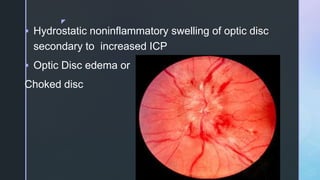
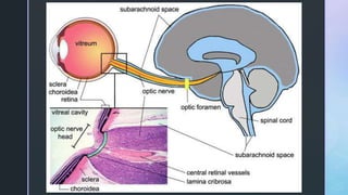
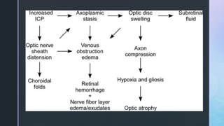
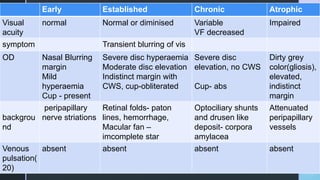
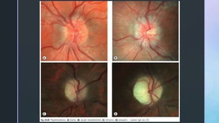
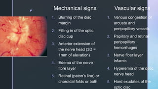
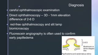
z
Papilledema
Shreeji Shrestha
z
ļ‚§ Hydrostatic noninflammatory swelling of optic disc
secondary to increased ICP
ļ‚§ Optic Disc edema or
Choked disc
z
z
ļ‚§ Subarachnoid communication between brain and ON
z
Axoplasmic transport
ļ‚§ ON axoplasmic transport ( protein, chemicals, mitochondria)
retrograde
IOP (11-21mmhg)
(Force)
Tissue pressure at optic nerve (6-8mmHg)
Retinal ganglion cell
LGB(degraded and returned to Cell body)
axons Orthograde
z
ļ‚§ Blood flow depends on
Vascular resistance,
BP,
IOP
ļ‚§ ONH blood flow - PP/R; PP is MABP-IOP
z
Etiology
Primary - Idiopathic ICH/pseudo tumor cererbi
Secondary - space-occupying lesion(midbrain, parietooccipital and cerebellum);
cerebral edema;
decrease in total volume within the cranial vault by
thickening of the skull;
Blockage of the flow of CSF (hydrocephalus);
Reduced absorption of CSF or increased production
z
Idiopathic intracranial hypertension
ļ‚§ Risk factors –
idiopathic,
Obese female,
pregnancy,
endocrine disorders,
drugs - tetracycline, nalidixic acid, amiodarone, lithium,
corticosteroid in high dose,
hypervitaminosis
z
Pathogenesis
ļ‚§ Mechanical theory - Hayreh theory – Inc
CSF - ON tissue pressure – block
axoplasmic transport (lamina) – venous
congestion – swelling of RGC axons
ļ‚§ Ischemic theory - Vasomotor theory –
inc ICP – Inc SVP – venous stasis –
reduced perfusion of axons
z
Other hypotheses includes,
ļ‚§ direct infiltration of the optic nerve head by CSF,
ļ‚§ swelling of glia,
ļ‚§ absence of the restraining influence of Muller cells in the
peripapillary retina,
ļ‚§ disequilibrium of hydrostatic pressure in the tissue and
bloodstream, and
ļ‚§ compression of the central retinal vein as it traverses the
subarachnoid space or cavernous sinus with elevated central
venous pressure
z
3 components
1. increased and fluctuating pressure in the distal optic
nerve sheath;
2. elevated central retinal venous pressure; and
3. impaired perfusion of the nerve fibers traversing the
lamina cribrosa
z
Pathology• neuronal swelling, venous and
capillary dilatation, abnormal
protrusion of the optic nerve
head toward the vitreous, lateral
displacement of the adjacent
retina, and folds of the posterior
retinal layers
• intra-axonal swelling is
accompanied by an increase in
mitochondria, disorganization of
neurofilaments, intracellular
membrane-enclosed bodies.
z
Development and resolution
ļ‚§ Papilledema takes over a week to develop,
2-8 hours (SAH) and resolve within hours, days or
weeks depending on the way ICP is lowered
Usually 6- 8 weeks after successful craniotomy
Less than a week after ON sheath decompression
2-3 months in IIH
z
Unilateral papilledema
ļ‚§ Sheltering uninvolved eye from elevated
pressure due to absence of subarachoid
space around anterior portion of ON
ļ‚§ Congenital or acquired (scaring)
ļ‚§ Causes Foster kennedy syndrome
Pseudo Foster kennedy syndrome
z
Pseudopapilloedema
ļ‚§ Hypermetropic eyes with small
lamina cribosa and crowded nerve
fibers
Swelling isnot >2D, no venous
engorgement, edema, blind spot not
enlarged
FA – no leakage
z
Features
ļ‚§ Headache - stretching of pain sensitive dura
and pial vessels
worse in awakening, Valsalva
ļ‚§ Transient blurring of vision – few sec, 20-30
times, ppt by bending posture
ļ‚§ Color vision – normal
ļ‚§ False localizing sign – lat rectus palsy due to
inc ICP
z
Stages
ļ‚§ Early
ļ‚§ Established
ļ‚§ Chronic
ļ‚§ Atrophic
z
z
Early Established Chronic Atrophic
Visual
acuity
normal Normal or diminised Variable
VF decreased
Impaired
symptom Transient blurring of vis
OD Nasal Blurring
margin
Mild
hyperaemia
Cup - present
Severe disc hyperaemia
Moderate disc elevation
Indistinct margin with
CWS, cup-obliterated
Severe disc
elevation, no CWS
Cup- abs
Dirty grey
color(gliosis),
elevated,
indistinct
margin
backgrou
nd
peripapillary
nerve striations
Retinal folds- paton
lines, hemorrhage,
Macular fan –
imcomplete star
Optociliary shunts
and drusen like
deposit- corpora
amylacea
Attenuated
peripapillary
vessels
Venous
pulsation(
20)
absent absent absent absent
z
z
ļ‚§ Visual Field Defects
 Concentric enlargement of the blind spot(30%) –
due to compression and lateral displacement of peripapillary
retina
Stiles - Crawford effect – peripapillary folds – light falls obliquely
in retina
 central and arcuate scotomas
z
z
Papilledema Grading System (Frisen Scale)
Stage 0 Normal Optic Disc
no elevation of Radial nerve fiber layer
obscuration of a major blood vessel on the upper pole
Stage 1 Very Early Papilledema
Obscuration of the nasal border of the disc
No elevation of the disc borders
Disruption of the normal radial NFL arrangement
Concentric or radial retrochoroidal folds
z
Stage 2 Early Papilledema
Obscuration of all borders
Elevation of the nasal border
Complete peripapillary halo
Stage 3 Moderate Papilledema
Obscurations of all borders
Increased diameter of optic nerve
head
Obscuration of one or more
segments of major blood vessels
leaving disc
Peripapillary halo irregular outer
fringe with finger-like
Stage 4 Marked Papilledema
Elevation of the entire nerve head
Obscuration of all borders
Peripapillary halo
Total obscuration on the disc of a
segment of a major blood vessel
Stage 5 Severe Papilledema
Dome-shaped protrusions representing
anterior expansion of the optic
nerve head
Peripapillary halo
Total obscuration of a segment of a
major blood vessel
Obliteration of the optic cup
z
z
z
Mechanical signs Vascular signs
1. Blurring of the disc
margin
2. Filling in of the optic
disc cup
3. Anterior extension of
the nerve head (3D =
1mm of elevation)
4. Edema of the nerve
fibre layer
5. Retinal (paton’s line) or
choroidal folds or both
1. Venous congestion of
arcuate and
peripapillary vessels
2. Papillary and retinal
peripapillary
hemorrhages
3. Nerve fiber layer
infarcts
4. Hyperemia of the optic
nerve head
5. Hard exudates of the
optic disc
z
Chronic Papilledema
z
Postpapilledema Atrophy
z
ļ‚§ Optic atrophy that results from chronic papilledema has a specific pattern
of axonal loss.
ļ‚§ Loss of peripheral axons with sparing of central axons has been
demonstrated in postmortem studies.
ļ‚§ Good central visual acuity despite severe papilledema and optic atrophy
is found in most patients with chronic papilledema
z
In the Foster Kennedy
syndrome, patients with
frontal lobe or olfactory
groove tumors develop the
triad of
(a) optic atrophy in one eye;
(b) papilledema in the other
eye; and
(c) anosmia
z
Diagnosis
ļ‚§ careful ophthalmoscopic examination
ļ‚§ Direct ophthalmoscopy – 3D - 1mm elevation
difference of 2-6 D
ļ‚§ red-free ophthalmoscopy and slit lamp
biomicroscopy
ļ‚§ Fluorescein angiography is often used to confirm
early papilledema
z
z
ļ‚§ CT scan – rule out IC lesions
If MRI is contraindicated, pacemaker, metallic clip
ļ‚§ MRI of orbits - subarachnoid space becomes distended, the nerve
sheath widens and there is flattening of the posterior sclera.
prelaminar optic nerve may enhance and protrude anteriorly.
ļ‚§ Confocal scanning laser tomography - gradually increasing
retinal surface elevation from the center of the disc to the disc
margin, with a steeper contour nasally than temporally.
z
Differential Diagnosis
1. Anomalously elevated optic disc
2. Intraocular inflammation
3. Asymptomatic nonarteritic anterior ischemic optic neuropathy (e.g.,
diabetic papillopathy)
4. Hypertensive disc edema
5. Optic neuritis, optic perineuritis (perioptic neuritis)
6. Infiltrative optic neuropathy (e.g., from leukemia)
7. Compressive optic neuropathy (e.g., from optic nerve sheath
meningioma)
z
z
LP and papilledema
ļ‚§ Usually contraindicated due to herniation of
brain through foramen magnum – pressure in
medulla - sudden death
ļ‚§ Guarded LP usually done
ļ‚§ Can be done in Pseudotumor cerebri
CSF for investigations
z
Treatment
ļ‚§ Treat the underlying cause
ļ‚§ IIH medical– acetazolamide, Mannitol, corticosteroid
surgery – repeated LP,
decompression - if failure of medical treatment,
progressive headache, progressive optic neuropathy;
Direct fenestration of ONSD,
Suboccipital craniectomy,
subtemporal decompression,
shunting procedure
z
Visual Prognosis
ļ‚§ more severe papilledema - worse visual prognosis
ļ‚§ Disc pallor with papilledema - indication of poor visual
prognosis, even if ICP is lowered immediately, because
the pallor is caused by loss of axons
ļ‚§ severe venous engorgement, retinal hemorrhages,
and hard and soft exudates have no prognostic
significance.
z
References
ļ‚§ Myron yanoff – neuroophthalomolgy
ļ‚§ Jacobiek - vol 3 neuroophthalmology
ļ‚§ Kanski
ļ‚§ Neuro-ophthalmology – A K khuruna

Papilledema - Optic Nerve Head Swelling

  • 1.
  • 2.
    z ļ‚§ Hydrostatic noninflammatoryswelling of optic disc secondary to increased ICP ļ‚§ Optic Disc edema or Choked disc
  • 3.
  • 4.
  • 5.
    z Axoplasmic transport ļ‚§ ONaxoplasmic transport ( protein, chemicals, mitochondria) retrograde IOP (11-21mmhg) (Force) Tissue pressure at optic nerve (6-8mmHg) Retinal ganglion cell LGB(degraded and returned to Cell body) axons Orthograde
  • 6.
    z ļ‚§ Blood flowdepends on Vascular resistance, BP, IOP ļ‚§ ONH blood flow - PP/R; PP is MABP-IOP
  • 7.
    z Etiology Primary - IdiopathicICH/pseudo tumor cererbi Secondary - space-occupying lesion(midbrain, parietooccipital and cerebellum); cerebral edema; decrease in total volume within the cranial vault by thickening of the skull; Blockage of the flow of CSF (hydrocephalus); Reduced absorption of CSF or increased production
  • 8.
    z Idiopathic intracranial hypertension ļ‚§Risk factors – idiopathic, Obese female, pregnancy, endocrine disorders, drugs - tetracycline, nalidixic acid, amiodarone, lithium, corticosteroid in high dose, hypervitaminosis
  • 9.
    z Pathogenesis ļ‚§ Mechanical theory- Hayreh theory – Inc CSF - ON tissue pressure – block axoplasmic transport (lamina) – venous congestion – swelling of RGC axons ļ‚§ Ischemic theory - Vasomotor theory – inc ICP – Inc SVP – venous stasis – reduced perfusion of axons
  • 10.
    z Other hypotheses includes, ļ‚§direct infiltration of the optic nerve head by CSF, ļ‚§ swelling of glia, ļ‚§ absence of the restraining influence of Muller cells in the peripapillary retina, ļ‚§ disequilibrium of hydrostatic pressure in the tissue and bloodstream, and ļ‚§ compression of the central retinal vein as it traverses the subarachnoid space or cavernous sinus with elevated central venous pressure
  • 11.
    z 3 components 1. increasedand fluctuating pressure in the distal optic nerve sheath; 2. elevated central retinal venous pressure; and 3. impaired perfusion of the nerve fibers traversing the lamina cribrosa
  • 12.
    z Pathology• neuronal swelling,venous and capillary dilatation, abnormal protrusion of the optic nerve head toward the vitreous, lateral displacement of the adjacent retina, and folds of the posterior retinal layers • intra-axonal swelling is accompanied by an increase in mitochondria, disorganization of neurofilaments, intracellular membrane-enclosed bodies.
  • 13.
    z Development and resolution ļ‚§Papilledema takes over a week to develop, 2-8 hours (SAH) and resolve within hours, days or weeks depending on the way ICP is lowered Usually 6- 8 weeks after successful craniotomy Less than a week after ON sheath decompression 2-3 months in IIH
  • 14.
    z Unilateral papilledema ļ‚§ Shelteringuninvolved eye from elevated pressure due to absence of subarachoid space around anterior portion of ON ļ‚§ Congenital or acquired (scaring) ļ‚§ Causes Foster kennedy syndrome Pseudo Foster kennedy syndrome
  • 15.
    z Pseudopapilloedema ļ‚§ Hypermetropic eyeswith small lamina cribosa and crowded nerve fibers Swelling isnot >2D, no venous engorgement, edema, blind spot not enlarged FA – no leakage
  • 16.
    z Features ļ‚§ Headache -stretching of pain sensitive dura and pial vessels worse in awakening, Valsalva ļ‚§ Transient blurring of vision – few sec, 20-30 times, ppt by bending posture ļ‚§ Color vision – normal ļ‚§ False localizing sign – lat rectus palsy due to inc ICP
  • 17.
  • 18.
  • 19.
    z Early Established ChronicAtrophic Visual acuity normal Normal or diminised Variable VF decreased Impaired symptom Transient blurring of vis OD Nasal Blurring margin Mild hyperaemia Cup - present Severe disc hyperaemia Moderate disc elevation Indistinct margin with CWS, cup-obliterated Severe disc elevation, no CWS Cup- abs Dirty grey color(gliosis), elevated, indistinct margin backgrou nd peripapillary nerve striations Retinal folds- paton lines, hemorrhage, Macular fan – imcomplete star Optociliary shunts and drusen like deposit- corpora amylacea Attenuated peripapillary vessels Venous pulsation( 20) absent absent absent absent
  • 20.
  • 21.
    z ļ‚§ Visual FieldDefects  Concentric enlargement of the blind spot(30%) – due to compression and lateral displacement of peripapillary retina Stiles - Crawford effect – peripapillary folds – light falls obliquely in retina  central and arcuate scotomas
  • 22.
  • 23.
    z Papilledema Grading System(Frisen Scale) Stage 0 Normal Optic Disc no elevation of Radial nerve fiber layer obscuration of a major blood vessel on the upper pole Stage 1 Very Early Papilledema Obscuration of the nasal border of the disc No elevation of the disc borders Disruption of the normal radial NFL arrangement Concentric or radial retrochoroidal folds
  • 24.
    z Stage 2 EarlyPapilledema Obscuration of all borders Elevation of the nasal border Complete peripapillary halo Stage 3 Moderate Papilledema Obscurations of all borders Increased diameter of optic nerve head Obscuration of one or more segments of major blood vessels leaving disc Peripapillary halo irregular outer fringe with finger-like Stage 4 Marked Papilledema Elevation of the entire nerve head Obscuration of all borders Peripapillary halo Total obscuration on the disc of a segment of a major blood vessel Stage 5 Severe Papilledema Dome-shaped protrusions representing anterior expansion of the optic nerve head Peripapillary halo Total obscuration of a segment of a major blood vessel Obliteration of the optic cup
  • 25.
  • 26.
  • 27.
    z Mechanical signs Vascularsigns 1. Blurring of the disc margin 2. Filling in of the optic disc cup 3. Anterior extension of the nerve head (3D = 1mm of elevation) 4. Edema of the nerve fibre layer 5. Retinal (paton’s line) or choroidal folds or both 1. Venous congestion of arcuate and peripapillary vessels 2. Papillary and retinal peripapillary hemorrhages 3. Nerve fiber layer infarcts 4. Hyperemia of the optic nerve head 5. Hard exudates of the optic disc
  • 28.
  • 29.
  • 30.
    z ļ‚§ Optic atrophythat results from chronic papilledema has a specific pattern of axonal loss. ļ‚§ Loss of peripheral axons with sparing of central axons has been demonstrated in postmortem studies. ļ‚§ Good central visual acuity despite severe papilledema and optic atrophy is found in most patients with chronic papilledema
  • 31.
    z In the FosterKennedy syndrome, patients with frontal lobe or olfactory groove tumors develop the triad of (a) optic atrophy in one eye; (b) papilledema in the other eye; and (c) anosmia
  • 32.
    z Diagnosis ļ‚§ careful ophthalmoscopicexamination ļ‚§ Direct ophthalmoscopy – 3D - 1mm elevation difference of 2-6 D ļ‚§ red-free ophthalmoscopy and slit lamp biomicroscopy ļ‚§ Fluorescein angiography is often used to confirm early papilledema
  • 33.
  • 34.
    z ļ‚§ CT scan– rule out IC lesions If MRI is contraindicated, pacemaker, metallic clip ļ‚§ MRI of orbits - subarachnoid space becomes distended, the nerve sheath widens and there is flattening of the posterior sclera. prelaminar optic nerve may enhance and protrude anteriorly. ļ‚§ Confocal scanning laser tomography - gradually increasing retinal surface elevation from the center of the disc to the disc margin, with a steeper contour nasally than temporally.
  • 35.
    z Differential Diagnosis 1. Anomalouslyelevated optic disc 2. Intraocular inflammation 3. Asymptomatic nonarteritic anterior ischemic optic neuropathy (e.g., diabetic papillopathy) 4. Hypertensive disc edema 5. Optic neuritis, optic perineuritis (perioptic neuritis) 6. Infiltrative optic neuropathy (e.g., from leukemia) 7. Compressive optic neuropathy (e.g., from optic nerve sheath meningioma)
  • 36.
  • 37.
    z LP and papilledema ļ‚§Usually contraindicated due to herniation of brain through foramen magnum – pressure in medulla - sudden death ļ‚§ Guarded LP usually done ļ‚§ Can be done in Pseudotumor cerebri CSF for investigations
  • 38.
    z Treatment ļ‚§ Treat theunderlying cause ļ‚§ IIH medical– acetazolamide, Mannitol, corticosteroid surgery – repeated LP, decompression - if failure of medical treatment, progressive headache, progressive optic neuropathy; Direct fenestration of ONSD, Suboccipital craniectomy, subtemporal decompression, shunting procedure
  • 39.
    z Visual Prognosis ļ‚§ moresevere papilledema - worse visual prognosis ļ‚§ Disc pallor with papilledema - indication of poor visual prognosis, even if ICP is lowered immediately, because the pallor is caused by loss of axons ļ‚§ severe venous engorgement, retinal hemorrhages, and hard and soft exudates have no prognostic significance.
  • 40.
    z References ļ‚§ Myron yanoff– neuroophthalomolgy ļ‚§ Jacobiek - vol 3 neuroophthalmology ļ‚§ Kanski ļ‚§ Neuro-ophthalmology – A K khuruna

Editor's Notes

  • #3Ā No ICP increase in ON meningioma, Ant ischemic Optic neuropathy
  • #4Ā Normal CSF = 20cm H2O
  • #5Ā Subarachnoid space of brain with subarachnoid space of OD Increase ICP – tumor, pseudotumor cerebri(ICH), hydrocephalous ON covered by PAD, myelinated proximal to lamina cribosa
  • #6Ā Stasis and accumulation of axoplasm Protein and enzyme – slow component – 3mm per day Intermediate – mitochondria Rapid - Subcellular organelles – 20cm - 1m
  • #8Ā Lesion – neoplasm, abscess, SAH, mass, AV malformation Edema = trauma, toxic, anoxia Craniosynostosis
  • #12Ā Despite the findings described above, numerous questions still exist regarding the pathogenesis of papilledema
  • #15Ā FK = Frontal lobe tumor, meningiomata of olfactory nerve with IL pressure OA and CL papilloedema PFK – Preevious OA with Papilloedema
  • #22Ā may experience brief, transient obscurations of vision. During these episodes, vision may vary from mild blurring to complete blindness.
  • #23Ā Difference in sensitivity of kinetic versus static perimetry in chronic papilledema. A and B, Right and left optic discs show chronic papilledema. C and D, Kinetic perimetry shows no abnormalities except for mildly enlarged blind spots. E and F, Static perimetry in the same patient shows nasal steps, inferior arcuate defects, and reduction in sensitivity in both eyes
  • #26Ā A, Very early papilledema-disc shows slight hyperemia and blurring of the peripapillary nerve fiber layer at the superior and inferior poles of the disc. B, Early papilledema-disc is hyperemic and mildly swollen. Note the inferior peripapillary nerve fiber hemorrhages. C, disc is moderately swollen with obscuration of all borders, a peripapillary halo, and several small splinter hemorrhages adjacent to the disc margins at 7 and 10 o'clock.
  • #27Ā A, Moderate papilledema-The nerve fiber layer has mild distortion of light reflexes and a muddy appearance obscuring small vessels traversing the disc margin. B, Marked papilledema-The reflexes from the peripapillary retinal nerve fiber layer are completely distorted and blurred. C, Pseudopapilledema-Although the disc is moderately elevated, the surface vessels appear normal and the nerve fiber layer reflexes are sharply defined.
  • #29Ā When papilledema persists, hemorrhages and exudates slowly resolve, and the disc develops a rounded appearance Over a period of months, the initial disc hyperemia changes to a milky gray appearance, with hard exudates becoming apparent in the superficial disc substance. These exudates resemble optic disc drusen (Fig. 5.14) and may result in a misdiagnosis of pseudopapilledema
  • #30Ā With time, untreated papilledema subsides, the disc becomes atrophic, and the retinal vessels become narrow and sheathed Some patients have persistent pigmentary changes or choroidal folds in the maculae
  • #32Ā Foster Kennedy syndrome. A, The right optic disc shows mild papilledema. B, The left disc is pale, The optic nerve ipsilateral to the tumor is generally atrophic because of compression. Optic nerve compression prevents elevated intrasheath pressure and produces ipsilateral atrophy of optic nerve fibers
  • #34Ā Fluorescein angiogram of mild papilledema (AĆ¢ā‚¬ā€œC) and pseudopapilledema B, In arteriovenous phase, fluorescein angiogram shows early leakage of dye into peripapillary region. C, Ten minutes after fluorescein injection, angiogram shows residual hyperfluorescence of disc and surrounding region. D, Optic disc shows tortuous vessels, indistinct disc margins and mild elevation. E, Fluorescein angiogram in early arteriovenous phase shows no disc fluorescence or leakage into the peripapillary region. F, Eight minutes after fluorescein injection shows no evidence of disc leakage or hyperfluorescence
  • #36Ā The differential diagnosis of papilledema (with normal visual acuity and bilaterality) includes anomalous elevation of one or both optic discs and true optic disc swelling caused by some etiologies other than increased ICP. In most cases, the patients have no neurologic or systemic symptoms or signs referable to increased ICP, and this lack of manifestations should help the physician focus on other possibilities