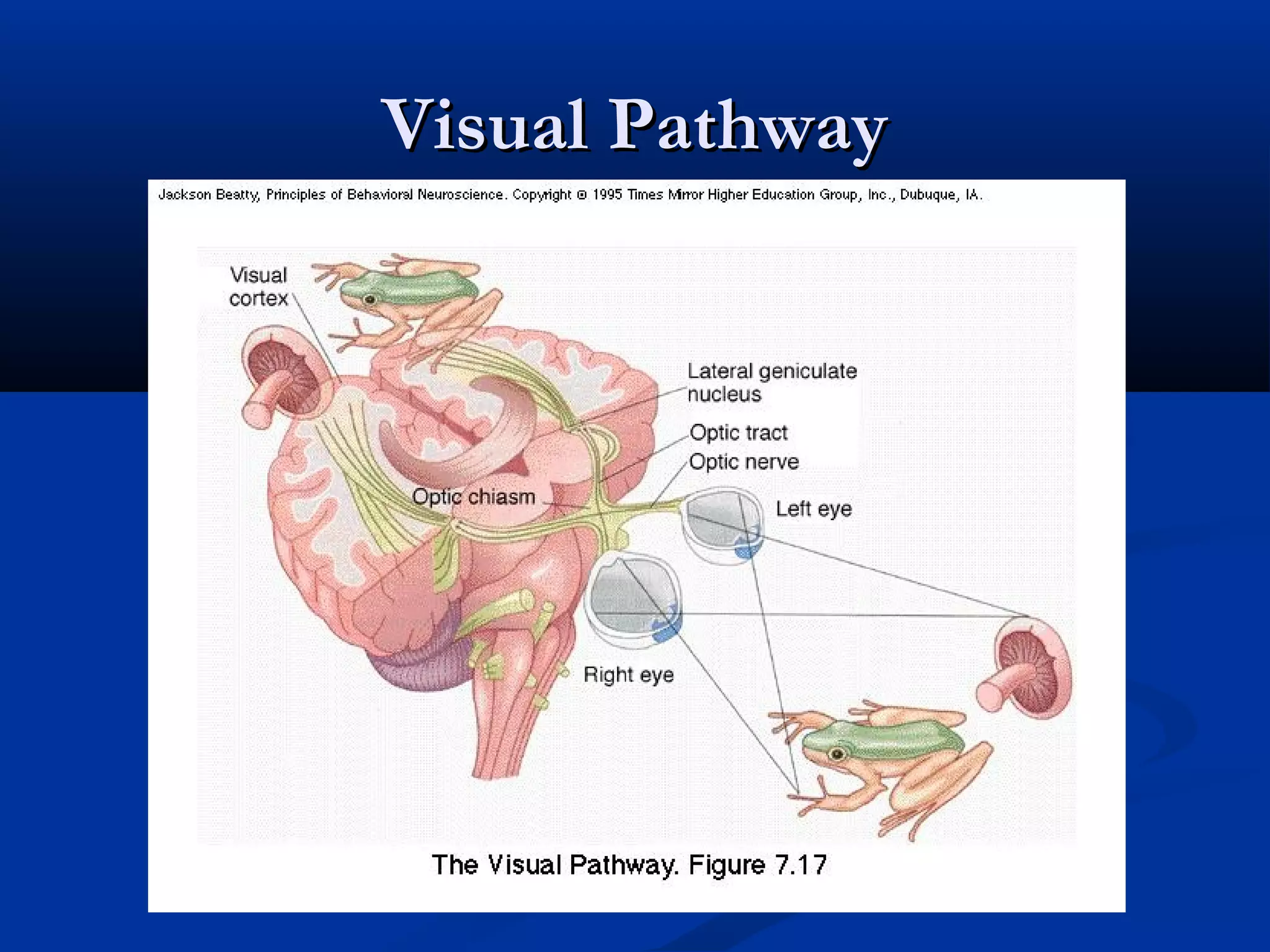
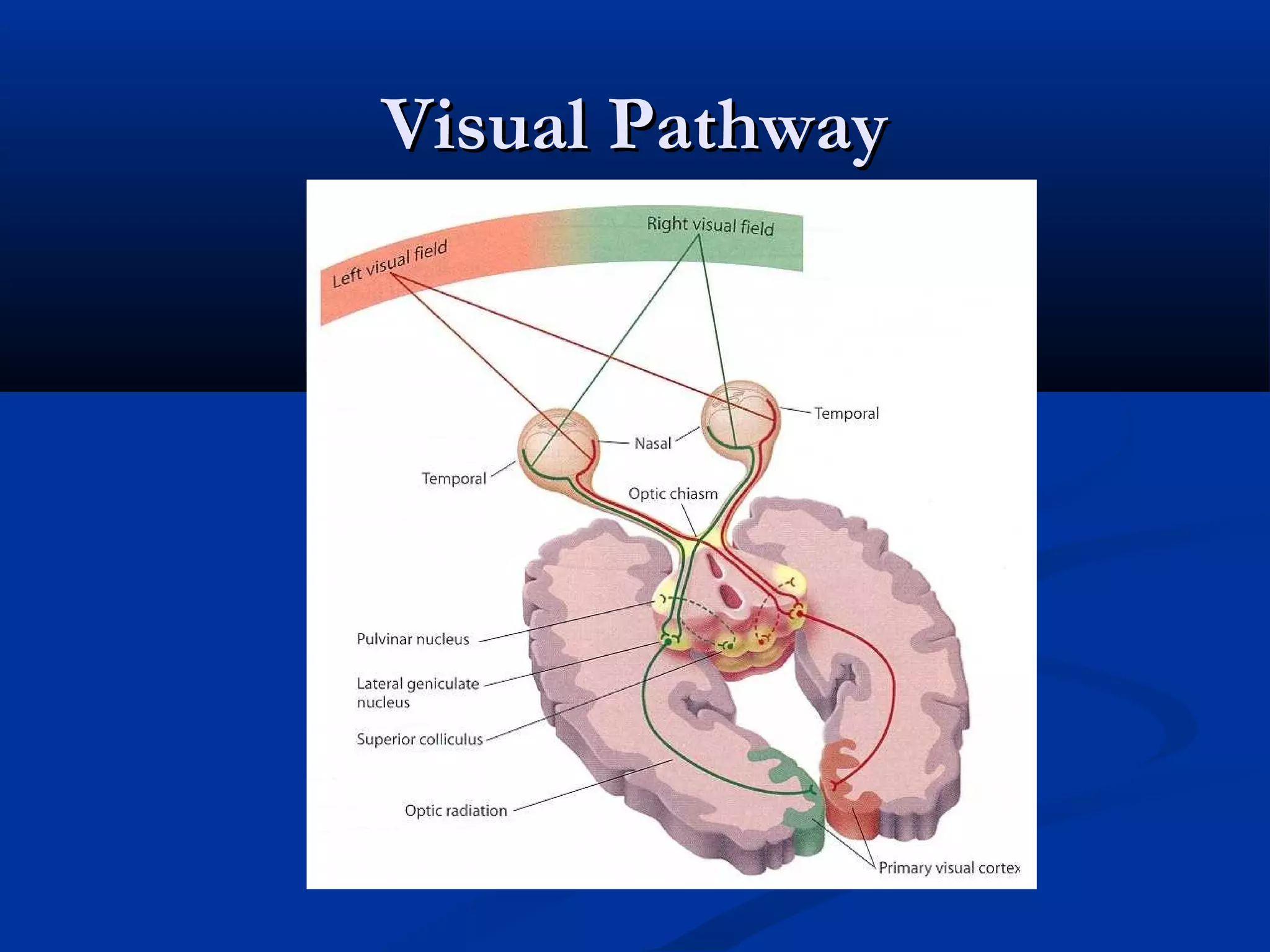
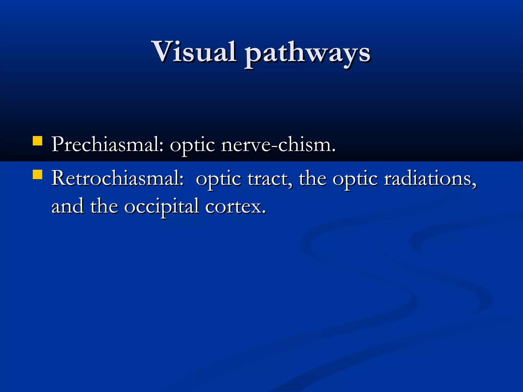
1. Visual pathway lesions can occur prechiasmally in the optic nerve or retrochiasmally in the optic tract, lateral geniculate nucleus, optic radiations, and occipital cortex.
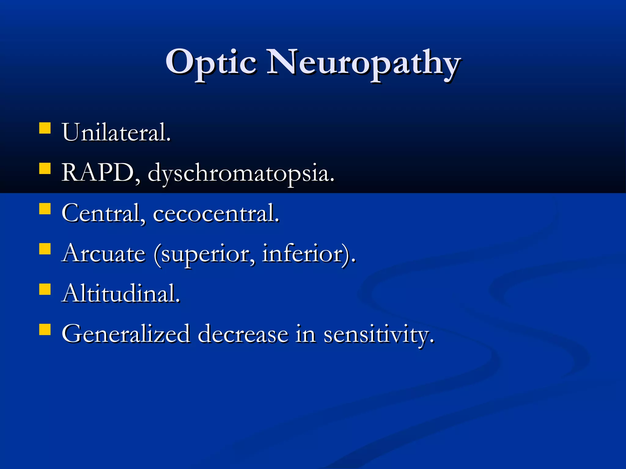
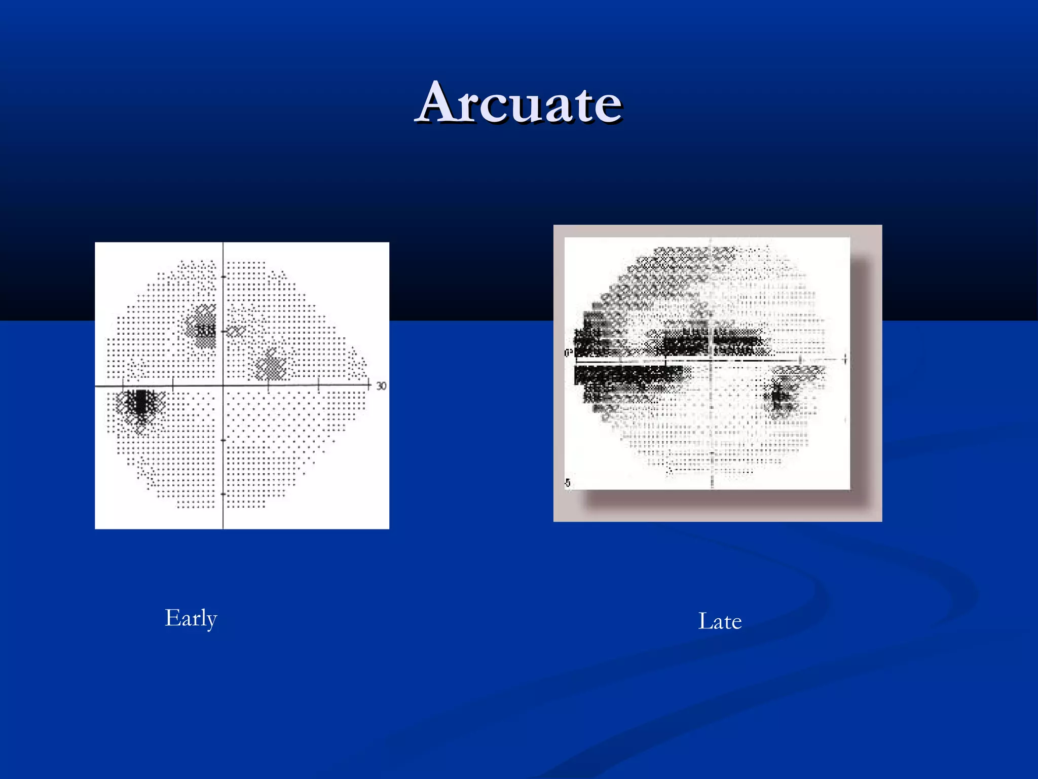
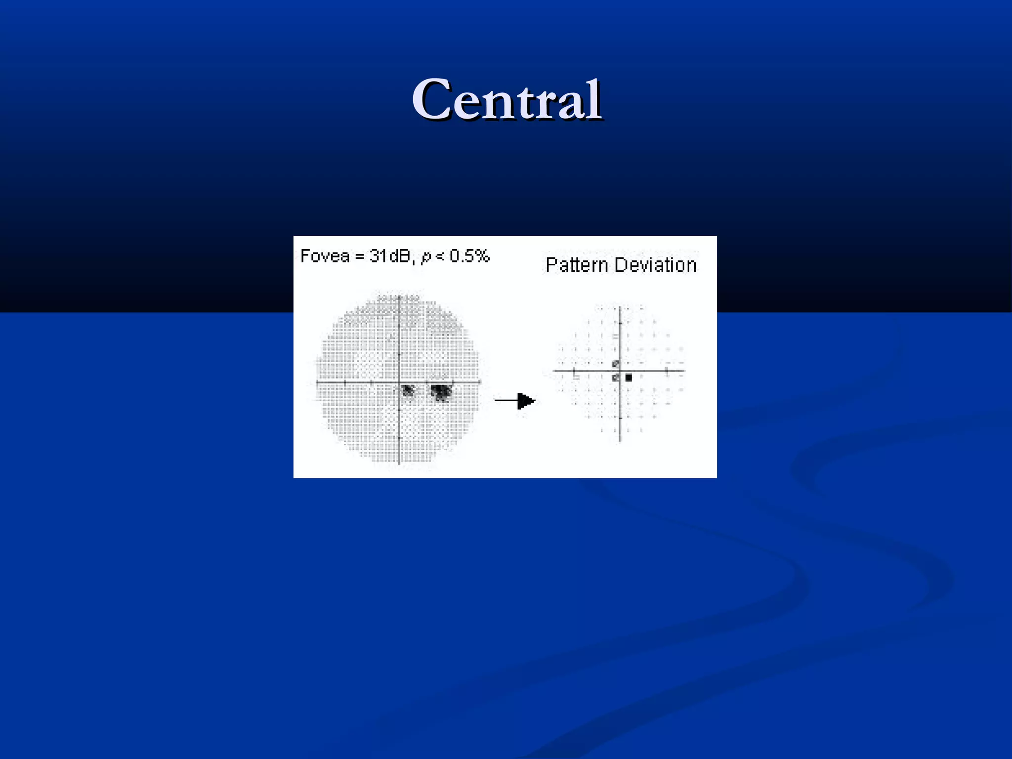
2. Optic neuropathy manifestations include visual field defects such as arcuate scotomas. Optic tract lesions cause incongruous homonymous hemianopsia.
3. Lateral geniculate nucleus lesions result in an incongruous wedge defect pointing toward fixation. Occipital cortex lesions cause homonymous field defects that can be paracentral or peripheral.
Introduction to visual pathways, their anatomy, and significance in understanding visual pathway lesions.
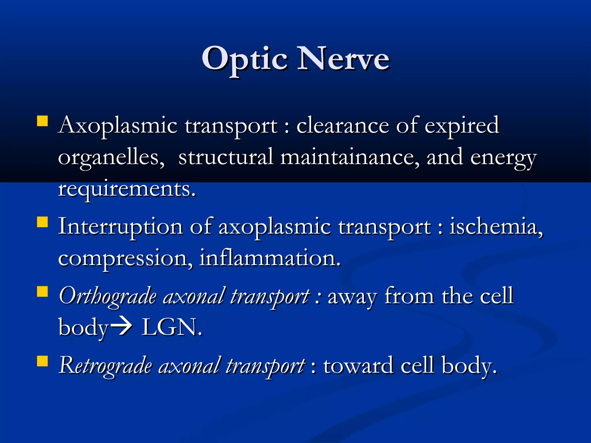
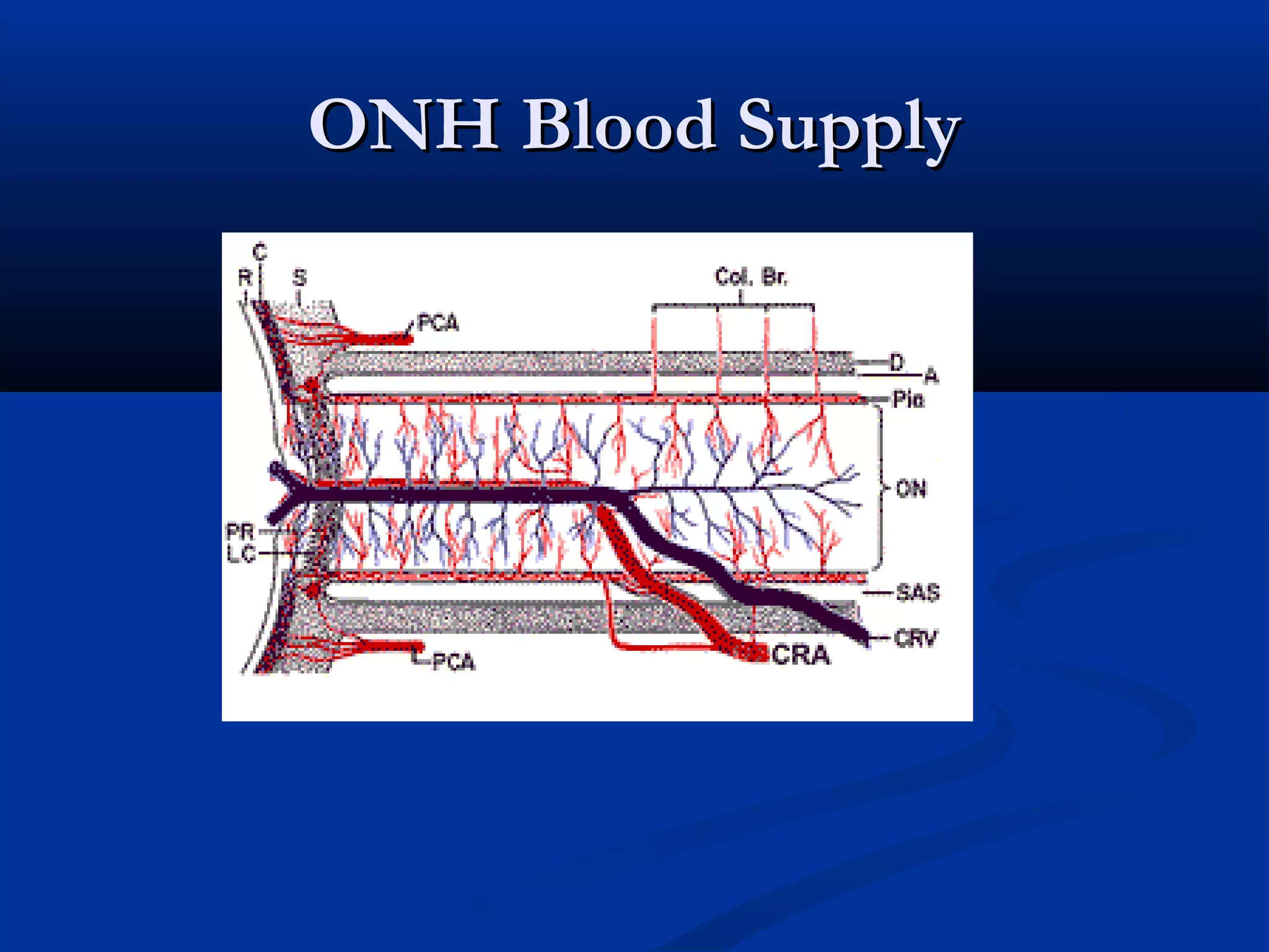
Discusses the components of visual pathways including prechiasmal, retrochiasmal segments, and details about optic nerve structure.
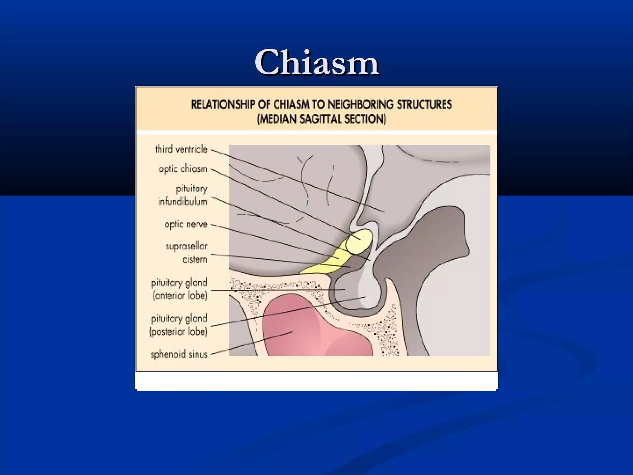
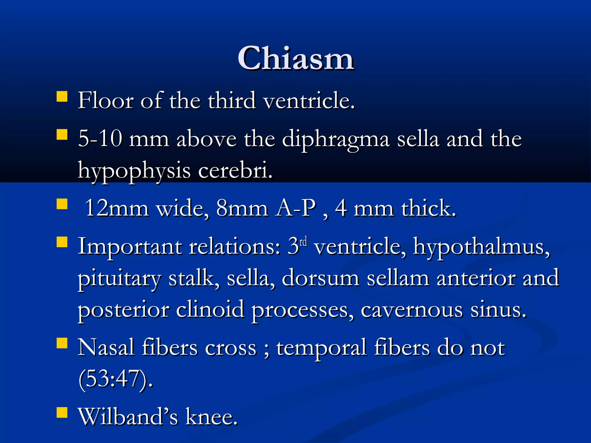
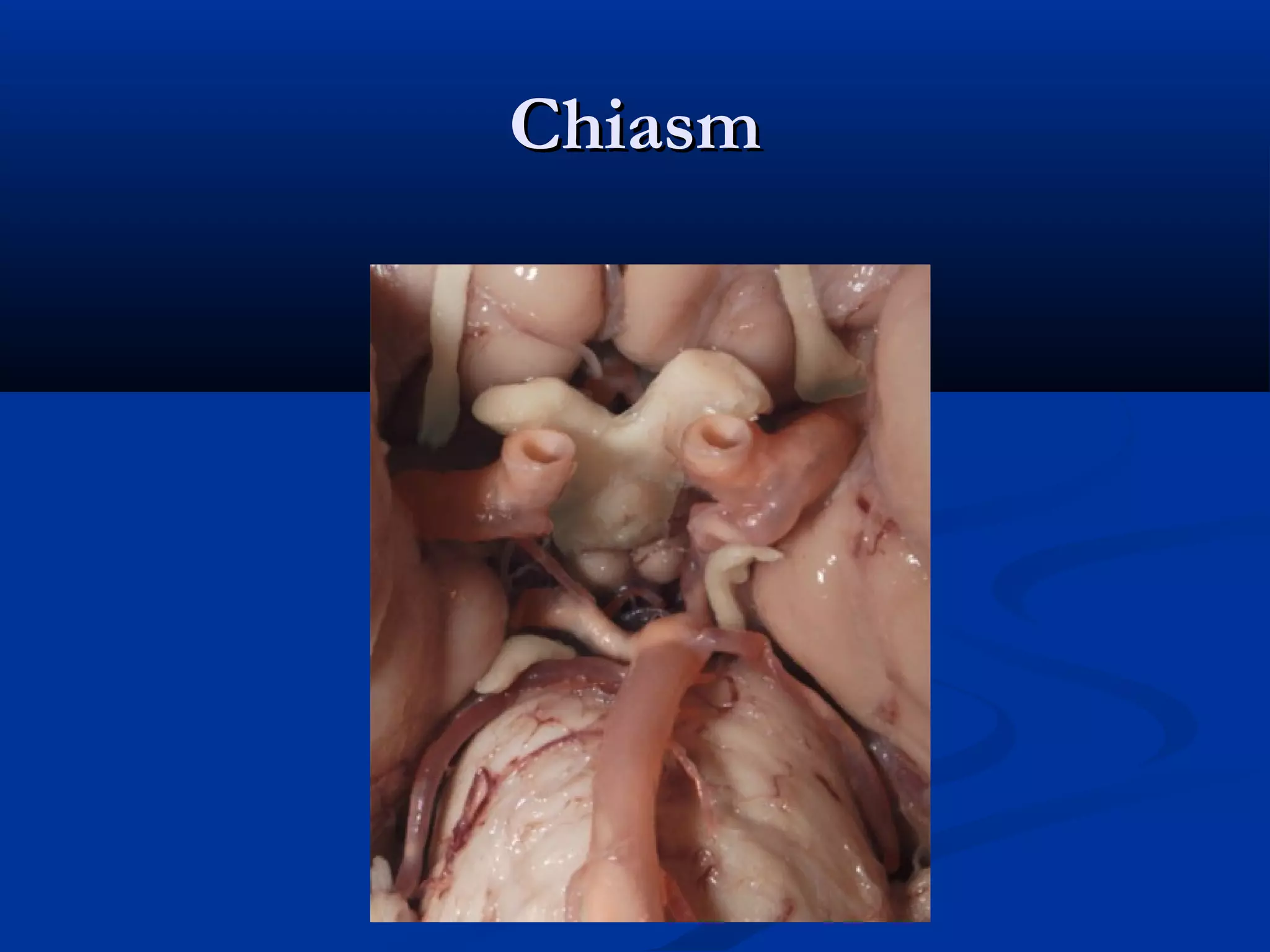
Describes chiasm anatomy, syndromes like junctional scotoma, bitemporal defects, and symptoms.
Lists various causes of chiasmal syndrome including tumors and uncommon conditions leading to visual impairments. Details on various visual field defects including junctional scotoma, bitemporal defect, and optic tract lesions.
Discussions about lesions affecting optic tract, lateral geniculate bodies, optic radiations, and the occipital lobe.
Describes the primary visual cortex, its layers, and distribution of visual fields.
Outlines visual association areas and the ‘what’ and ‘where’ pathways involved in processing vision.
Describes conditions like cortical blindness, dyschromatopsia, and their underlying causes.
Optic NerveOptic Nerve
Axoplasmic transport : clearance of expiredAxoplasmic transport : clearance of expired
organelles, structural maintainance, and energyorganelles, structural maintainance, and energy
requirements.requirements.
Interruption of axoplasmic transport : ischemia,Interruption of axoplasmic transport : ischemia,
compression, inflammation.compression, inflammation.
Orthograde axonal transport :Orthograde axonal transport : away from the cellaway from the cell
bodybody LGN.LGN.
Retrograde axonal transportRetrograde axonal transport : toward cell body.: toward cell body.
Intra-orbital Optic NerveIntra-orbitalOptic Nerve
Myelination (oligodendrocytes).Myelination (oligodendrocytes).
20-30 mm Long.20-30 mm Long.
Axons: mylein and glial cell (metabolic supportAxons: mylein and glial cell (metabolic support
at the nodes of Ranvier).at the nodes of Ranvier).
10.
Intracranalicular Optic NerveIntracranalicularOptic Nerve
Within the two bases of the LWS.Within the two bases of the LWS.
Medial wall of canal forms lateral wall ofMedial wall of canal forms lateral wall of
sphenoid sinus (can be absent !).sphenoid sinus (can be absent !).
Within canal : meninges, ophthalmic artery andWithin canal : meninges, ophthalmic artery and
sympathetic plexus.sympathetic plexus.
10 mm length.10 mm length.
Tight space !Tight space !
Internal carotid artery.Internal carotid artery.
11.
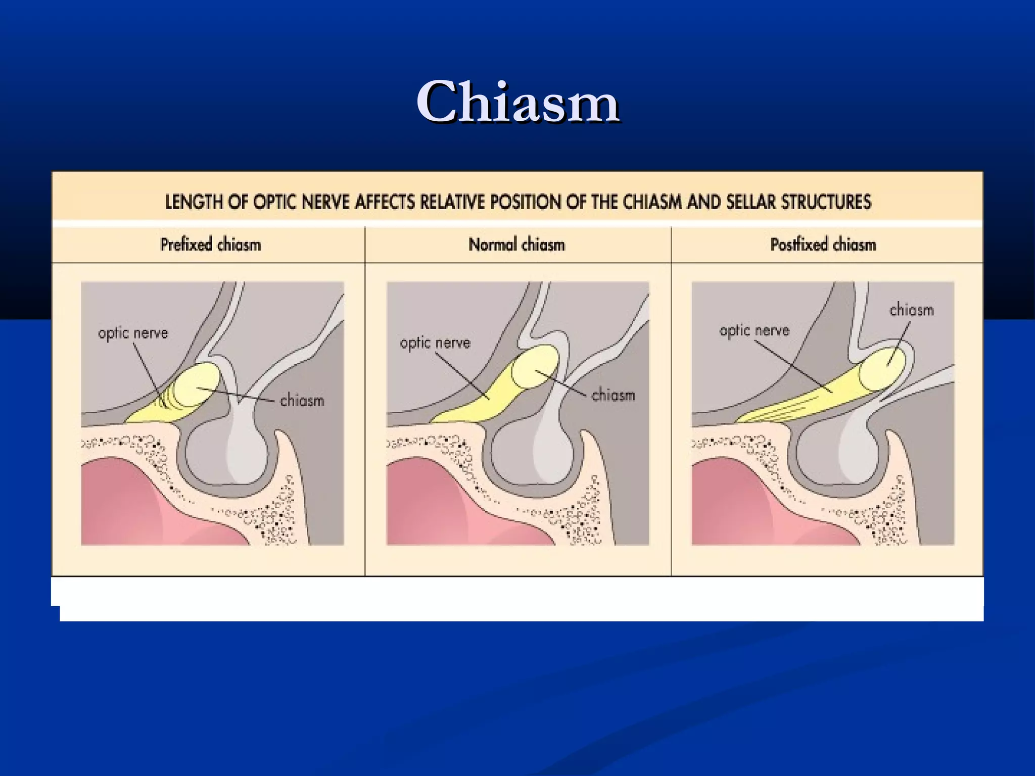
Intracranial Optic NerveIntracranialOptic Nerve
Leaves the cranial end of the optic canalLeaves the cranial end of the optic canal
(medially, backwards, upwards).(medially, backwards, upwards).
4-15 m (depending on the position of chiasm).4-15 m (depending on the position of chiasm).
Upward 45 degree-angle.Upward 45 degree-angle.
Anterior cerebral and anterior comunicatingAnterior cerebral and anterior comunicating
artery lie superior.artery lie superior.
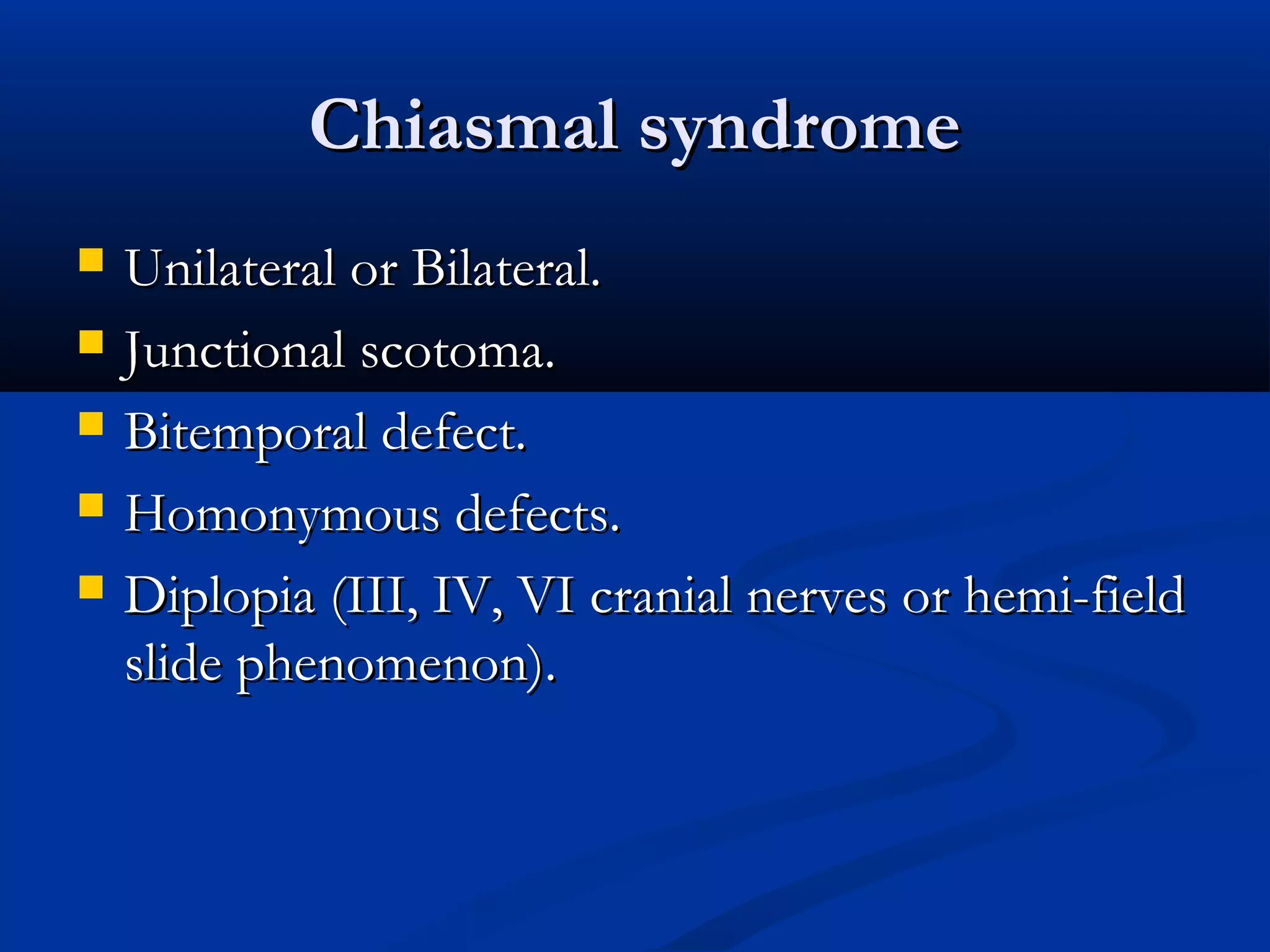
Chiasmal syndromeChiasmal syndrome
Unilateral or Bilateral.Unilateral or Bilateral.
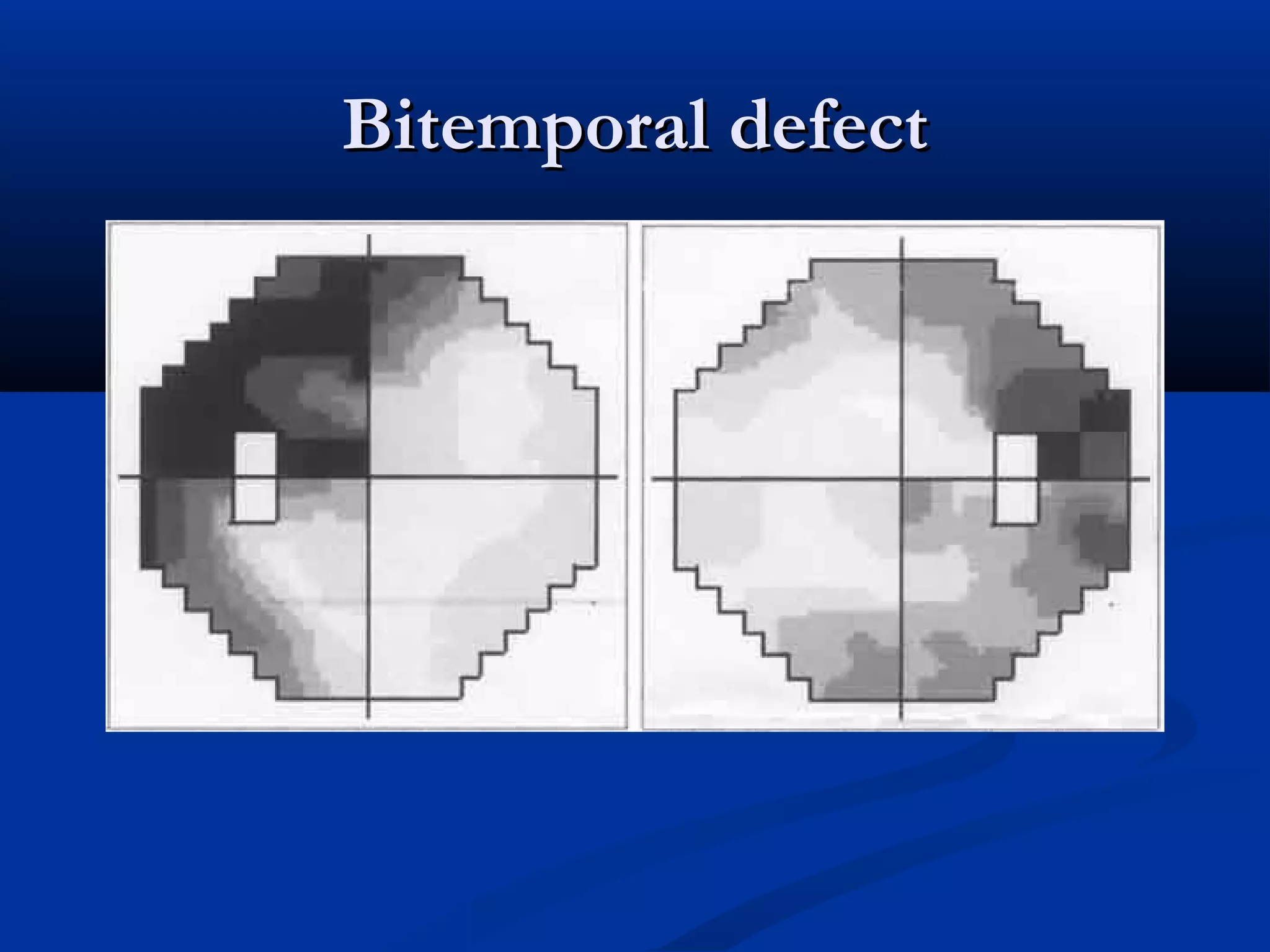
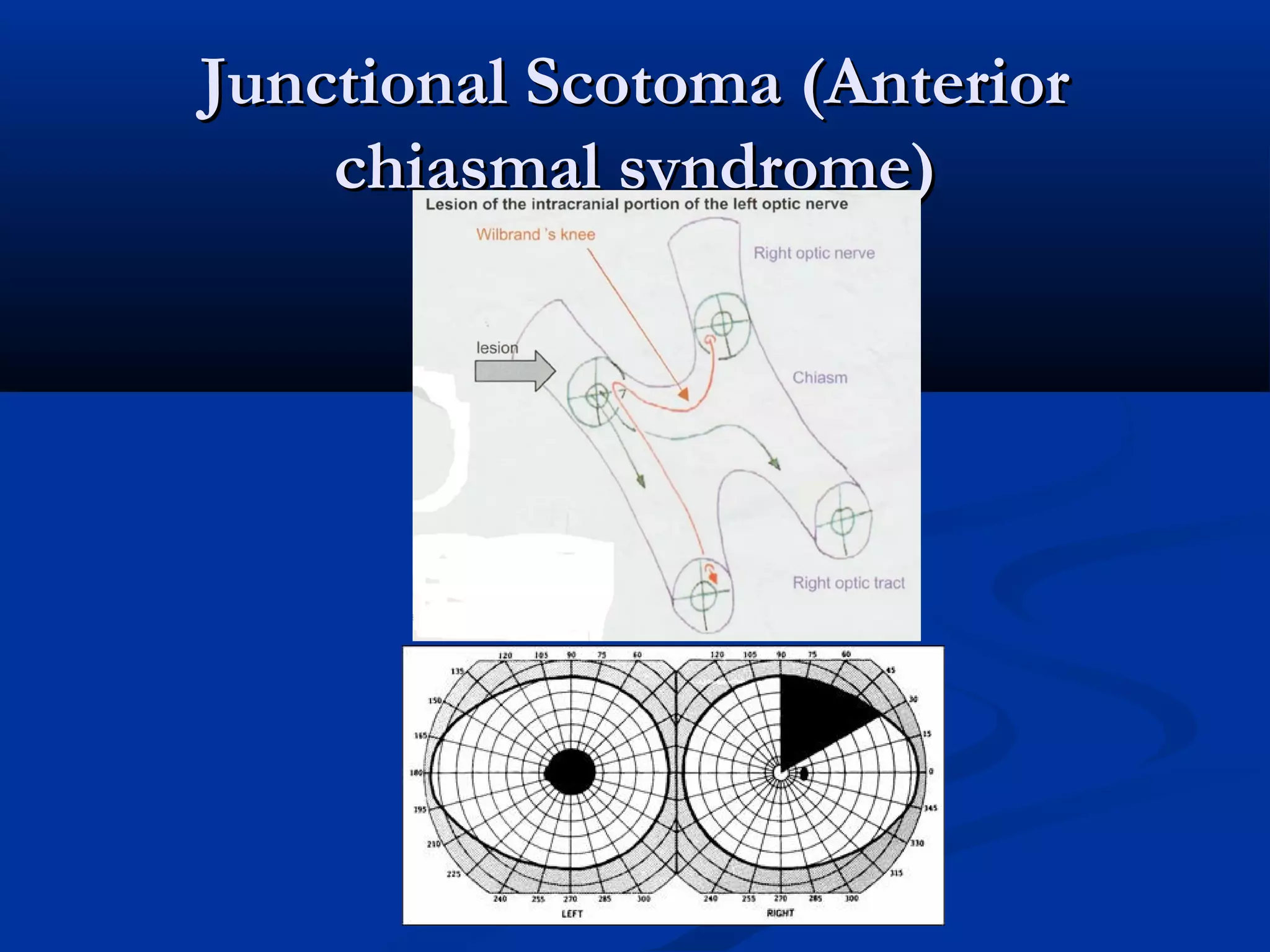
Junctional scotoma.Junctional scotoma.
Bitemporal defect.Bitemporal defect.
Homonymous defects.Homonymous defects.
Diplopia (III, IV, VI cranial nerves or hemi-fieldDiplopia (III, IV, VI cranial nerves or hemi-field
slide phenomenon).slide phenomenon).
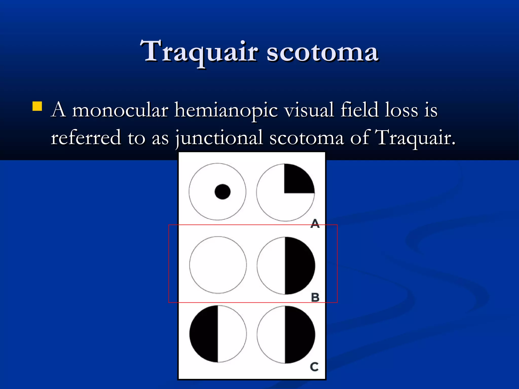
Traquair scotomaTraquair scotoma
A monocular hemianopic visual field loss isA monocular hemianopic visual field loss is
referred to as junctional scotoma of Traquair.referred to as junctional scotoma of Traquair.
23.
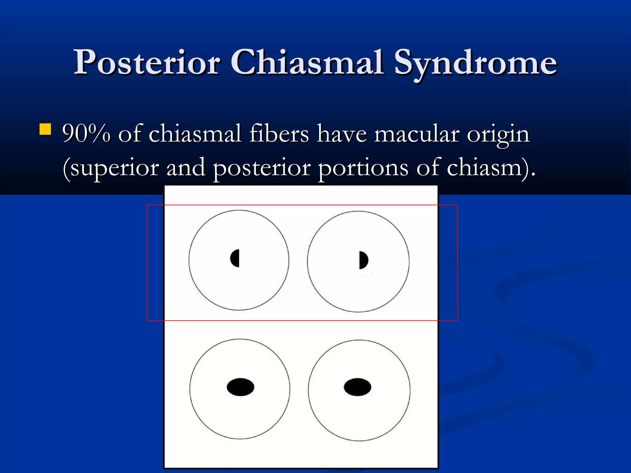
Posterior Chiasmal SyndromePosteriorChiasmal Syndrome
90% of chiasmal fibers have macular origin90% of chiasmal fibers have macular origin
(superior and posterior portions of chiasm).(superior and posterior portions of chiasm).
Retrochiasmal Visual PathwayRetrochiasmalVisual Pathway
LesionsLesions
Bilateral.Bilateral.
Homonymous.Homonymous.
Complete or incomplete.Complete or incomplete.
Congrous or incongrous.Congrous or incongrous.
27.
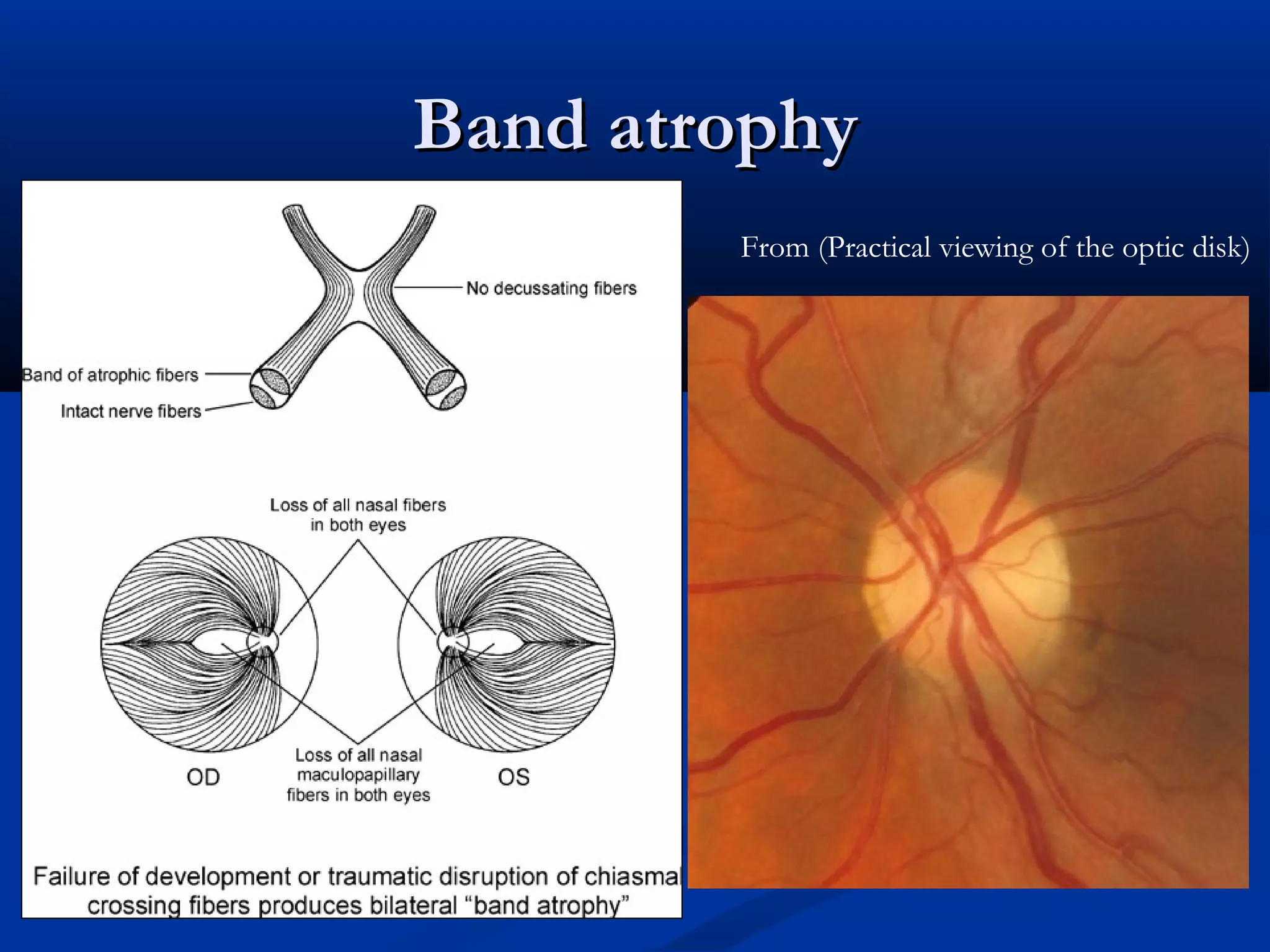
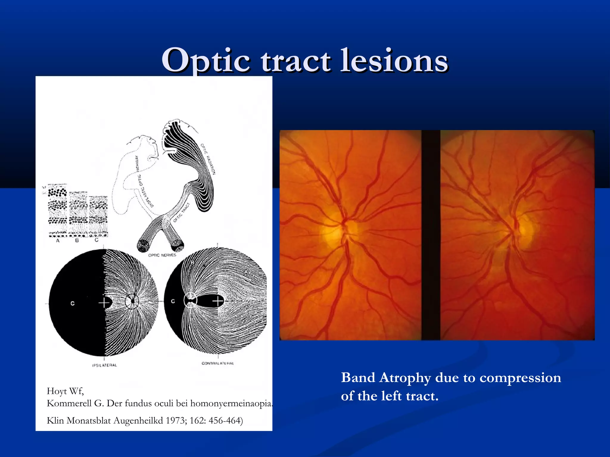
Optic Tract LesionsOpticTract Lesions
Contralateral RAPDContralateral RAPD ((may be an ipsilateralmay be an ipsilateral
afferent pupillary defect if a concomitant opticafferent pupillary defect if a concomitant optic
neuropathy existsneuropathy exists))
A specific form of optic atrophy (band atrophy)A specific form of optic atrophy (band atrophy)
due to the involvement of nasal fibers (temporaldue to the involvement of nasal fibers (temporal
field) in the contralateral eyefield) in the contralateral eye
An incongruous homonymous hemianopsia.An incongruous homonymous hemianopsia.
28.
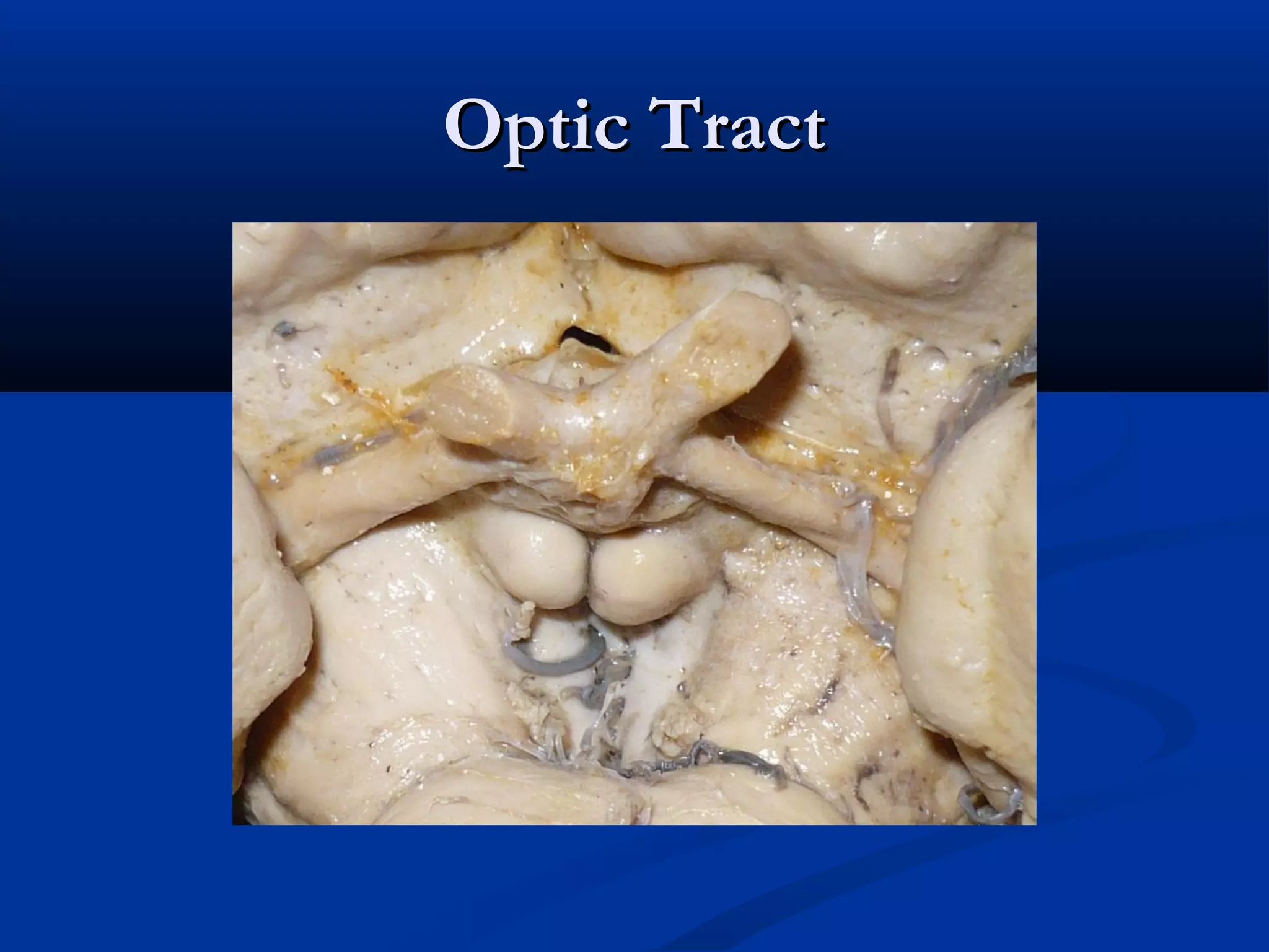
Optic TractOptic Tract
Travel around the cerebral peduncles at dorsalTravel around the cerebral peduncles at dorsal
midbrain.midbrain.
Divides into lateral rootDivides into lateral root LGN , and a smallerLGN , and a smaller
medial rootmedial root pretectal area (pupillary lightpretectal area (pupillary light
reflex)reflex)
Optic tract lesionsOptictract lesions
Band Atrophy due to compression
of the left tract.Hoyt Wf,
Kommerell G. Der fundus oculi bei homonyermeinaopia.
Klin Monatsblat Augenheilkd 1973; 162: 456-464)
31.
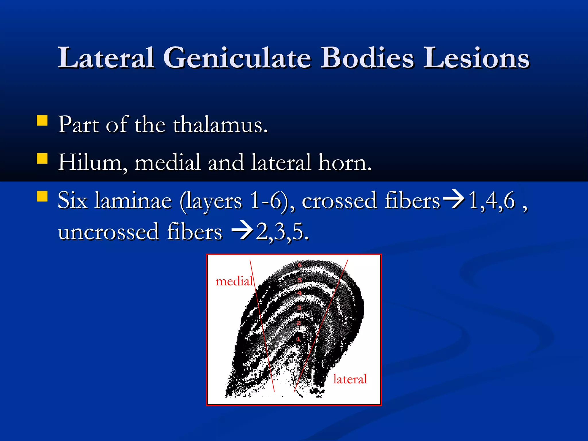
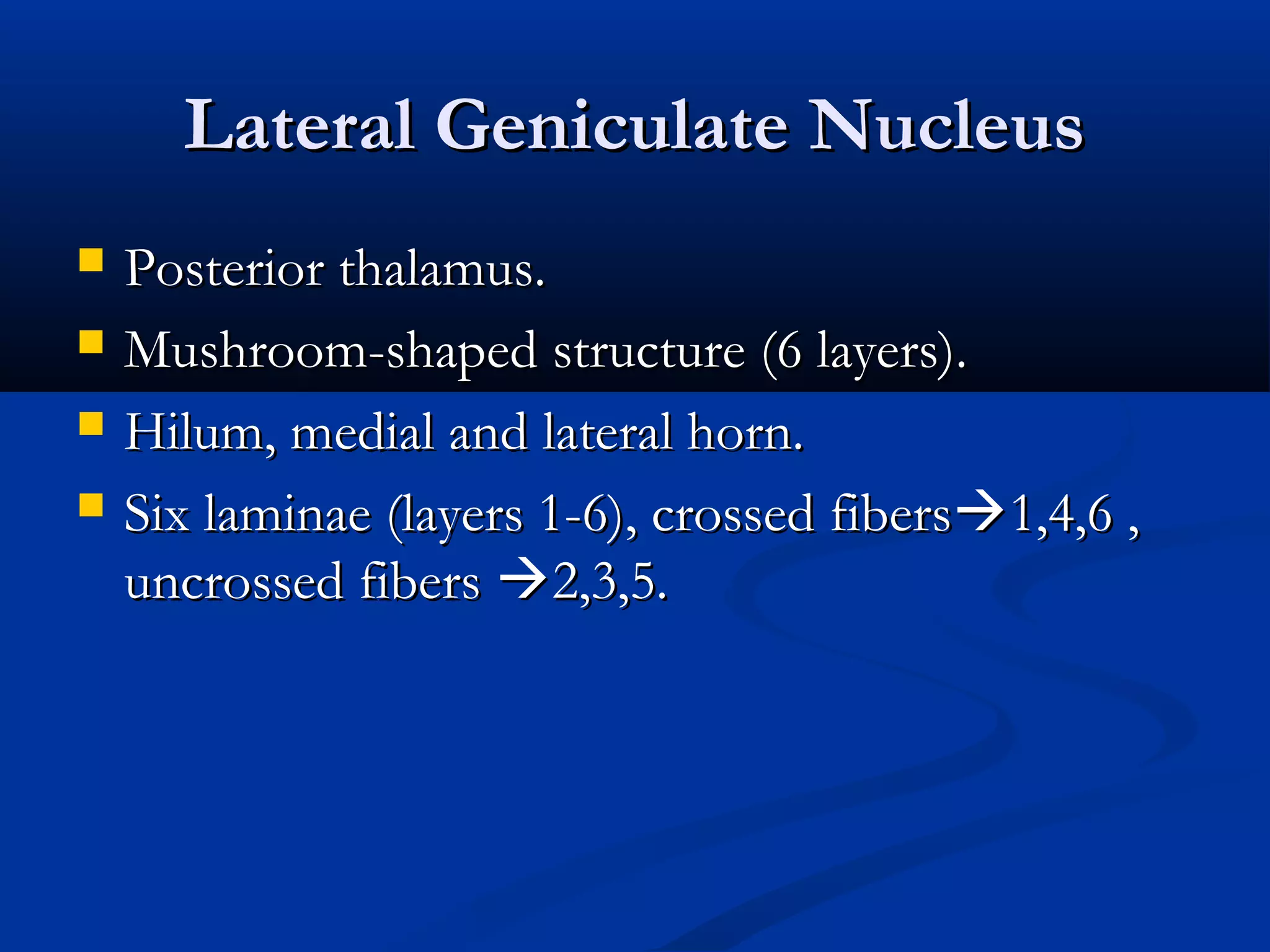
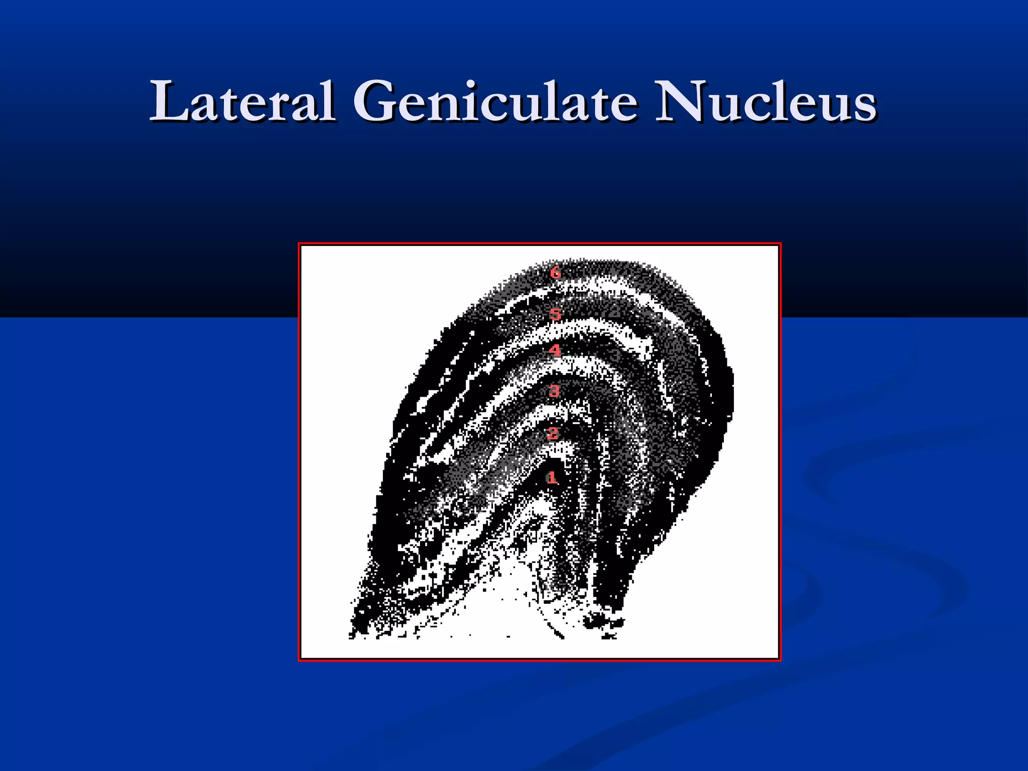
Lateral Geniculate BodiesLesionsLateral Geniculate Bodies Lesions
Part of the thalamus.Part of the thalamus.
Hilum, medial and lateral horn.Hilum, medial and lateral horn.
Six laminae (layers 1-6), crossed fibersSix laminae (layers 1-6), crossed fibers1,4,6 ,1,4,6 ,
uncrossed fibersuncrossed fibers 2,3,5.2,3,5.
medial
lateral
32.
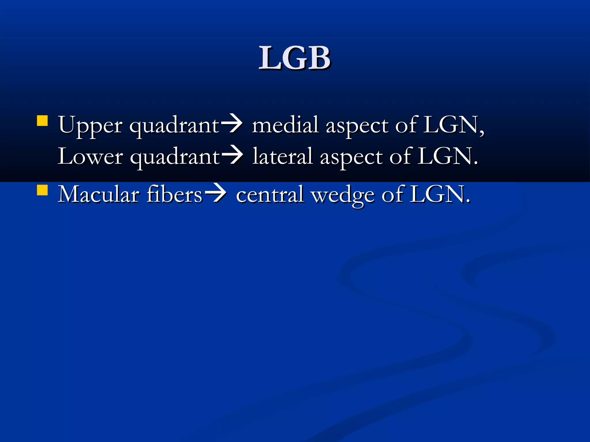
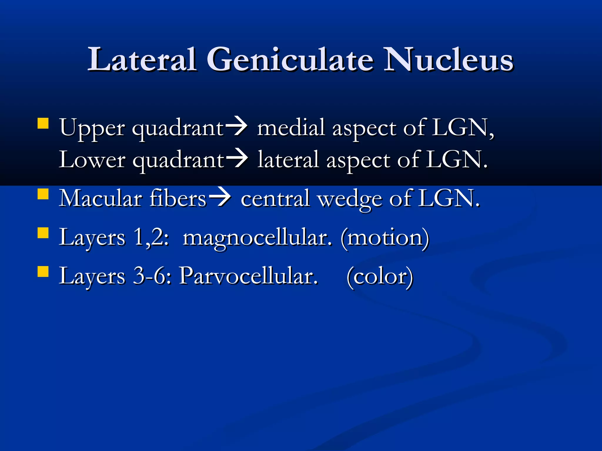
LGBLGB
Upper quadrantUpperquadrant medial aspect of LGN,medial aspect of LGN,
Lower quadrantLower quadrant lateral aspect of LGN.lateral aspect of LGN.
Macular fibersMacular fibers central wedge of LGN.central wedge of LGN.
33.
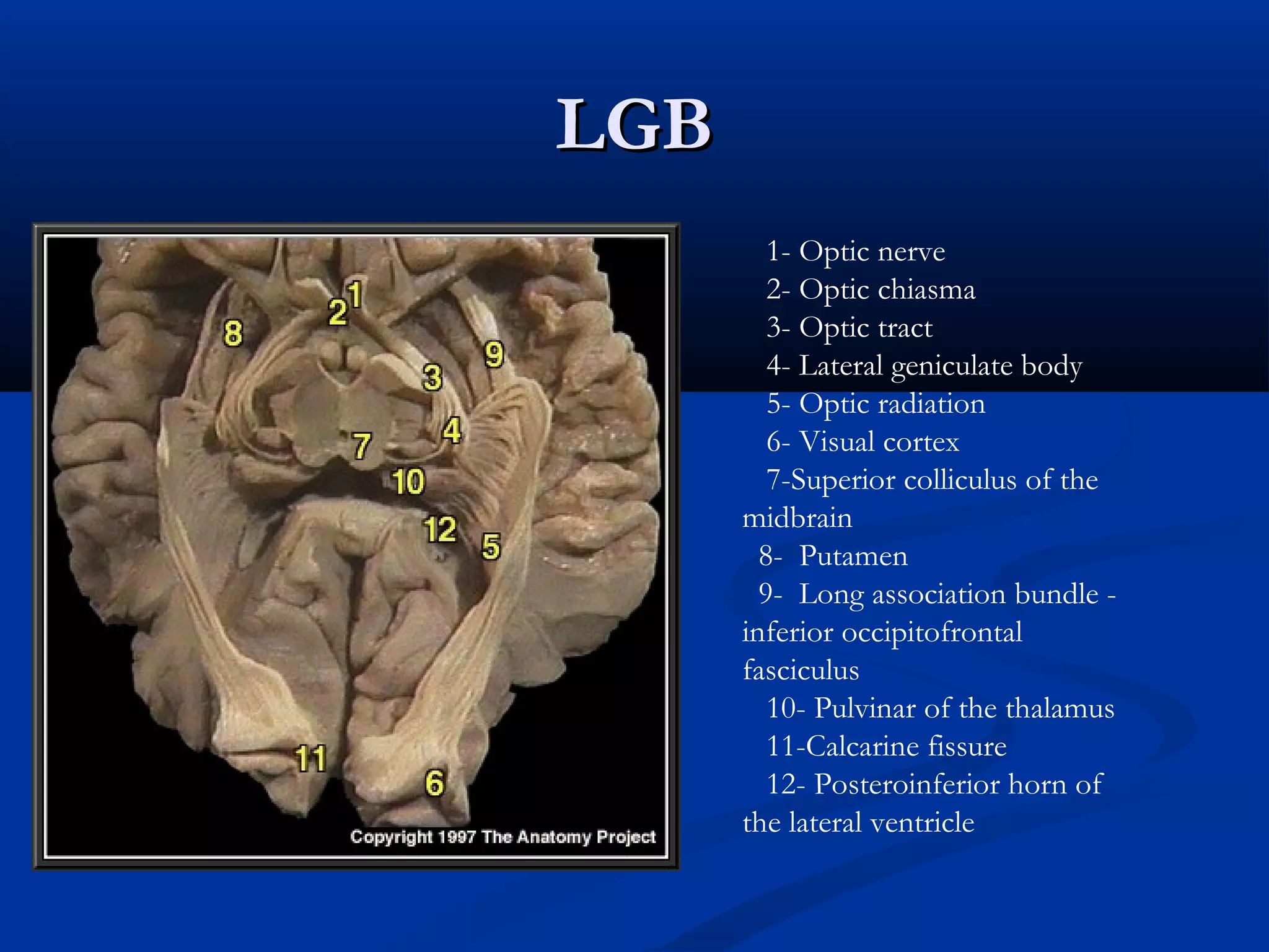
LGBLGB
1- Optic nerve
2-Optic chiasma
3- Optic tract
4- Lateral geniculate body
5- Optic radiation
6- Visual cortex
7-Superior colliculus of the
midbrain
8- Putamen
9- Long association bundle -
inferior occipitofrontal
fasciculus
10- Pulvinar of the thalamus
11-Calcarine fissure
12- Posteroinferior horn of
the lateral ventricle
Lateral Geniculate NucleusLateralGeniculate Nucleus
Upper quadrantUpper quadrant medial aspect of LGN,medial aspect of LGN,
Lower quadrantLower quadrant lateral aspect of LGN.lateral aspect of LGN.
Macular fibersMacular fibers central wedge of LGN.central wedge of LGN.
Layers 1,2: magnocellular. (motion)Layers 1,2: magnocellular. (motion)
Layers 3-6: Parvocellular. (color)Layers 3-6: Parvocellular. (color)
37.
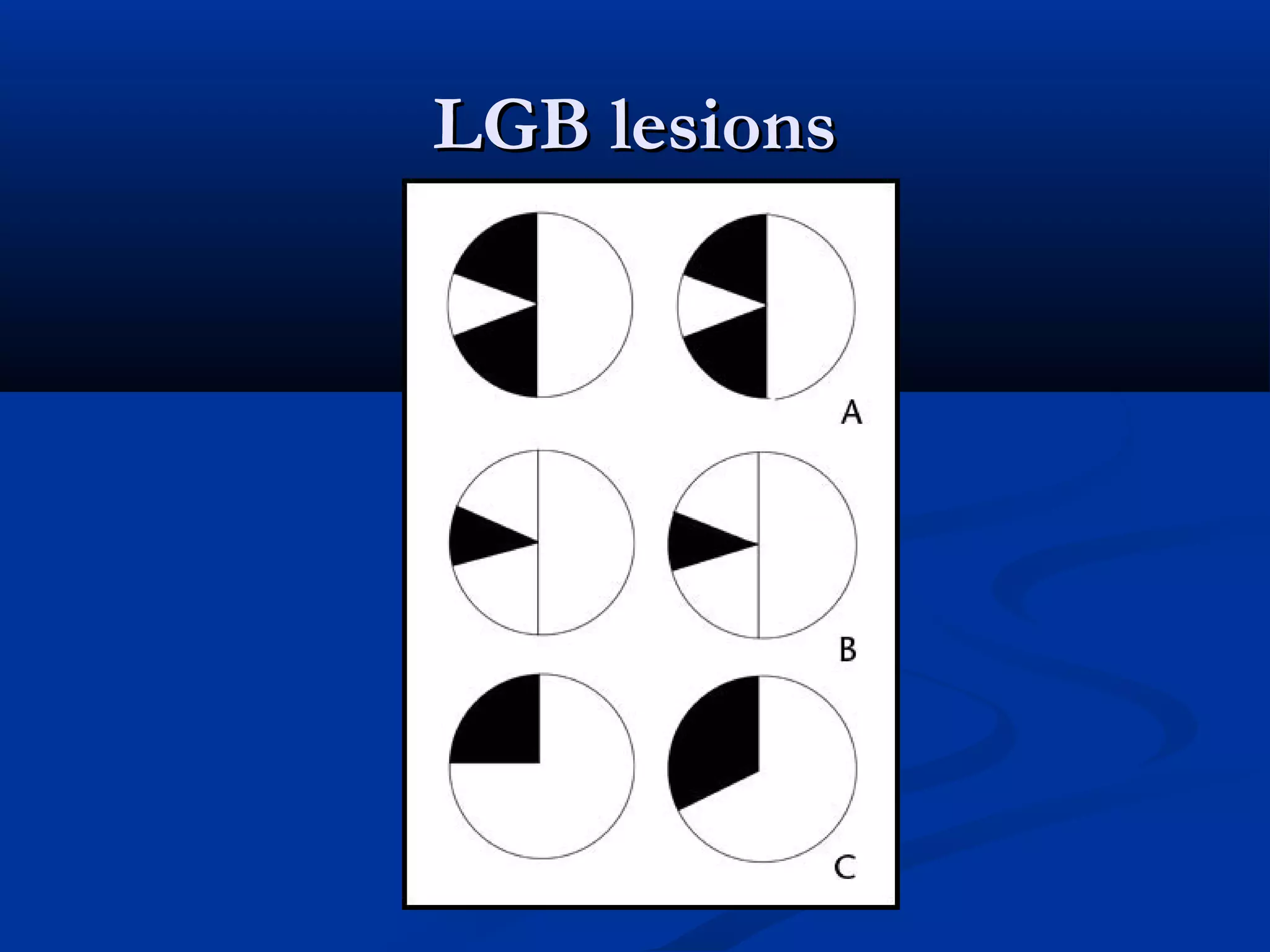
LGB lesionsLGB lesions
An incongruous wedge defect tending to pointAn incongruous wedge defect tending to point
toward fixationtoward fixation ((spears to fixationspears to fixation))
Usually complete or nearly complete fieldUsually complete or nearly complete field
homonymous defect.homonymous defect.
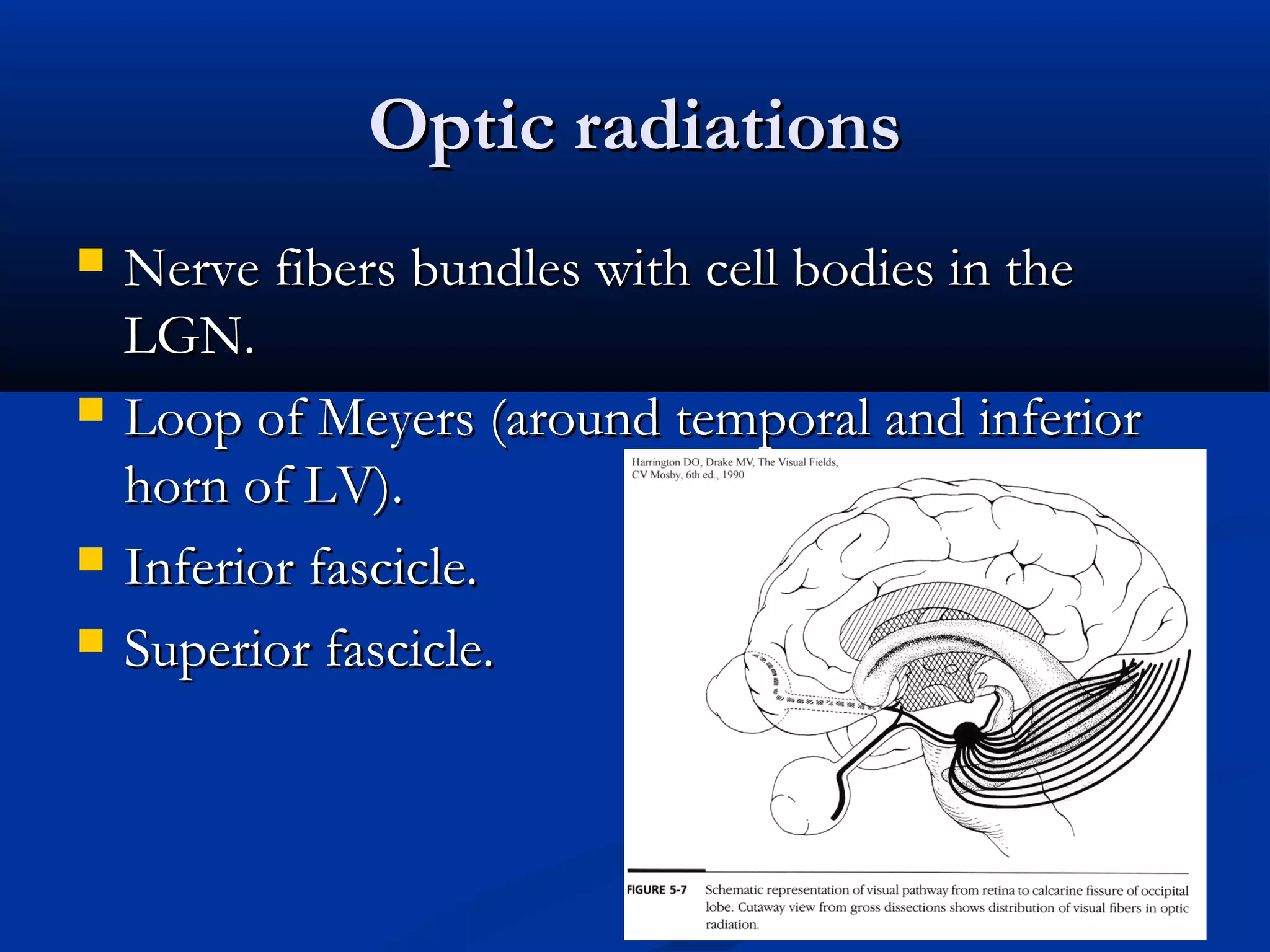
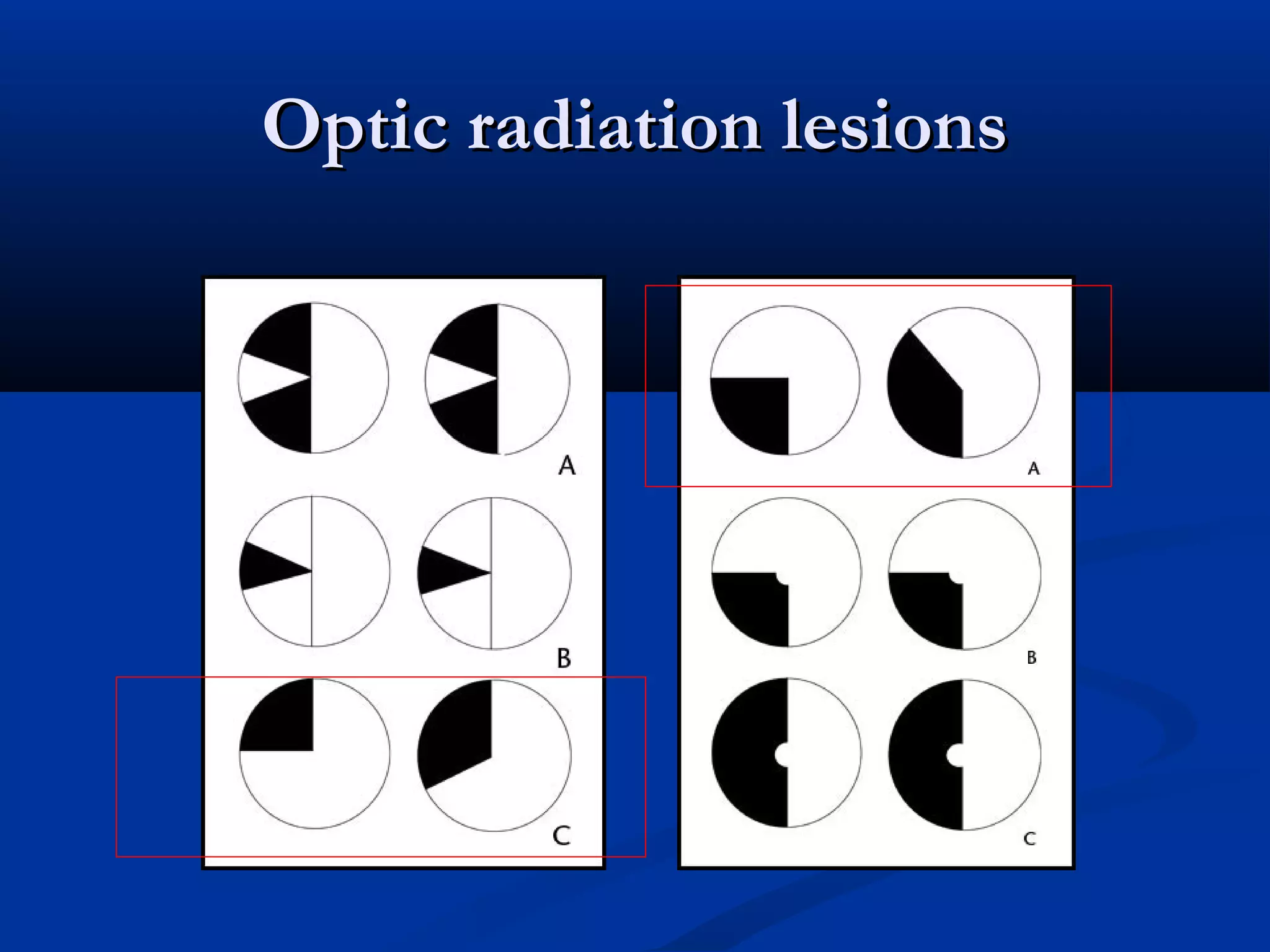
Optic radiationsOptic radiations
Nerve fibers bundles with cell bodies in theNerve fibers bundles with cell bodies in the
LGN.LGN.
Loop of Meyers (around temporal and inferiorLoop of Meyers (around temporal and inferior
horn of LV).horn of LV).
Inferior fascicle.Inferior fascicle.
Superior fascicle.Superior fascicle.
40.
Optic radiationsOptic radiations
Inferior fascicleInferior fascicle anterior pole of temporalanterior pole of temporal
lobelobe lower calcarine cortex.lower calcarine cortex.
Superior fascicleSuperior fascicle parietal lobeparietal lobe upperupper
calacrine cortex.calacrine cortex.
41.
Parietal lesionsParietal lesions
““Pie on the floor” homonynous defect.Pie on the floor” homonynous defect.
Associated neurologic signs and symptomsAssociated neurologic signs and symptoms
(e.g., hemiplegia, hemisensory loss, visual, or(e.g., hemiplegia, hemisensory loss, visual, or
neglect) may be present .neglect) may be present .
42.
Anterior temporal lobeAnteriortemporal lobe
““Pie on the sky” homonymous.Pie on the sky” homonymous.
Often incongrous.Often incongrous.
Seizures, hemiparesis, hemianesthesia.Seizures, hemiparesis, hemianesthesia.
Contralateral neglect (Non-dominant).Contralateral neglect (Non-dominant).
Aphasia (Dominant).Aphasia (Dominant).
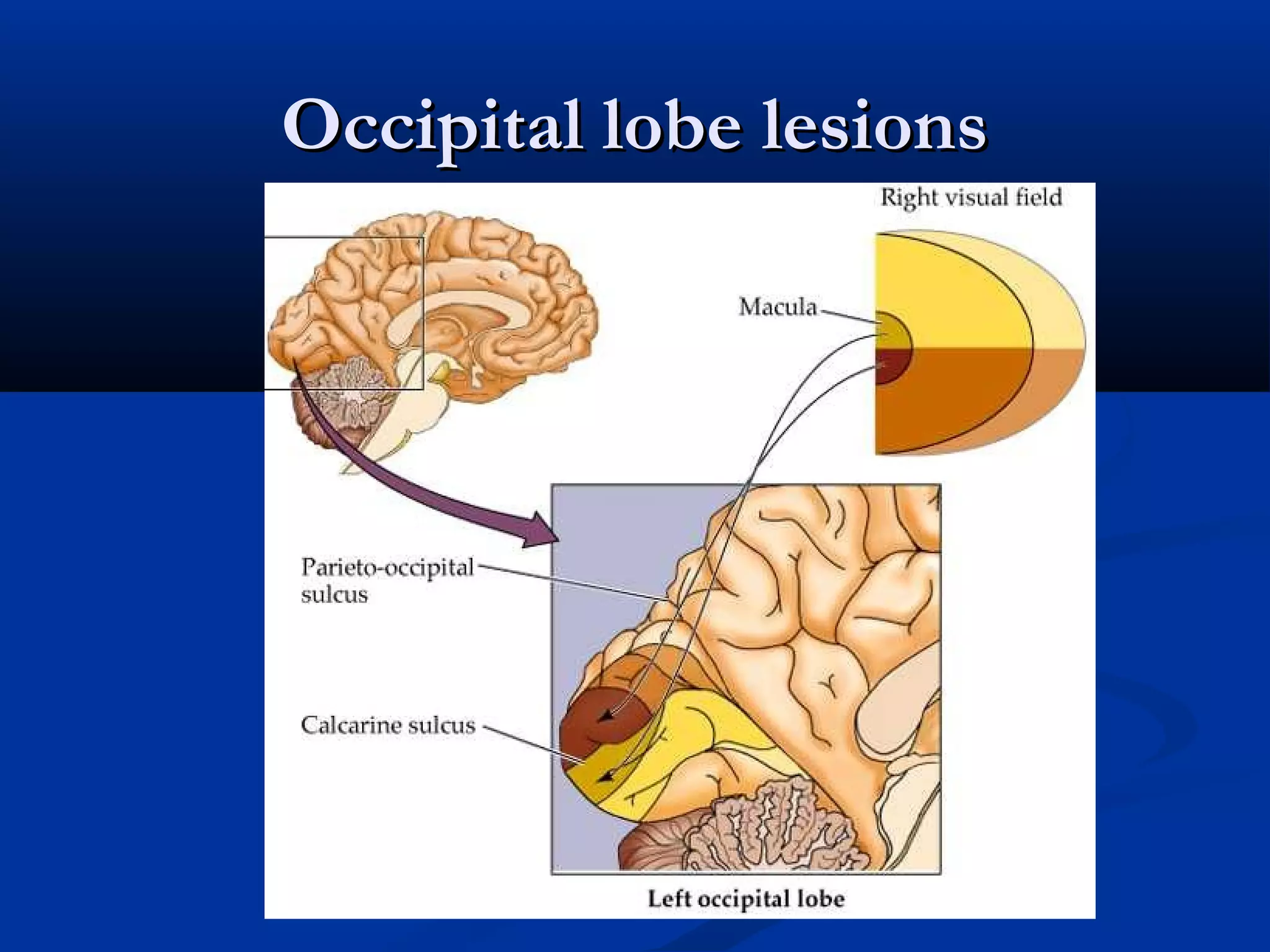
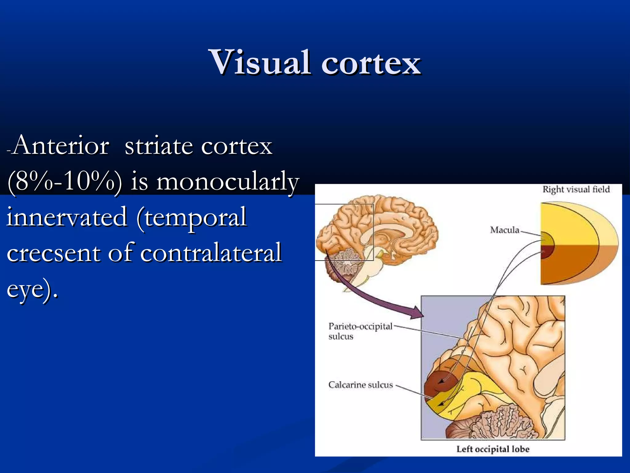
Primary Visual Cortex( V1)Primary Visual Cortex ( V1)
Upper bank and lower bank (Calcarine fissure).Upper bank and lower bank (Calcarine fissure).
Inferior visual filed (upper bank) , SuperiorInferior visual filed (upper bank) , Superior
visual field (lower bank).visual field (lower bank).
Macular projections represented by 50%-60% ofMacular projections represented by 50%-60% of
the area of the calcarine cortex.the area of the calcarine cortex.
Occipital tip is for foveal vision.Occipital tip is for foveal vision.
47.
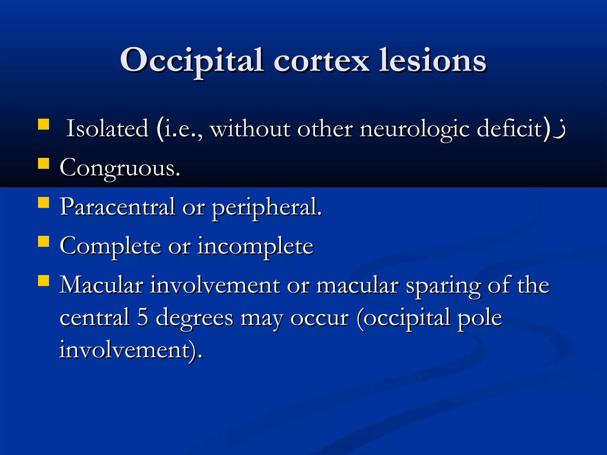
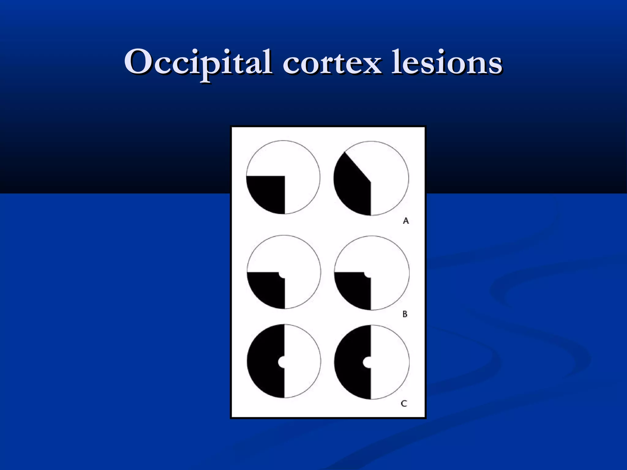
Occipital cortex lesionsOccipitalcortex lesions
IsolatedIsolated ((ii..ee.., without other neurologic deficit, without other neurologic deficit))زز
Congruous.Congruous.
Paracentral or peripheral.Paracentral or peripheral.
Complete or incompleteComplete or incomplete
Macular involvement or macular sparing of theMacular involvement or macular sparing of the
central 5 degrees may occur (occipital polecentral 5 degrees may occur (occipital pole
involvement).involvement).
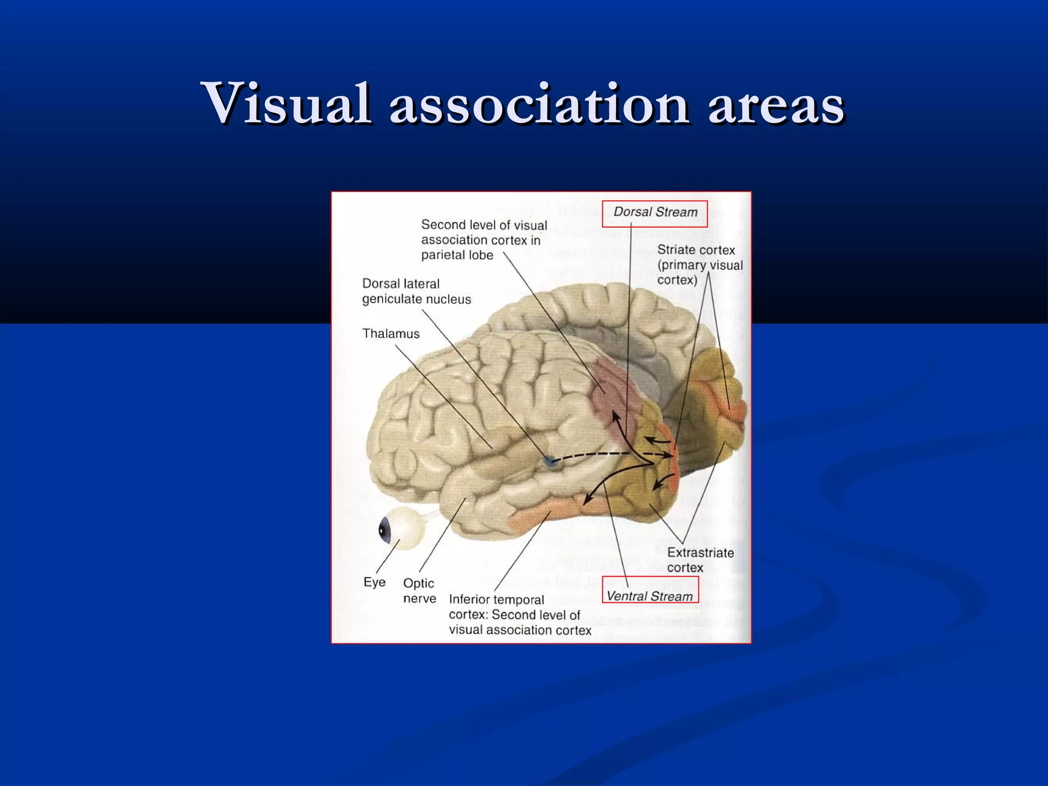
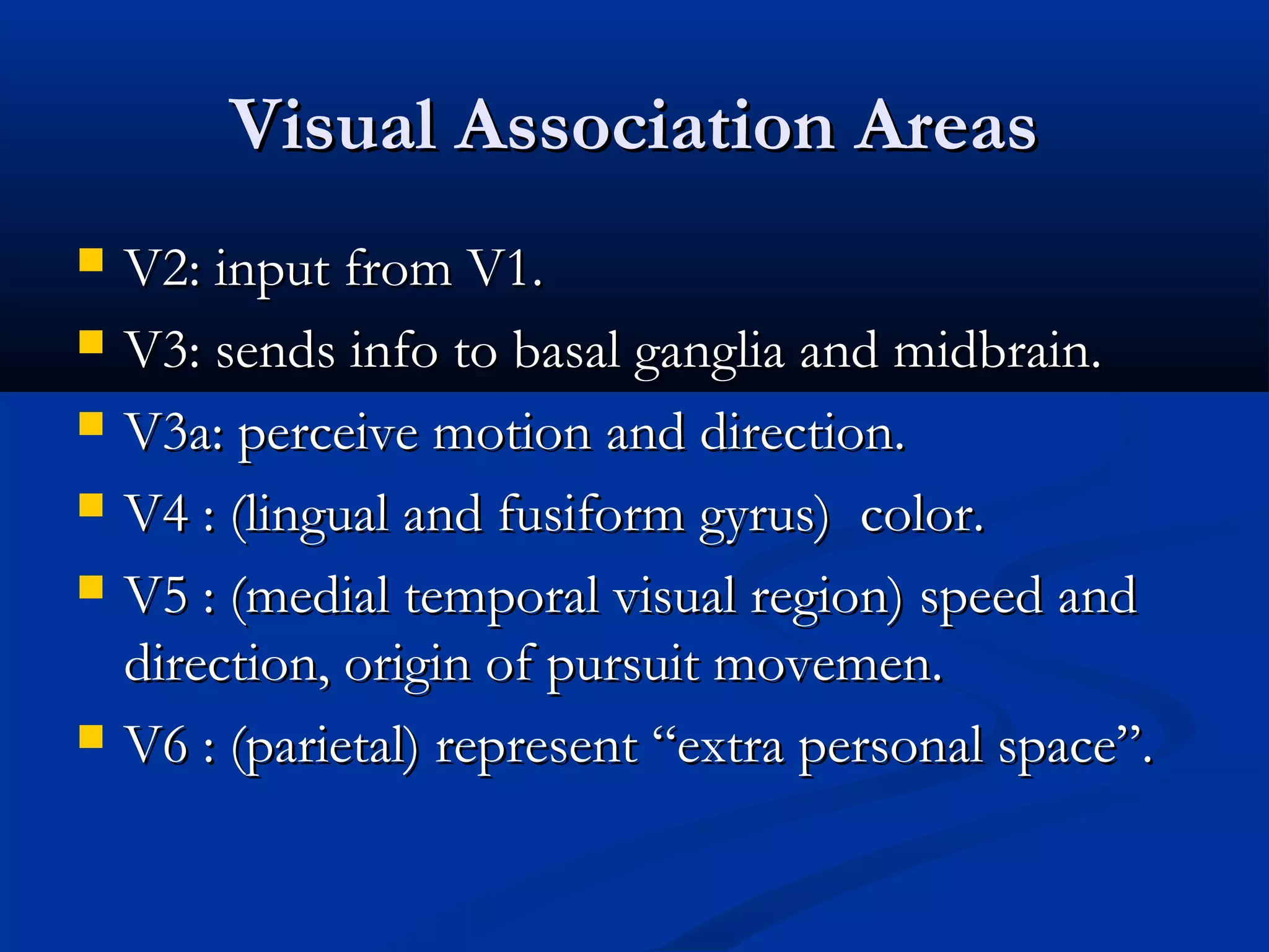
Visual Association AreasVisualAssociation Areas
V2: input from V1.V2: input from V1.
V3: sends info to basal ganglia and midbrain.V3: sends info to basal ganglia and midbrain.
V3a: perceive motion and direction.V3a: perceive motion and direction.
V4 : (lingual and fusiform gyrus) color.V4 : (lingual and fusiform gyrus) color.
V5 : (medial temporal visual region) speed andV5 : (medial temporal visual region) speed and
direction, origin of pursuit movemen.direction, origin of pursuit movemen.
V6 : (parietal) represent “extra personal space”.V6 : (parietal) represent “extra personal space”.
52.
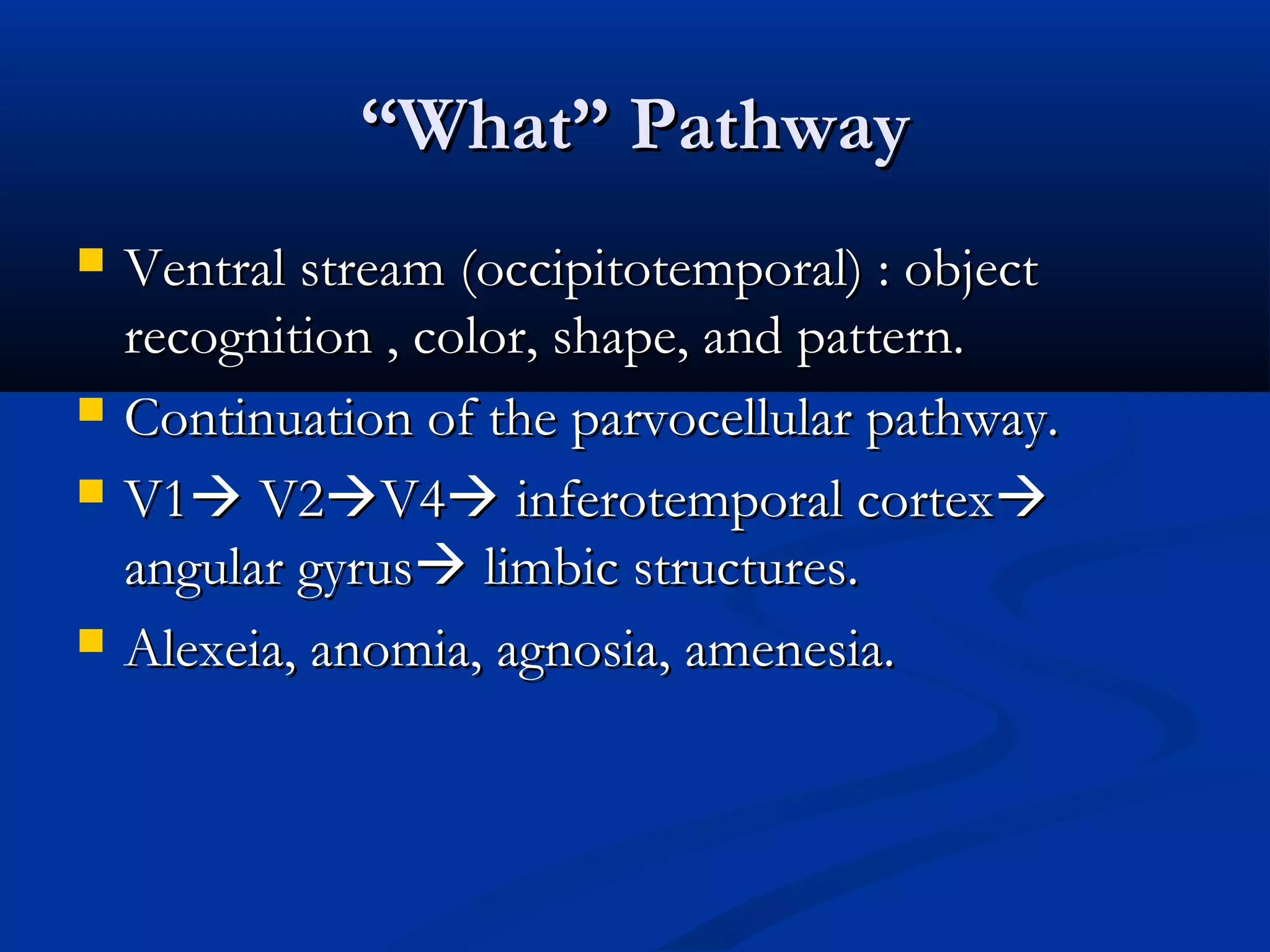
““What” PathwayWhat” Pathway
Ventral stream (occipitotemporal) : objectVentral stream (occipitotemporal) : object
recognition , color, shape, and pattern.recognition , color, shape, and pattern.
Continuation of the parvocellular pathway.Continuation of the parvocellular pathway.
V1V1 V2V2V4V4 inferotemporal cortexinferotemporal cortex
angular gyrusangular gyrus limbic structures.limbic structures.
Alexeia, anomia, agnosia, amenesia.Alexeia, anomia, agnosia, amenesia.
53.
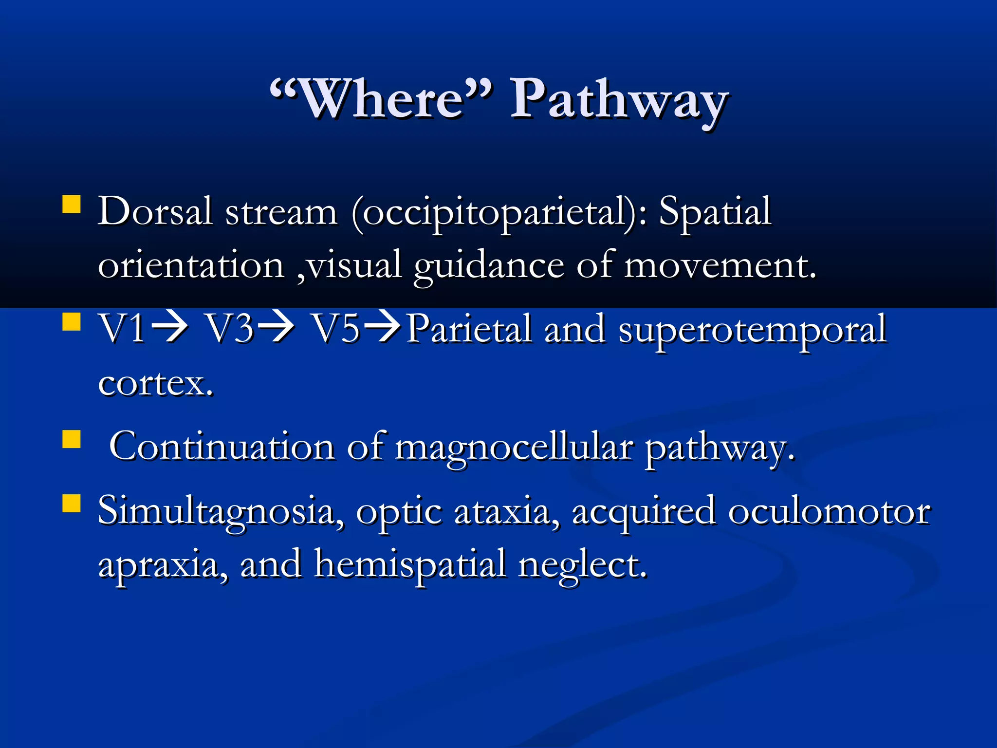
““Where” PathwayWhere” Pathway
Dorsal stream (occipitoparietal): SpatialDorsal stream (occipitoparietal): Spatial
orientation ,visual guidance of movement.orientation ,visual guidance of movement.
V1V1 V3V3 V5V5Parietal and superotemporalParietal and superotemporal
cortex.cortex.
Continuation of magnocellular pathway.Continuation of magnocellular pathway.
Simultagnosia, optic ataxia, acquired oculomotorSimultagnosia, optic ataxia, acquired oculomotor
apraxia, and hemispatial neglect.apraxia, and hemispatial neglect.
54.
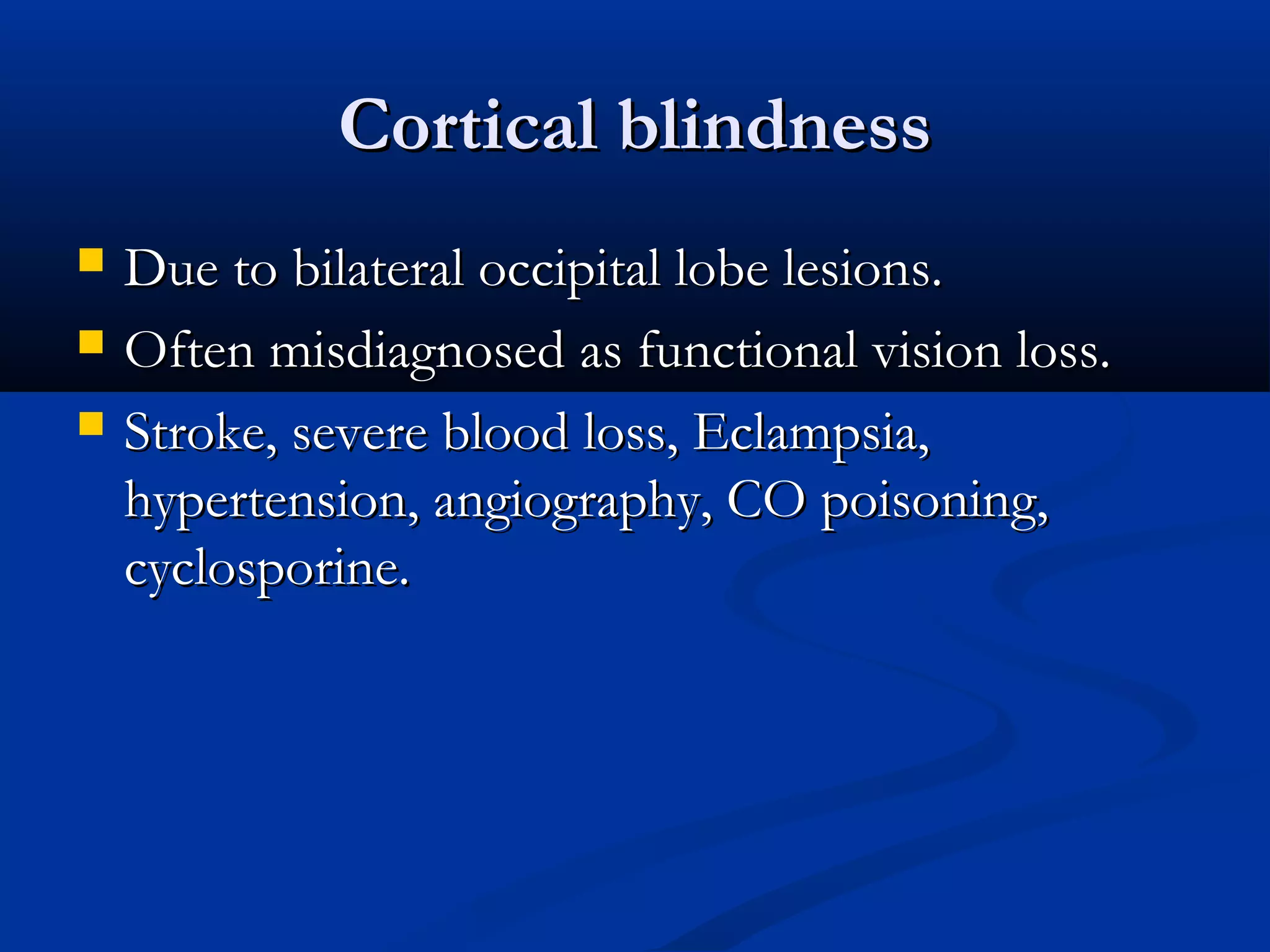
Cortical blindnessCortical blindness
Due to bilateral occipital lobe lesions.Due to bilateral occipital lobe lesions.
Often misdiagnosed as functional vision loss.Often misdiagnosed as functional vision loss.
Stroke, severe blood loss, Eclampsia,Stroke, severe blood loss, Eclampsia,
hypertension, angiography, CO poisoning,hypertension, angiography, CO poisoning,
cyclosporine.cyclosporine.
55.
DyschromatopsiaDyschromatopsia
Bilateral occipitallobe lesions in the lingual orBilateral occipital lobe lesions in the lingual or
fusiform gyri of the medial occipital lobe (medialfusiform gyri of the medial occipital lobe (medial
occipito-temporal lobe).occipito-temporal lobe).
Rarely no field defect.Rarely no field defect.
Unilateral involvement may causeUnilateral involvement may cause
hemidyschromatopsia.hemidyschromatopsia.
56.
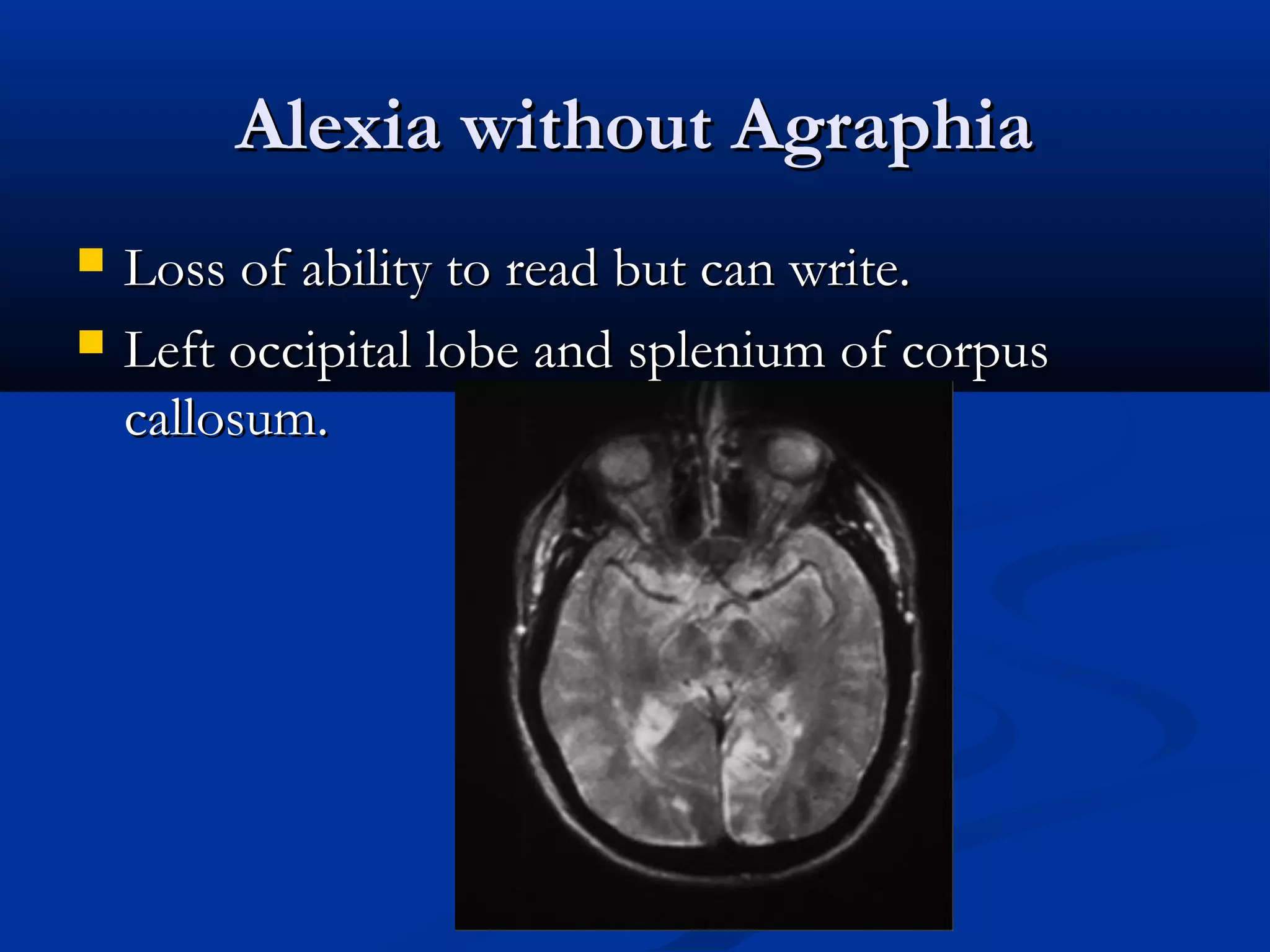
Alexia without AgraphiaAlexiawithout Agraphia
Loss of ability to read but can write.Loss of ability to read but can write.
Left occipital lobe and splenium of corpusLeft occipital lobe and splenium of corpus
callosum.callosum.
57.
PalinopsiaPalinopsia
Persistant orrecurrence of visual stimulus afterPersistant or recurrence of visual stimulus after
it has been removed.it has been removed.