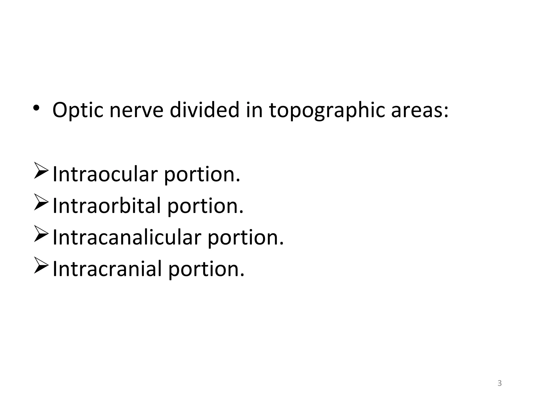
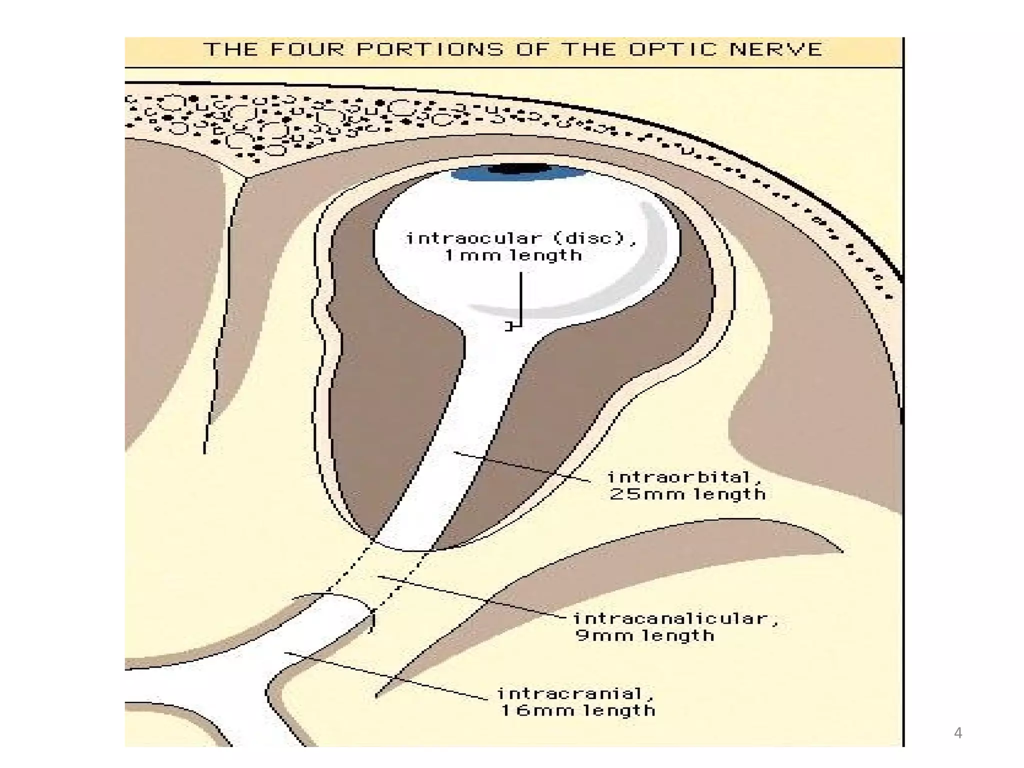
E-mail: Shaf3y_h@hotmail.com
facebook/shafie eye clinic
Symptoms & Signs in Clinical Medicine
By/Mohamed Ahmed El –Shafie
Assistant Lecturer in ophthalmology department KafrELShiekh University
Anatomy of optic nerve
• Optic nerve- more than 1 million axons.
• Consisting of axons originating from ganglion
cells.
• Starts from optic disc upto optic chiasma.
• Contains the afferent fibers of light reflex.
• Elongated tract of white matter
• Not covered by neurilemma.
2
• Optic nerve divided in topographic areas:
Intraocular portion.
Intraorbital portion.
Intracanalicular portion.
Intracranial portion.
3
4
Intraocular optic nerve
• 1 mm in length.
• 1.5 mm diameter.
• Which expands approximately 3-4 mm behind
the sclera.
5
Intraorbital optic nerve
Relation of ophthalmic artery
At the optic foramen: inferior and lateral
Lateral to optic nerve (in posterior orbit)
Inferior division of 3rd
nerve-Sixth Nerve
Ciliary ganglion
Nasociliary artery
6
At the orbital apex – optic nerve surrounded
by annulus of Zinn.
Blood supply: Ophthalmic artery with
meningeal branches
7
Intracanalicular optic nerve
• 9 mm
• Tightly fixed within the canal (compressive
optic neuropathy Optic nerve edema)
• Blunt trauma
Blood supply: Pial branches from ophthalmic
artery. 8
Intracranial optic nerve
• Length-10mm
• Diameter-4.5mm
Extends post & medially ascending at an angle
of 45º to join the chiasma
Blood supply: pial vessels arising from ICA
branches from ant cerebral and anterior
communicating artery 9
Normal optic disc
- Color: Orange or pink
- Margin
- Counter
- Crescent
- Distribution of veins.
Why the normal disc is pink?
• Thickness and the
cytoarchitecture of fiber
bundles passing
between glial columns
containing capillaries
12
• Pathologies of the optic nerve, even though
not always detected on ophthalmoscopic
exam, may compromise its function and
cause the following sign;
Visual Acuity
 Near vision
- Reading from a book
 Far vision
- Snellen chart.
 - Ishihara charts
Dyschromatopsia
• Impaired color vision = 94%
• Desaturation
• – eg: red = dark/ beached
• Distinguishes b/w macular lesion
confrontation
Kinetic perimetry Static perimetryKinetic perimetry Static perimetry
• all patients show V.F abnormalities in acute phase
Central ⁺⁺⁺, peripheral⁺, particular region⁺
• If Vn severely impaired – confrontation test
• Vision improved –
• kinetic Goldman Perimetry ( only central scotoma)
• Automated static Perimetry
• Focal defect –
• Altitudinal
• Arcaute
• Nasal step defect
• Stimulus
• Receptors
• Afferent
• Center
• Efferent
• Effector organ
• Effect
Pup. light reflex
VEDIO
VEDIO
Afferent pupillary defect
• In the absence of an optic nerve lesion in the fellow eye, RAPD
can be demonstrated by swinging flash light
• -ve RAPD in recurrent attacks
Causes:Causes:
• Optic neuropathyOptic neuropathy
• Total retinal detachmentTotal retinal detachment
• Dense vitreous haemorrhageDense vitreous haemorrhage
• Dense amblyopiaDense amblyopia
VEDIO
Contrast sensitivity
• - good for subclinical ON
Optic Disc SwellingOptic Disc Swelling
How do you approach?How do you approach?
PapilloedemaPapilloedema vsvs Optic NeuritisOptic Neuritis
Papilloeodema
 Definition ,
Non-inflammatory swelling of Optic disc
 Causes
1. Raised Intracranial Pressure:
- Space-occupying lesions
-Occlusion of :retinal veins
cavernous sinus
-Other cause.
Optic Neuritis
(ON)• Inflammation of optic nerve - ON
• Associated with swollen disc – papillitis
• Normal disc – retro bulbar ON
Papilledema Optic neuritis AION
increased ICP Inflammatory swelling Vascular accident (occlusion of
short posterior ciliary artery
causing infarction
Brain tumors , hematomas,
meningitis
Multiple Sclerosis is highly
associated
Hypertension, giant cell arteritis,
hypercoagulable state
Bilateral , may be
asymmetric
Unilateral Unilateral
28
Headache, nausea, vomittingHeadache, nausea, vomitting
No visual loss usually,No visual loss usually,
only enlarged blind spotonly enlarged blind spot
Retrobulbar pain on ocularRetrobulbar pain on ocular
movement, early centralmovement, early central
scotoma, decreased acuity,scotoma, decreased acuity,
impaired color vision,impaired color vision,
presence of APDpresence of APD
Acute painless visual loss, usuallyAcute painless visual loss, usually
hemialtitudinal defect involvinghemialtitudinal defect involving
the lower visual fieldthe lower visual field
Variable degree of disc
swelling, hemorrhages
Fewer hemorrhages and
cotton wool spots
Pale segmental swelling and
splinter hemorrhages at its
margins
Prognosis usually good if
primary cause of increased
ICP is treated
Vision usually returns to
normal
Poorer prognosis, permanent loss.
Second eye is involved in one
third of cases.
OPTIC ATROPHY
Degeneration of the optic nerve
occurs as an end result of any pathologic process
that damages axons
Ophthalmoscopic classification
 Primary optic atrophy
Secondary optic atrophy
Consecutive optic atrophy
Glaucomatous optic atrophy
Primary optic
atrophy
Consecutive
optic atrophy
Secondary optic
atrophy
Glaucomatous
optic atrophy
chalky white or
white
Disc appears
yellow waxy
dirty white in colour Pale disc
Margins are sharply
outlined
edges are not
so sharply
defined
Edges are blurred,. Edges well defined
Lamina cribrosa is
clearly seen at the
bottom of the
physiological cup
physiological cup is
obliterated
deep and wide
cupping of the optic
disc and nasal shift of
the blood vessels
Major retinal vessels
and surrounding
retina are normal
Retinal vessels
are attenuated
vessels are
attenuated and
perivascular
sheathing
Normal
32

Optic nerve Clinical significance

  • 1.
    E-mail: Shaf3y_h@hotmail.com facebook/shafie eyeclinic Symptoms & Signs in Clinical Medicine By/Mohamed Ahmed El –Shafie Assistant Lecturer in ophthalmology department KafrELShiekh University
  • 2.
    Anatomy of opticnerve • Optic nerve- more than 1 million axons. • Consisting of axons originating from ganglion cells. • Starts from optic disc upto optic chiasma. • Contains the afferent fibers of light reflex. • Elongated tract of white matter • Not covered by neurilemma. 2
  • 3.
    • Optic nervedivided in topographic areas: Intraocular portion. Intraorbital portion. Intracanalicular portion. Intracranial portion. 3
  • 4.
  • 5.
    Intraocular optic nerve •1 mm in length. • 1.5 mm diameter. • Which expands approximately 3-4 mm behind the sclera. 5
  • 6.
    Intraorbital optic nerve Relationof ophthalmic artery At the optic foramen: inferior and lateral Lateral to optic nerve (in posterior orbit) Inferior division of 3rd nerve-Sixth Nerve Ciliary ganglion Nasociliary artery 6
  • 7.
    At the orbitalapex – optic nerve surrounded by annulus of Zinn. Blood supply: Ophthalmic artery with meningeal branches 7
  • 8.
    Intracanalicular optic nerve •9 mm • Tightly fixed within the canal (compressive optic neuropathy Optic nerve edema) • Blunt trauma Blood supply: Pial branches from ophthalmic artery. 8
  • 9.
    Intracranial optic nerve •Length-10mm • Diameter-4.5mm Extends post & medially ascending at an angle of 45º to join the chiasma Blood supply: pial vessels arising from ICA branches from ant cerebral and anterior communicating artery 9
  • 11.
    Normal optic disc -Color: Orange or pink - Margin - Counter - Crescent - Distribution of veins.
  • 12.
    Why the normaldisc is pink? • Thickness and the cytoarchitecture of fiber bundles passing between glial columns containing capillaries 12
  • 13.
    • Pathologies ofthe optic nerve, even though not always detected on ophthalmoscopic exam, may compromise its function and cause the following sign;
  • 14.
    Visual Acuity  Nearvision - Reading from a book  Far vision - Snellen chart.  - Ishihara charts
  • 15.
    Dyschromatopsia • Impaired colorvision = 94% • Desaturation • – eg: red = dark/ beached • Distinguishes b/w macular lesion
  • 16.
  • 17.
    Kinetic perimetry StaticperimetryKinetic perimetry Static perimetry
  • 18.
    • all patientsshow V.F abnormalities in acute phase Central ⁺⁺⁺, peripheral⁺, particular region⁺ • If Vn severely impaired – confrontation test • Vision improved – • kinetic Goldman Perimetry ( only central scotoma) • Automated static Perimetry • Focal defect – • Altitudinal • Arcaute • Nasal step defect
  • 19.
    • Stimulus • Receptors •Afferent • Center • Efferent • Effector organ • Effect Pup. light reflex
  • 20.
  • 21.
    Afferent pupillary defect •In the absence of an optic nerve lesion in the fellow eye, RAPD can be demonstrated by swinging flash light • -ve RAPD in recurrent attacks Causes:Causes: • Optic neuropathyOptic neuropathy • Total retinal detachmentTotal retinal detachment • Dense vitreous haemorrhageDense vitreous haemorrhage • Dense amblyopiaDense amblyopia
  • 22.
  • 23.
    Contrast sensitivity • -good for subclinical ON
  • 24.
    Optic Disc SwellingOpticDisc Swelling How do you approach?How do you approach?
  • 25.
  • 26.
    Papilloeodema  Definition , Non-inflammatoryswelling of Optic disc  Causes 1. Raised Intracranial Pressure: - Space-occupying lesions -Occlusion of :retinal veins cavernous sinus -Other cause.
  • 27.
    Optic Neuritis (ON)• Inflammationof optic nerve - ON • Associated with swollen disc – papillitis • Normal disc – retro bulbar ON
  • 28.
    Papilledema Optic neuritisAION increased ICP Inflammatory swelling Vascular accident (occlusion of short posterior ciliary artery causing infarction Brain tumors , hematomas, meningitis Multiple Sclerosis is highly associated Hypertension, giant cell arteritis, hypercoagulable state Bilateral , may be asymmetric Unilateral Unilateral 28 Headache, nausea, vomittingHeadache, nausea, vomitting No visual loss usually,No visual loss usually, only enlarged blind spotonly enlarged blind spot Retrobulbar pain on ocularRetrobulbar pain on ocular movement, early centralmovement, early central scotoma, decreased acuity,scotoma, decreased acuity, impaired color vision,impaired color vision, presence of APDpresence of APD Acute painless visual loss, usuallyAcute painless visual loss, usually hemialtitudinal defect involvinghemialtitudinal defect involving the lower visual fieldthe lower visual field Variable degree of disc swelling, hemorrhages Fewer hemorrhages and cotton wool spots Pale segmental swelling and splinter hemorrhages at its margins Prognosis usually good if primary cause of increased ICP is treated Vision usually returns to normal Poorer prognosis, permanent loss. Second eye is involved in one third of cases.
  • 29.
    OPTIC ATROPHY Degeneration ofthe optic nerve occurs as an end result of any pathologic process that damages axons Ophthalmoscopic classification  Primary optic atrophy Secondary optic atrophy Consecutive optic atrophy Glaucomatous optic atrophy
  • 30.
    Primary optic atrophy Consecutive optic atrophy Secondaryoptic atrophy Glaucomatous optic atrophy chalky white or white Disc appears yellow waxy dirty white in colour Pale disc Margins are sharply outlined edges are not so sharply defined Edges are blurred,. Edges well defined Lamina cribrosa is clearly seen at the bottom of the physiological cup physiological cup is obliterated deep and wide cupping of the optic disc and nasal shift of the blood vessels Major retinal vessels and surrounding retina are normal Retinal vessels are attenuated vessels are attenuated and perivascular sheathing Normal
  • 32.