Optical Coherence Tomography in Multiple Sclerosis
Optical coherence tomography (OCT) is a non-invasive imaging technique used to monitor neurodegeneration in multiple sclerosis (MS) by providing detailed images of retinal structures, particularly the retinal nerve fiber layer (RNFL). Research demonstrates that OCT can detect axonal loss in MS patients, including in asymptomatic cases, and its measurements correlate with visual function and disease progression. OCT's reproducibility and ability to assess deeper retinal layers, alongside its use in clinical trials, indicate its increasing role in understanding and treating MS.
Optical Coherence Tomography in Multiple Sclerosis
1.
The Utility ofOptical
Coherence Tomography
in Multiple Sclerosis
Raed Behbehani , MD FRCSC
2.
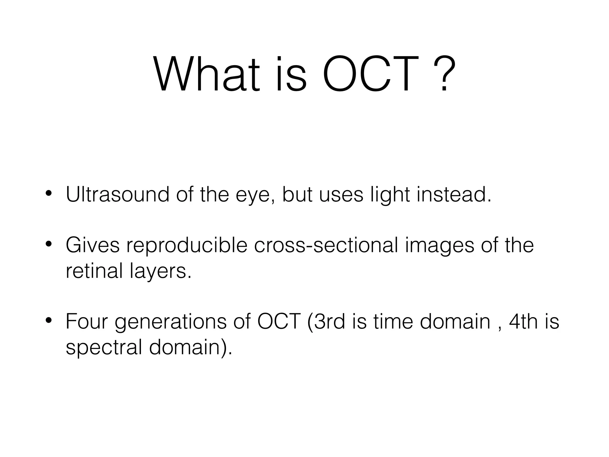
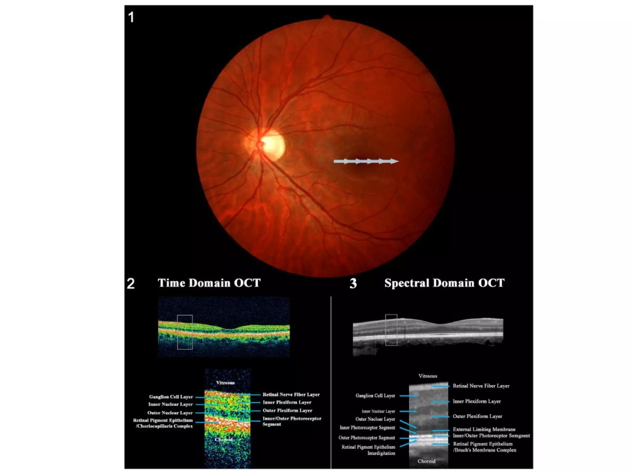
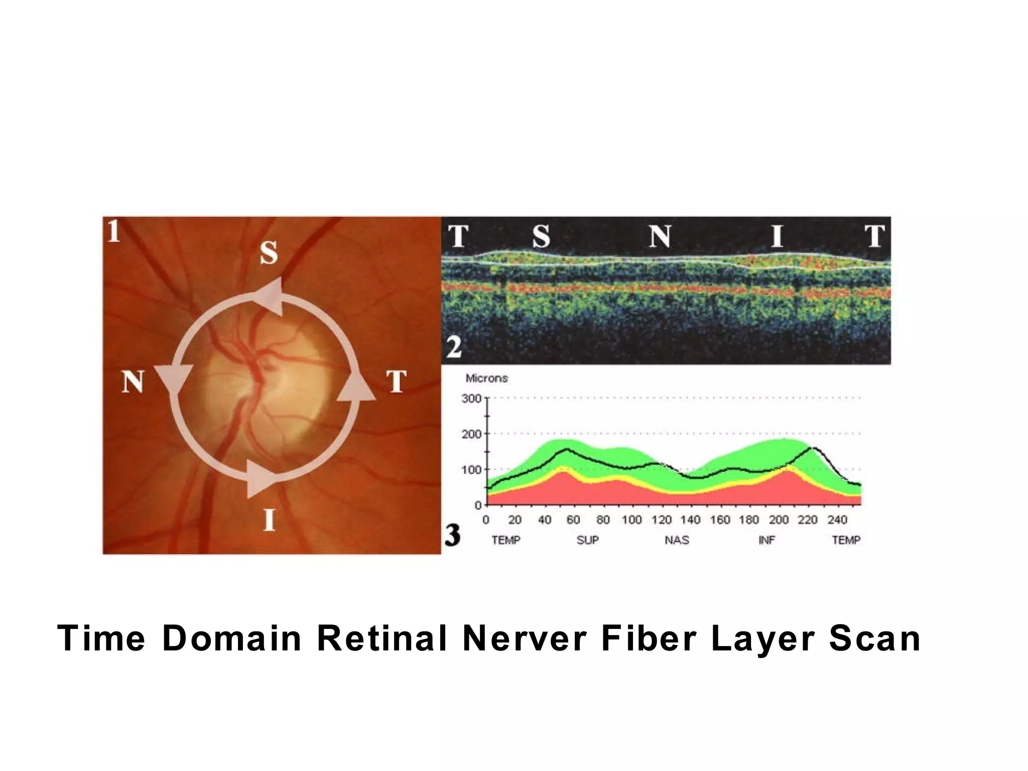
What is OCT?
• Ultrasound of the eye, but uses light instead.
• Gives reproducible cross-sectional images of the
retinal layers.
• Four generations of OCT (3rd is time domain , 4th is
spectral domain).
3.
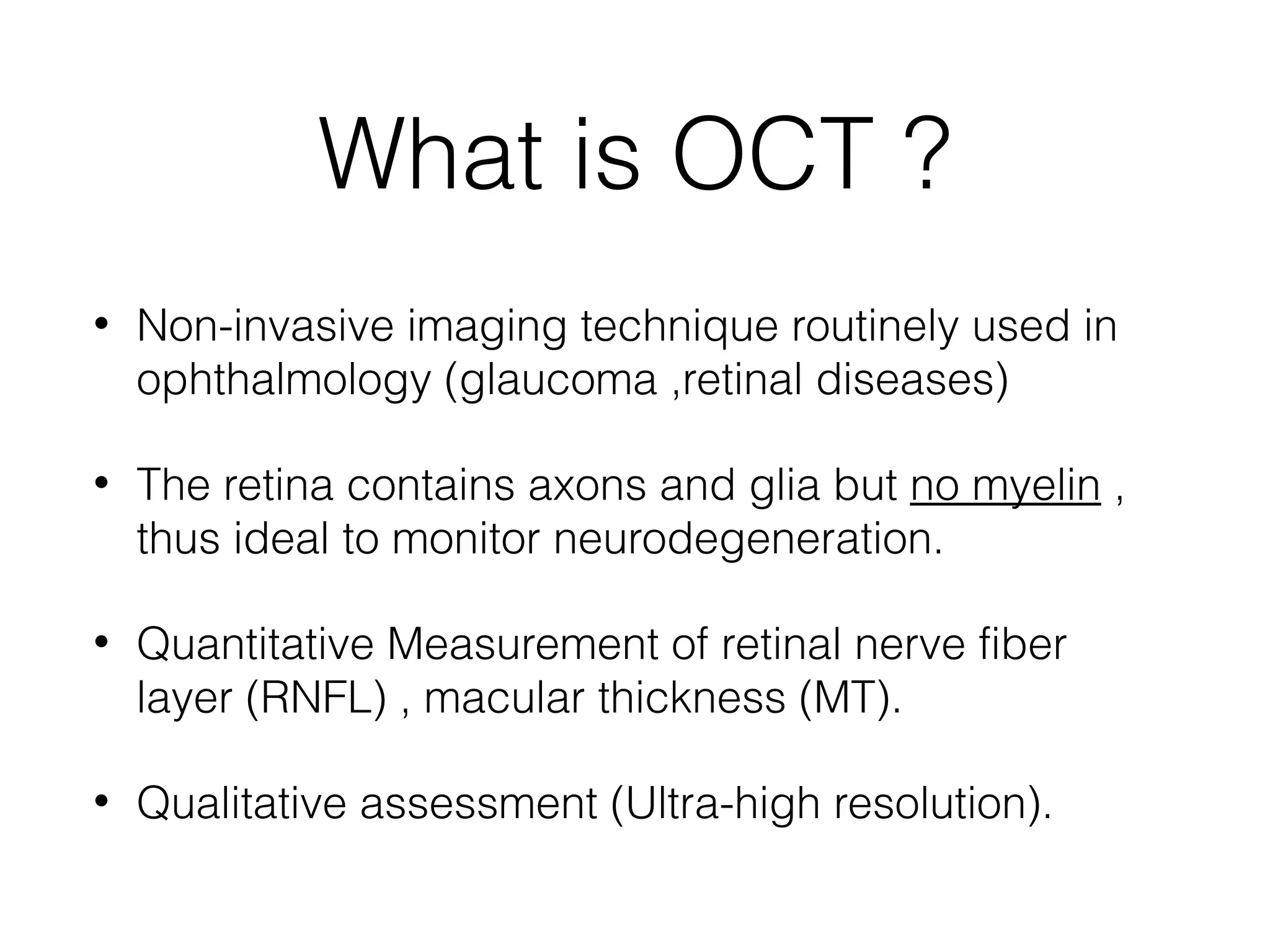
What is OCT?
• Non-invasive imaging technique routinely used in
ophthalmology (glaucoma ,retinal diseases)
• The retina contains axons and glia but no myelin ,
thus ideal to monitor neurodegeneration.
• Quantitative Measurement of retinal nerve fiber
layer (RNFL) , macular thickness (MT).
• Qualitative assessment (Ultra-high resolution).
4.
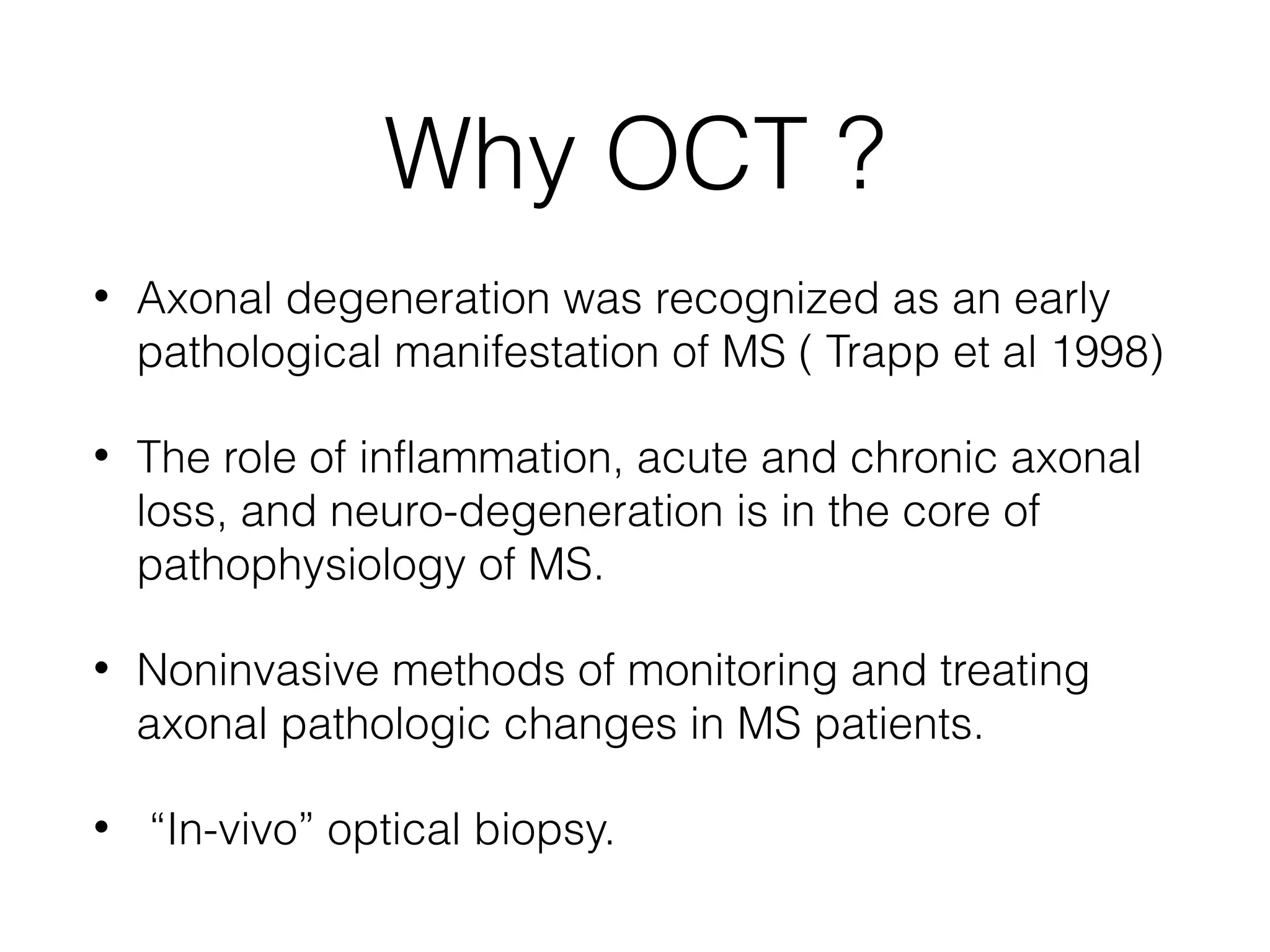
Why OCT ?
• Axonal degeneration was recognized as an early
pathological manifestation of MS ( Trapp et al 1998)
• The role of inflammation, acute and chronic axonal
loss, and neuro-degeneration is in the core of
pathophysiology of MS.
• Noninvasive methods of monitoring and treating
axonal pathologic changes in MS patients.
• “In-vivo” optical biopsy.
5.
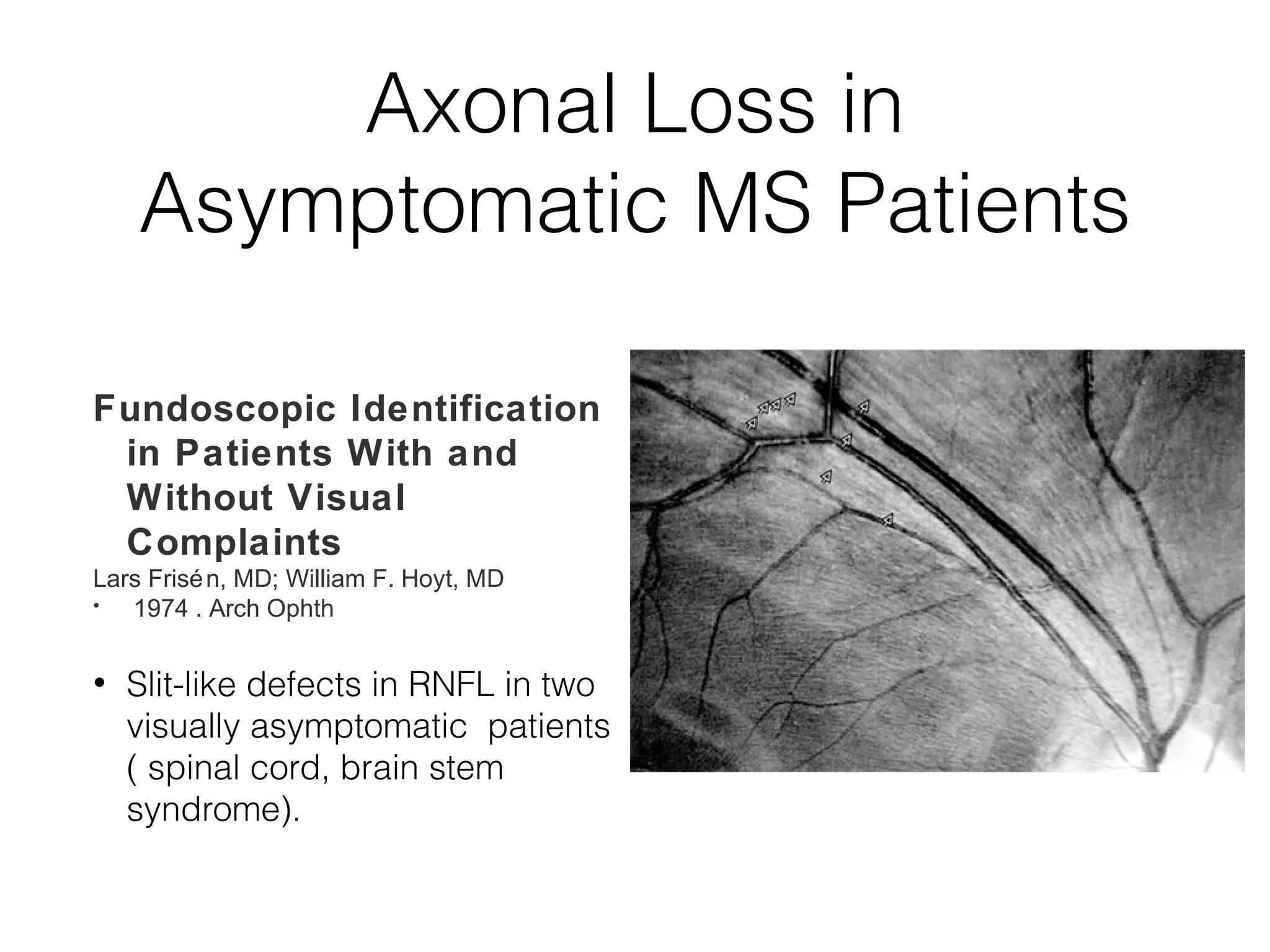
Axonal Loss in
Asymptomatic MS Patients
Fundoscopic Identification
in Patients With and
Without Visual
Complaints
Lars Frisén, MD; William F. Hoyt, MD
• 1974 . Arch Ophth
• Slit-like defects in RNFL in two
visually asymptomatic patients
( spinal cord, brain stem
syndrome).
6.
Axonal Loss inMS
• Post-mortem analysis showed that most MS were
found to have changes in the optic nerve and
RNFL, regardless of whether they had optic neuritis
(Ikuta and Zimmerman, 1976; Toussaint et al., 1983 , Green et al. 2010)
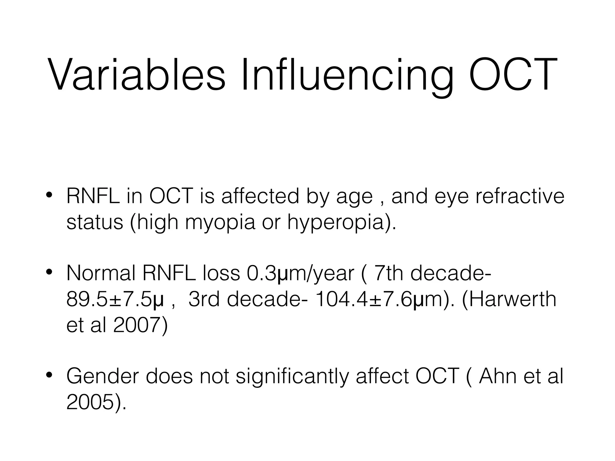
Variables Influencing OCT
• RNFL in OCT is affected by age , and eye refractive
status (high myopia or hyperopia).
• Normal RNFL loss 0.3μm/year ( 7th decade-
89.5±7.5μ , 3rd decade- 104.4±7.6μm). (Harwerth
et al 2007)
• Gender does not significantly affect OCT ( Ahn et al
2005).
11.
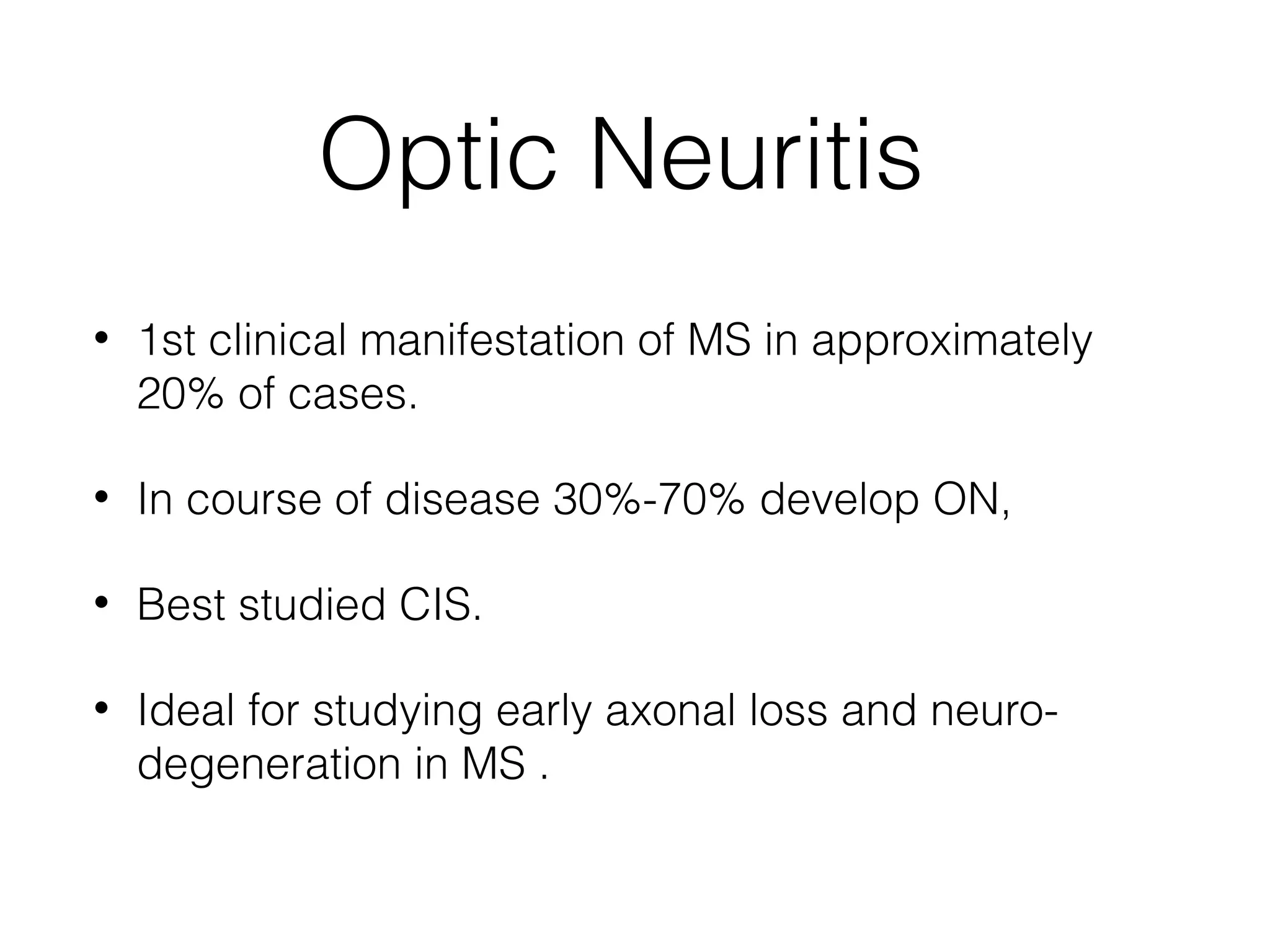
Optic Neuritis
•1st clinical manifestation of MS in approximately
20% of cases.
• In course of disease 30%-70% develop ON,
• Best studied CIS.
• Ideal for studying early axonal loss and neuro-degeneration
in MS .
12.
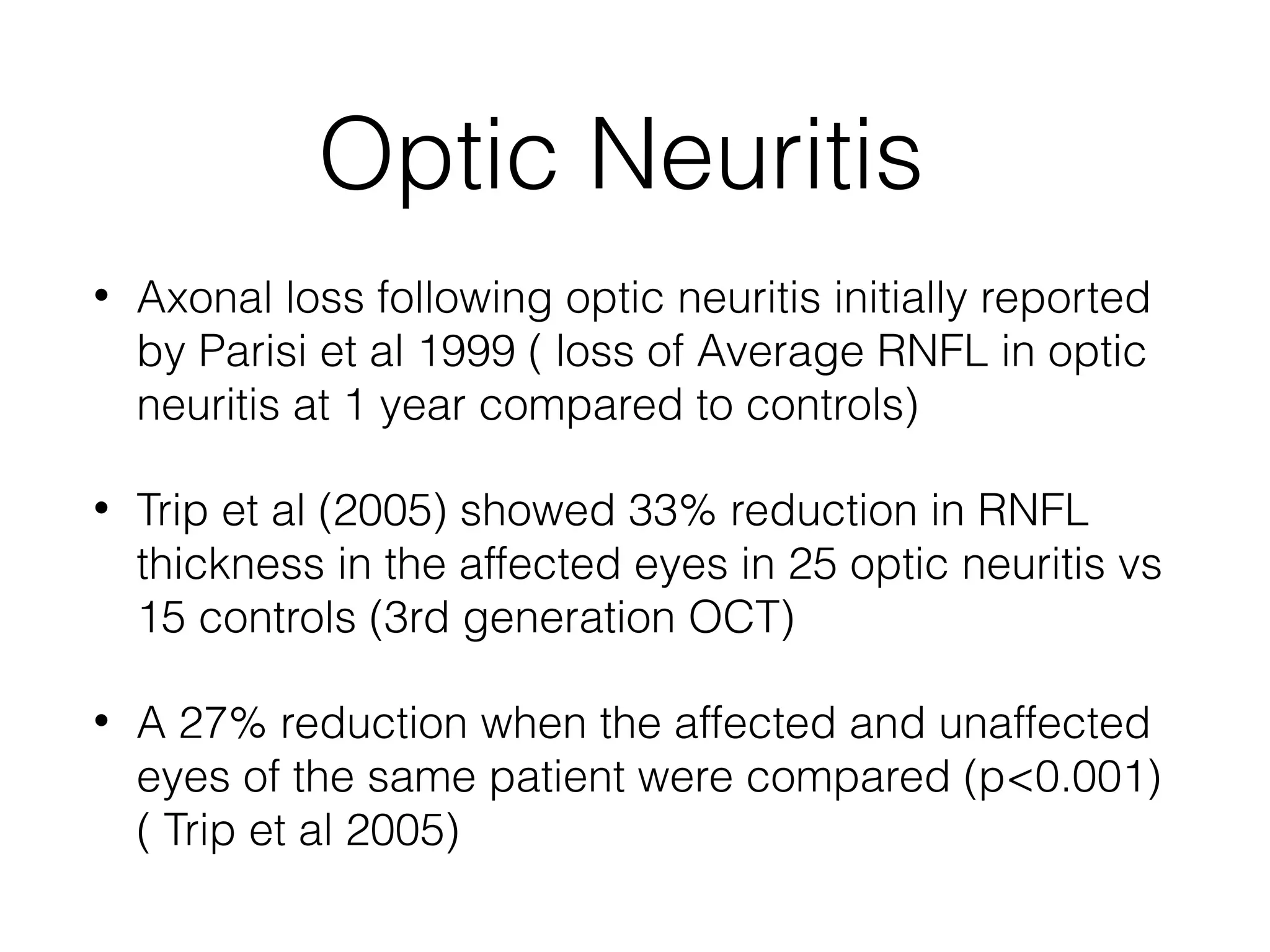
Optic Neuritis
•Axonal loss following optic neuritis initially reported
by Parisi et al 1999 ( loss of Average RNFL in optic
neuritis at 1 year compared to controls)
• Trip et al (2005) showed 33% reduction in RNFL
thickness in the affected eyes in 25 optic neuritis vs
15 controls (3rd generation OCT)
• A 27% reduction when the affected and unaffected
eyes of the same patient were compared (p<0.001)
( Trip et al 2005)
13.
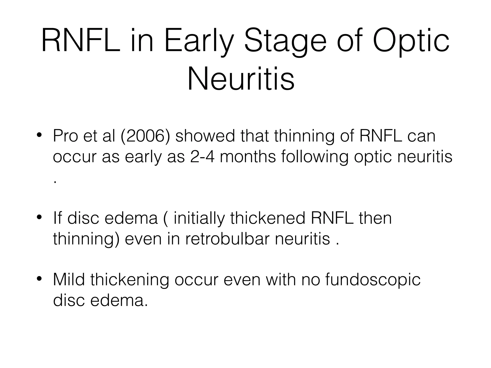
RNFL in EarlyStage of Optic
Neuritis
• Pro et al (2006) showed that thinning of RNFL can
occur as early as 2-4 months following optic neuritis
.
• If disc edema ( initially thickened RNFL then
thinning) even in retrobulbar neuritis .
• Mild thickening occur even with no fundoscopic
disc edema.
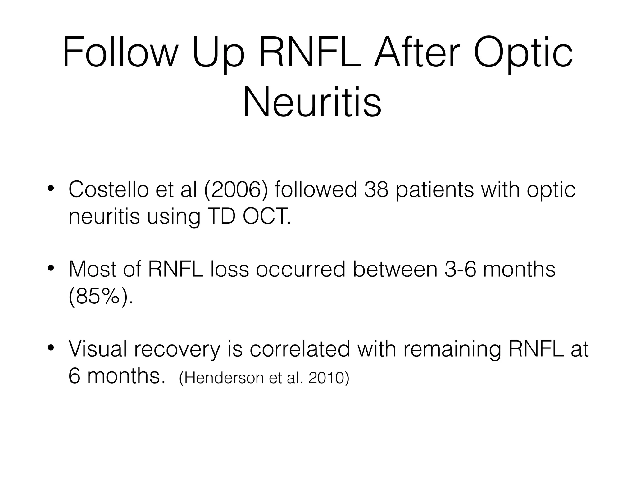
Follow Up RNFLAfter Optic
Neuritis
• Costello et al (2006) followed 38 patients with optic
neuritis using TD OCT.
• Most of RNFL loss occurred between 3-6 months
(85%).
• Visual recovery is correlated with remaining RNFL at
6 months. (Henderson et al. 2010)
17.
Follow Up RNFLin Optic
Neuritis
• Follow up 78 patients for 1 year post-neuritis .
(Costello et al. 2008)
• RNFL thinning starts at 2-3 months , progressed till
6 months and then stabilized up to 2 years (Costello et
al. 2009)
• A meta-analysis (14 studies) showed that RNFL
values are reduced from 5 to 40 μm (averaging 10
to 20 μm) in eyes with MS and ON. (Petzold et al. 2010)
18.
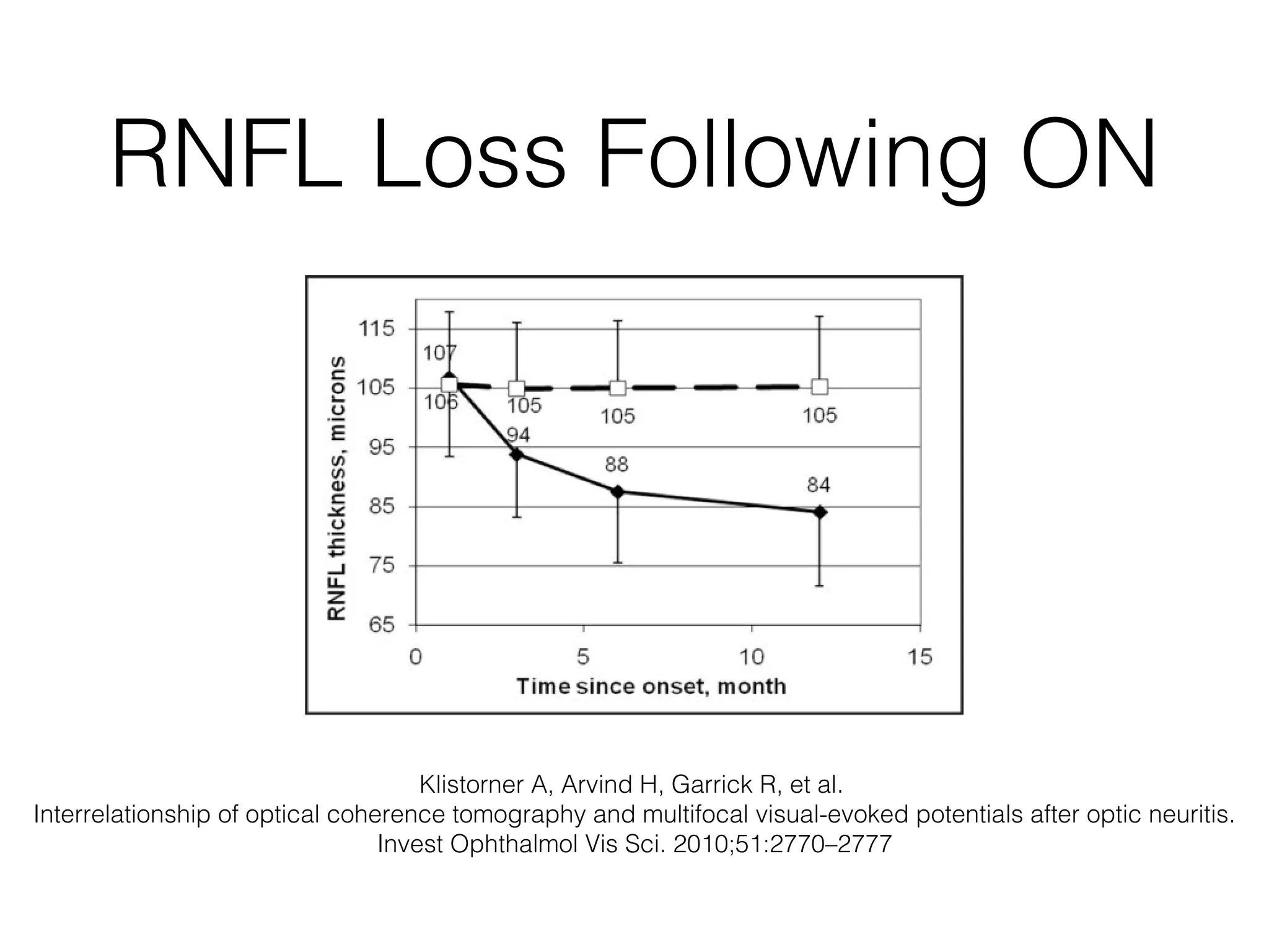
RNFL Loss FollowingON
Klistorner A, Arvind H, Garrick R, et al.
Interrelationship of optical coherence tomography and multifocal visual-evoked potentials after optic neuritis.
Invest Ophthalmol Vis Sci. 2010;51:2770–2777
19.
RNFL of theContralateral
Eye in Optic Neuritis
• Many studies showed that RNFL loss occurs
also in the asymptomatic affected eye in
optic neuritis. (Fisher et al., 2006; Henderson et
al., 2008; Jeanjean et al., 2008; Pueyo et al., 2009;
Pueyo et al., 2008; Pulicken et al., 2007; Sepulcre et
al., 2007).
20.
RNFL in CIS
• No RNFL thinning in CIS patients without optic
neuritis compared to controls over 1 year, but tend
towards temporal RNFL loss. ( Outteryck O et al, 2009)
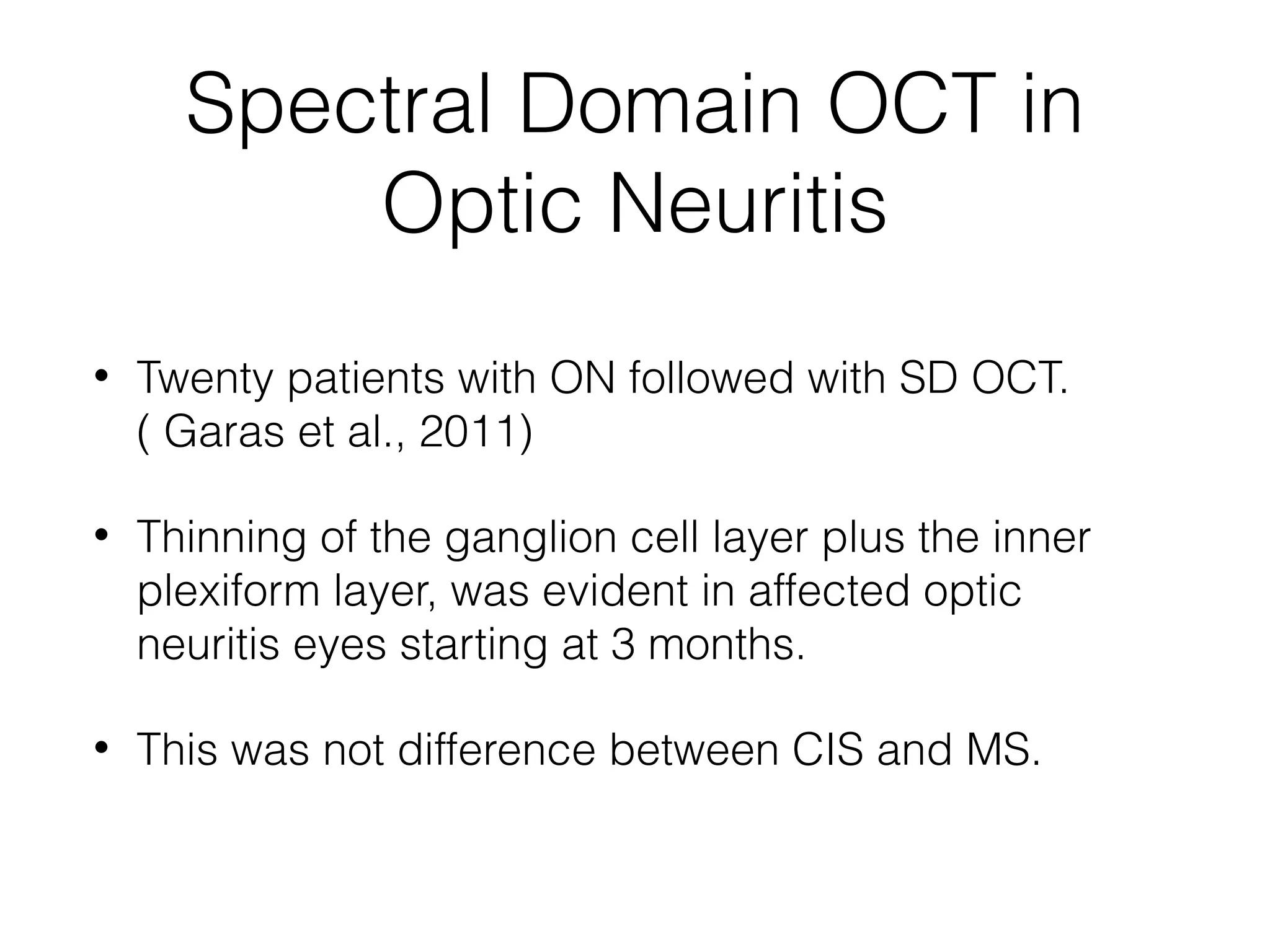
Spectral Domain OCTin
Optic Neuritis
• Twenty patients with ON followed with SD OCT.
( Garas et al., 2011)
• Thinning of the ganglion cell layer plus the inner
plexiform layer, was evident in affected optic
neuritis eyes starting at 3 months.
• This was not difference between CIS and MS.
23.
Spectral Domain OCTin
Optic Neuritis
• Ganglion cell layer thickness decreased after the
baseline visit in affected acute optic neuritis eyes
and was not influenced by the presence of initial
disc or retinal nerve fibre layer oedema (Garas et
al., 2011)
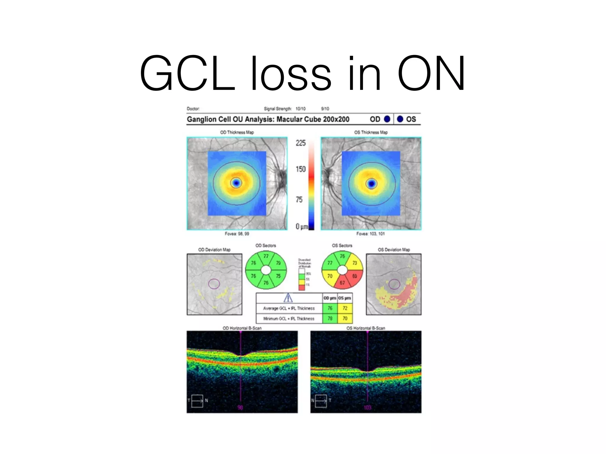
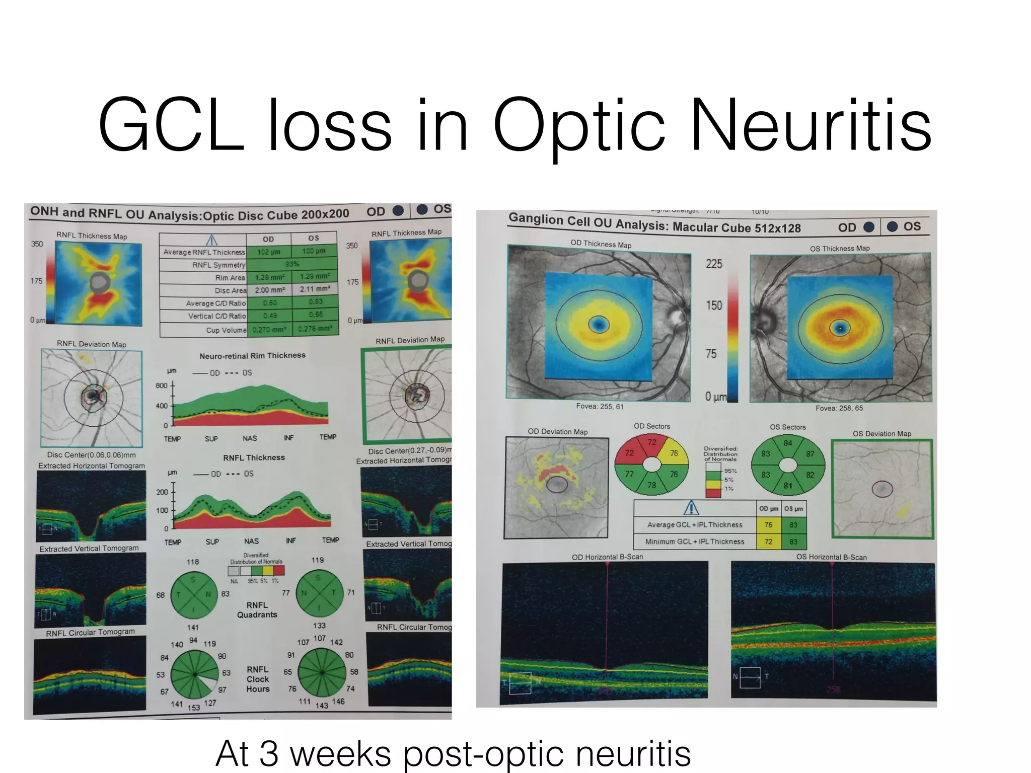
GCL loss inOptic Neuritis
At 3 weeks post-optic neuritis
26.
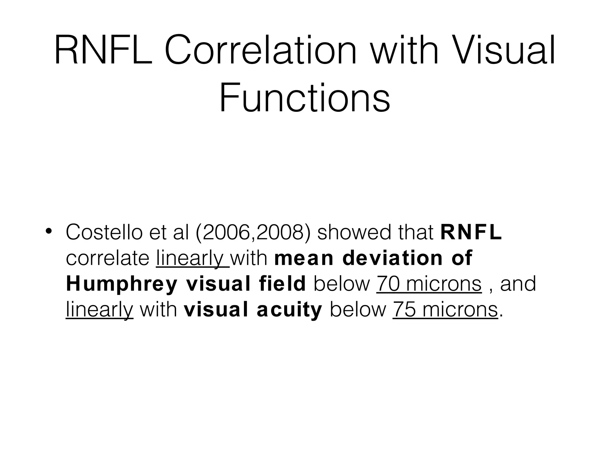
RNFL Correlation withVisual
Functions
• Costello et al (2006,2008) showed that RNFL
correlate linearly with mean deviation of
Humphrey visual field below 70 microns , and
linearly with visual acuity below 75 microns.
27.
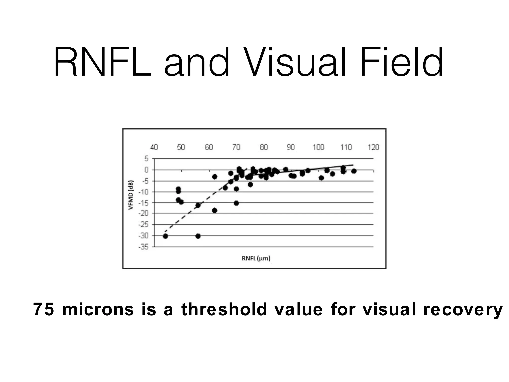
RNFL and VisualField
75 microns is a threshold value for visual recovery
28.
Predictive Value ofOCT
• No significant differences in RNFL thickness in
either ON eyes or non-ON eyes between patients
who developed clinically definite MS (42%) and
those who did not develop MS (58%) during the 2-
year study period. (Costello et al. 2008)
• OCT does not predict conversion to MS at 6 months
in CIS patients.
29.
Correlation between MSand
MRI
• No link between RNFL and (1) MRI evidence of
CNS inflammation at baseline; (2) disseminated
CNS inflammation according to the revised
McDonald criteria; (3) gadolinium enhancement on
initial MRI. (Outteryck O et al. 2009)
30.
Ongoing Axonal Lossin MS
• MS and ON and non-ON eyes each year of follow-up
was associated with an average 2-μm decrease in
RNFL (P < .001) (Talman LS et al.2010)
• Progressive sub-clinical axonal loss in MS.
• Gives a case to early aggressive treatment to prevent
axonal loss.
• Longitudinal studies with high-resolution SD-OCT to
minimize repeat measurement variability are needed.
31.
Macular Volume andMS
• Macula is 60% Ganglion cells.
• MV is a good index to assess neuro-degeneration.
• Not influenced by edema in acute stage of ON.
• Reductions of volume in the macula (approximately 34% neuronal
cells by average thickness) accompany RNFL axonal loss.
• Peripapillary RNFL thinning and inner macular volume loss are
more strongly linked in eyes of MS patients with a history of ON,
which suggests an alternative mechanism for neurodegeneration.
(Burkholder 2009).
32.
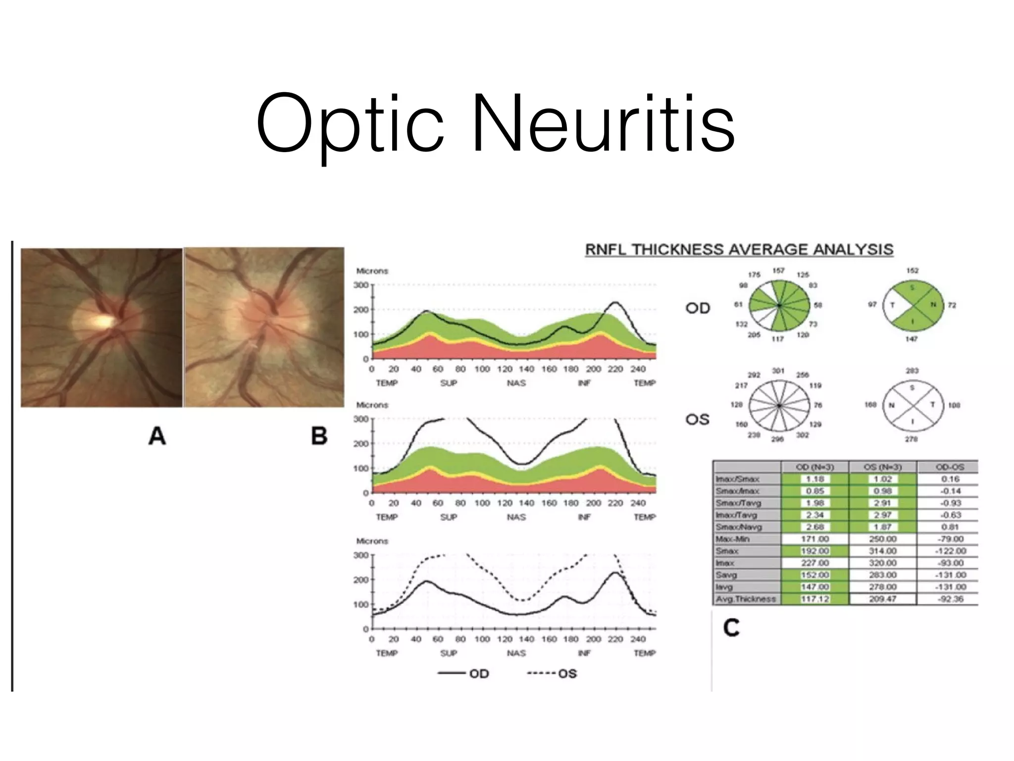
RNFL Loss andMS Severity
• Baseline temporal RNFL atrophy was associated with the
presence of new relapses and EDSS changes (P < .05) at 2
years, (Sepulcre et al. 2007, Spain et al 2009) and recent progression and
disease activity (Toledo et al. 2008)
• PPMS had temporal RNFL loss while SPMS had overall mean,
superior and temporal RNFL loss (Henderson et al 2008).
• Greater RNFL loss in PPMS or SPMS compared to RRMS (Pulicken
et al., 2007).
• RNFL thickness (particularly the temporal quadrant) in the eye
with no prior history of optic neuritis of MS patients may be
helpful in differentiating MS subtypes.
33.
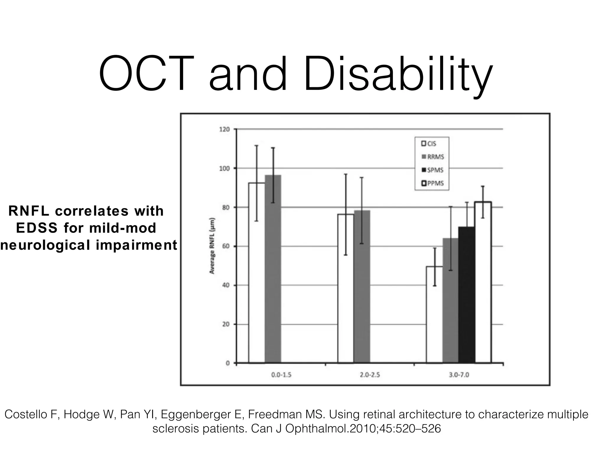
OCT and Disability
RNFL correlates with
EDSS for mild-mod
neurological impairment
Costello F, Hodge W, Pan YI, Eggenberger E, Freedman MS. Using retinal architecture to characterize multiple
sclerosis patients. Can J Ophthalmol.2010;45:520–526
34.
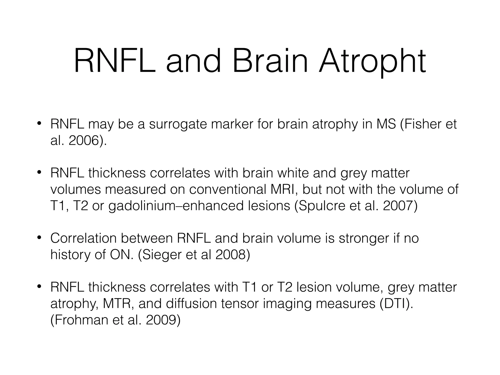
RNFL and BrainAtropht
• RNFL may be a surrogate marker for brain atrophy in MS (Fisher et
al. 2006).
• RNFL thickness correlates with brain white and grey matter
volumes measured on conventional MRI, but not with the volume of
T1, T2 or gadolinium–enhanced lesions (Spulcre et al. 2007)
• Correlation between RNFL and brain volume is stronger if no
history of ON. (Sieger et al 2008)
• RNFL thickness correlates with T1 or T2 lesion volume, grey matter
atrophy, MTR, and diffusion tensor imaging measures (DTI).
(Frohman et al. 2009)
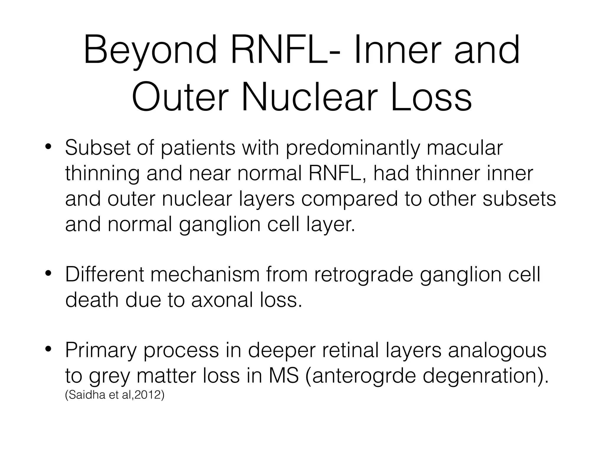
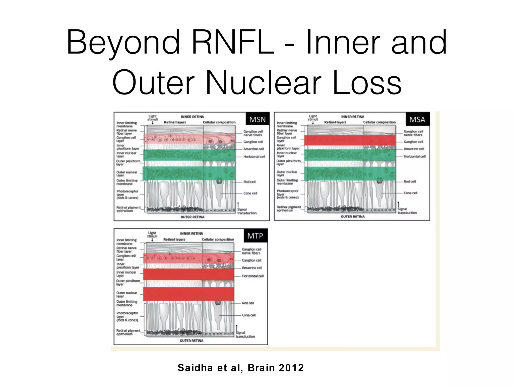
Beyond RNFL- Innerand
Outer Nuclear Loss
• Subset of patients with predominantly macular
thinning and near normal RNFL, had thinner inner
and outer nuclear layers compared to other subsets
and normal ganglion cell layer.
• Different mechanism from retrograde ganglion cell
death due to axonal loss.
• Primary process in deeper retinal layers analogous
to grey matter loss in MS (anterogrde degenration).
(Saidha et al,2012)
37.
Beyond RNFL -Inner and
Outer Nuclear Loss
Saidha et al, Brain 2012
38.
Beyond RNFL -Inner and
Outer Nuclear Loss
• Patients with thin INL and ONL had more
progressive disease.
• Unique visual symptoms (photophobia , glare, poor
night vision)
• Retina may serve as model to understand the
heterogeneity of the inflammatory and
demyelinating mechanisms of MS.
39.
Inner and OuterNuclear
Layer
• RRMS patients INL thickness were not different from
controls and they did not have predominantly
macular thinning.
• Inner and Outer Nuclear Layer loss does not
exclude a primary process in retina.
40.
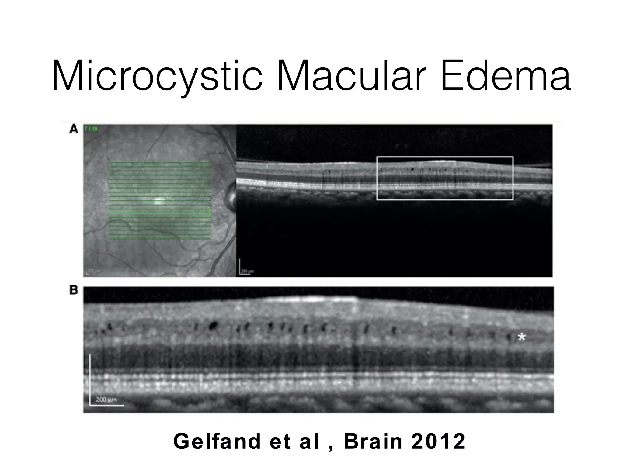
Beyond RNFL -Microcystic
Macular Edema
• Microcystic Edema of the inner nuclear layer in a subset of patients
with MS. (Gelfand et al, Brain 2012).
• Subset had higher EDSS and MSSS (Gelfand 2012, Saidha et al 2012) .
• Predicted the development of contrast- enhancing lesions
(p=0·007), new T2 lesions (p=0·015), EDSS progression (p=0·034),
and relapses ( Saidha et al 2012)
• More common in patients with prior optic neuritis (50 versus 27%).
• Mechanism : ? Patients did use Fingolimod or ? had uveitis.
• Breakdown of the retinal-blood barrier
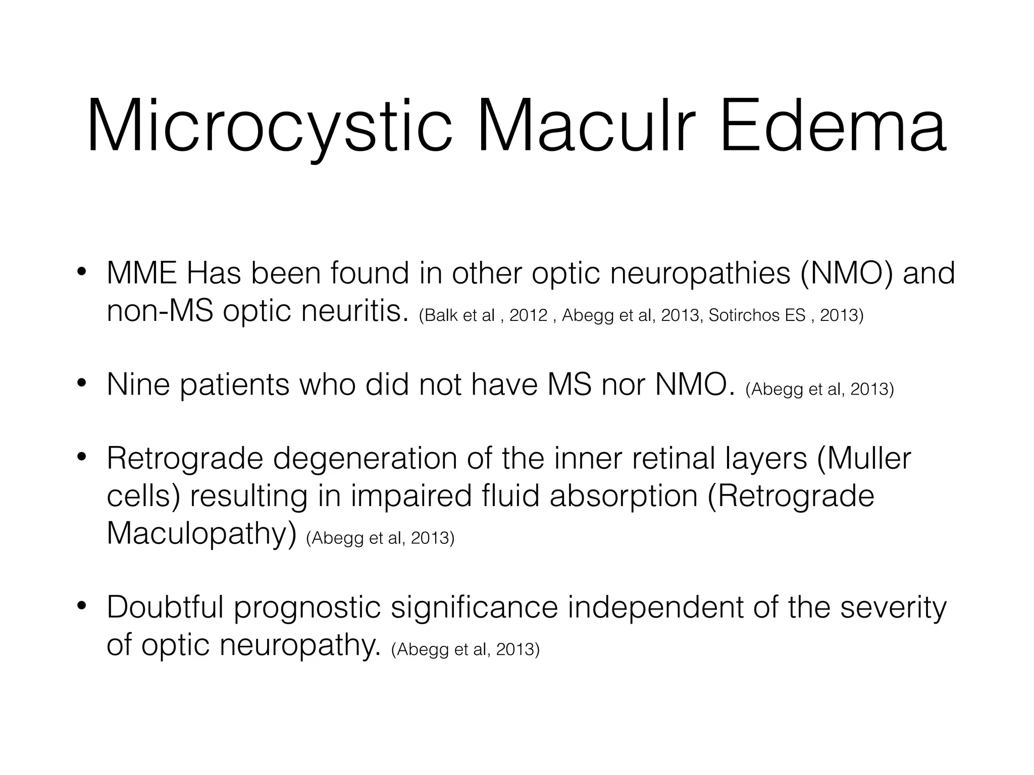
Microcystic Maculr Edema
• MME Has been found in other optic neuropathies (NMO) and
non-MS optic neuritis. (Balk et al , 2012 , Abegg et al, 2013, Sotirchos ES , 2013)
• Nine patients who did not have MS nor NMO. (Abegg et al, 2013)
• Retrograde degeneration of the inner retinal layers (Muller
cells) resulting in impaired fluid absorption (Retrograde
Maculopathy) (Abegg et al, 2013)
• Doubtful prognostic significance independent of the severity
of optic neuropathy. (Abegg et al, 2013)
43.
NMO
• NMOis a distinct disease from MS
(Pathophysiology and Treatment)
• Need more ways to distinguish NMO from MS.
• Visual acuity and RNFL thickness were significantly
worse in NMO and CRION eyes than in RRMS (Bichuetti
et al, 2013)
• RNFL 41 um thickness is 100% specific for NMO
and CRION. (Bichuetti et al, 2013)
44.
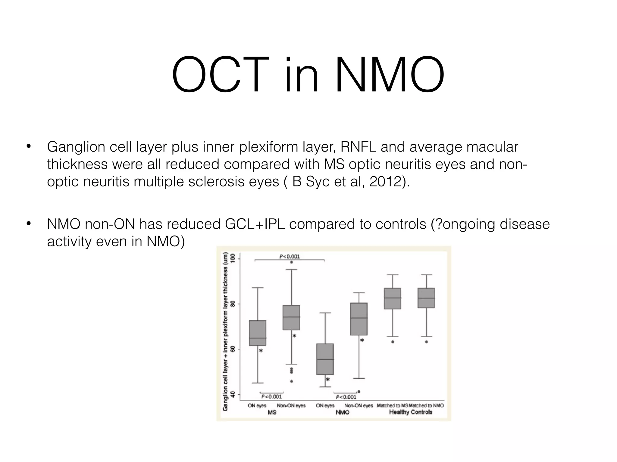
OCT in NMO
• Ganglion cell layer plus inner plexiform layer, RNFL and average macular
thickness were all reduced compared with MS optic neuritis eyes and non-optic
neuritis multiple sclerosis eyes ( B Syc et al, 2012).
• NMO non-ON has reduced GCL+IPL compared to controls (?ongoing disease
activity even in NMO)
45.
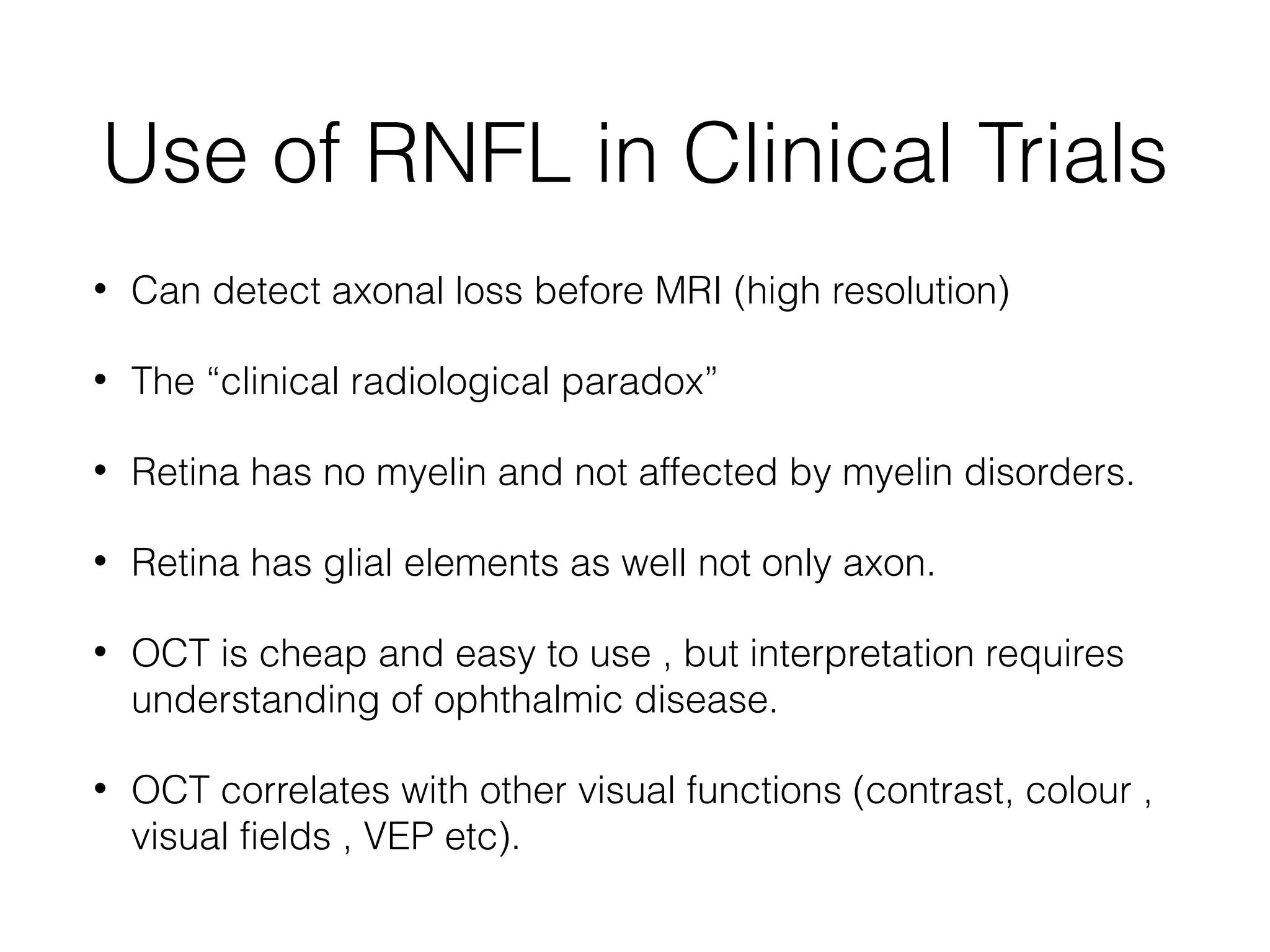
Use of RNFLin Clinical Trials
• Can detect axonal loss before MRI (high resolution)
• The “clinical radiological paradox”
• Retina has no myelin and not affected by myelin disorders.
• Retina has glial elements as well not only axon.
• OCT is cheap and easy to use , but interpretation requires
understanding of ophthalmic disease.
• OCT correlates with other visual functions (contrast, colour ,
visual fields , VEP etc).
46.
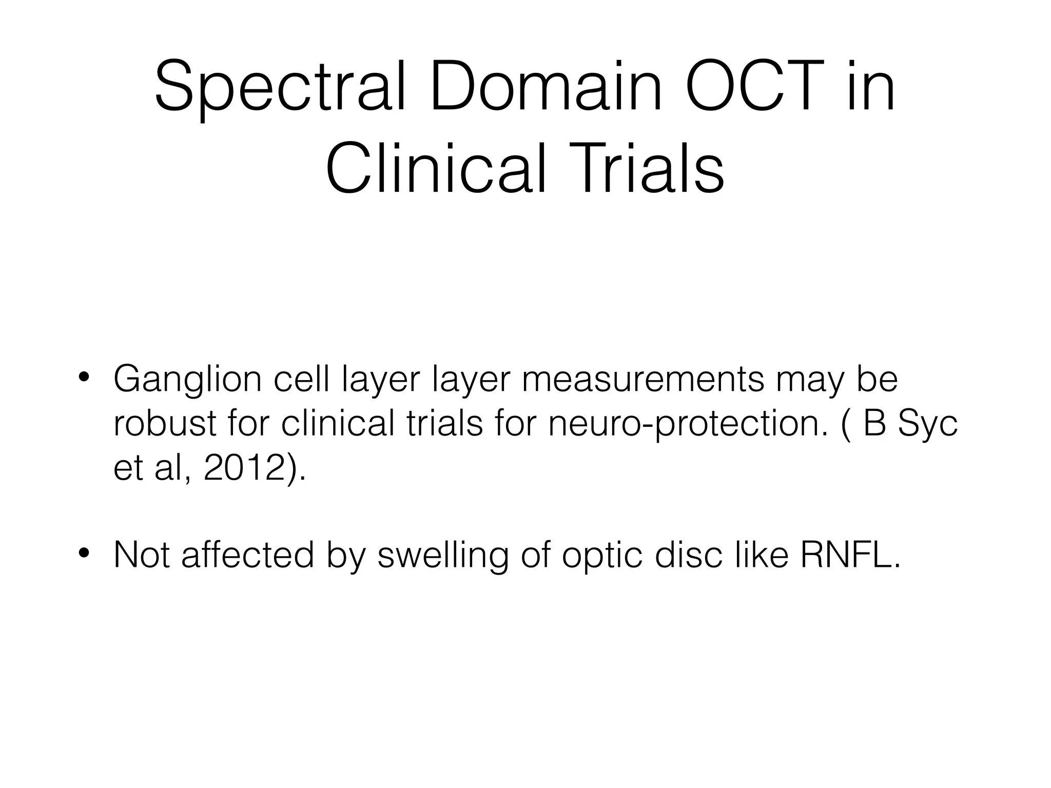
Spectral Domain OCTin
Clinical Trials
• Ganglion cell layer layer measurements may be
robust for clinical trials for neuro-protection. ( B Syc
et al, 2012).
• Not affected by swelling of optic disc like RNFL.
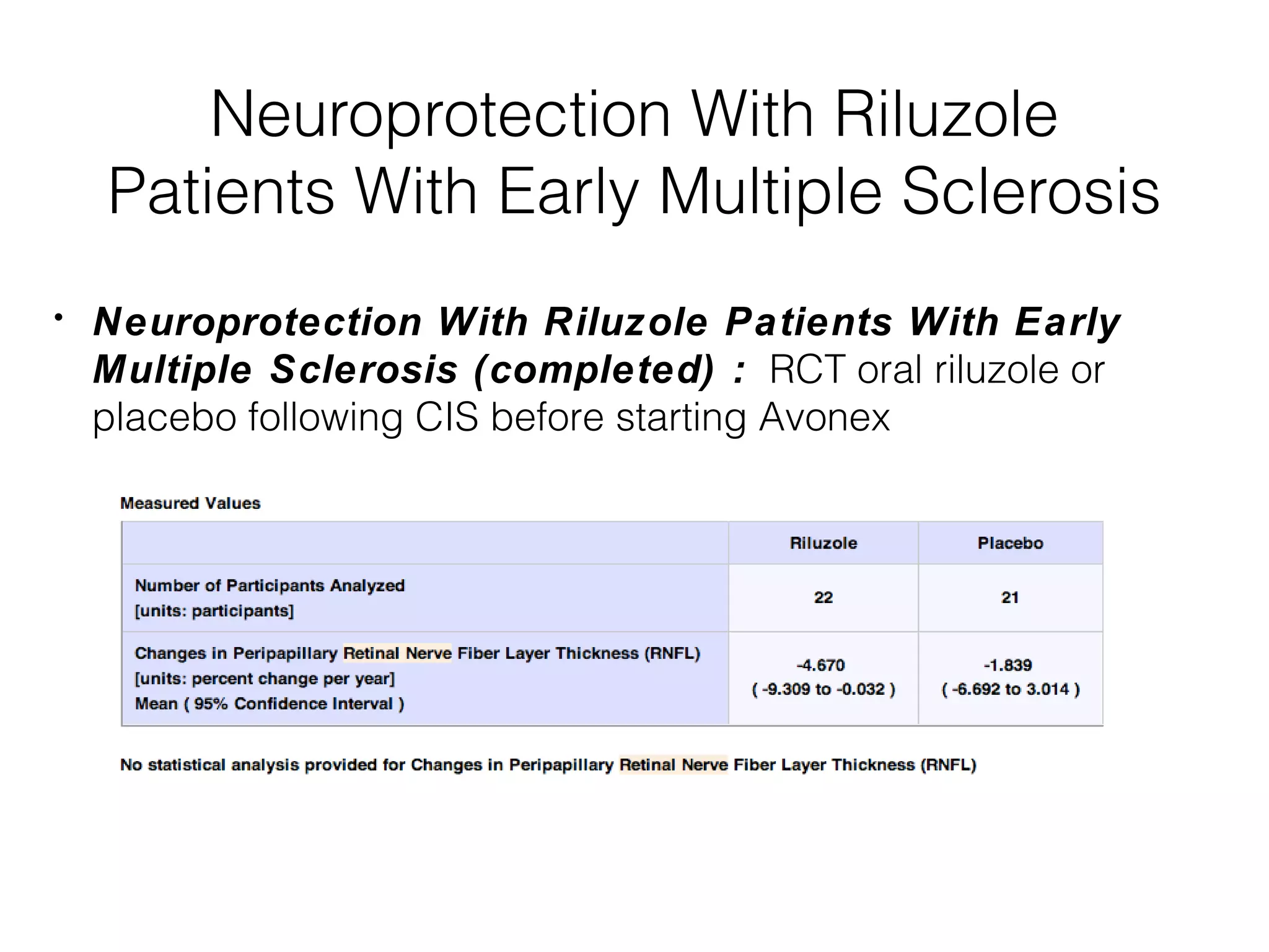
Neuroprotection With Riluzole
Patients With Early Multiple Sclerosis
• Neuroprotection With Riluzole Patients With Early
Multiple Sclerosis (completed) : RCT oral riluzole or
placebo following CIS before starting Avonex
49.
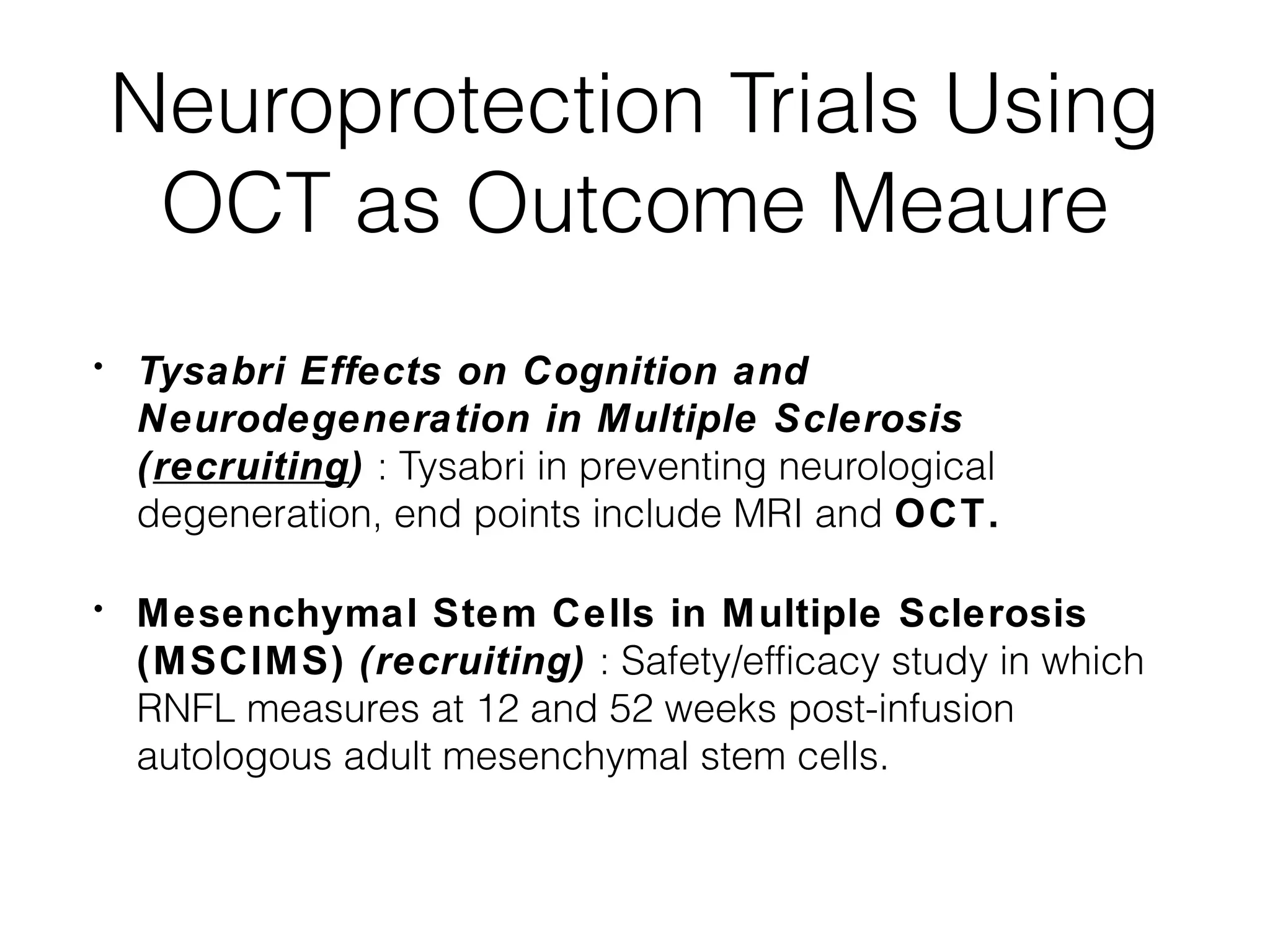
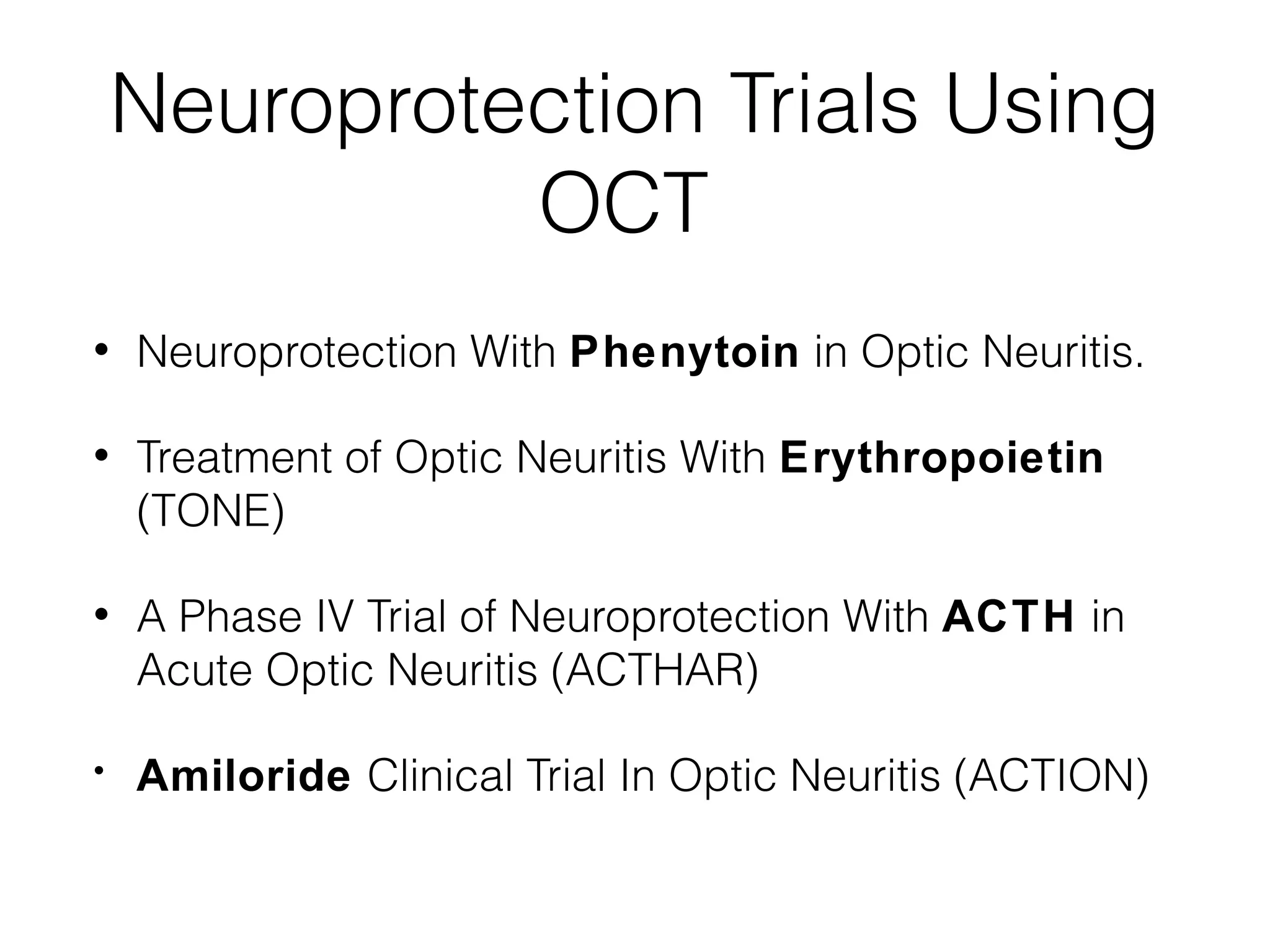
Neuroprotection Trials Using
OCT as Outcome Meaure
• Tysabri Effects on Cognition and
Neurodegeneration in Multiple Sclerosis
(recruiting) : Tysabri in preventing neurological
degeneration, end points include MRI and OCT.
• Mesenchymal Stem Cells in Multiple Sclerosis
(MSCIMS) (recruiting) : Safety/efficacy study in which
RNFL measures at 12 and 52 weeks post-infusion
autologous adult mesenchymal stem cells.
50.
Neuroprotection Trials Using
OCT
• Neuroprotection With Phenytoin in Optic Neuritis.
• Treatment of Optic Neuritis With Erythropoietin
(TONE)
• A Phase IV Trial of Neuroprotection With ACTH in
Acute Optic Neuritis (ACTHAR)
• Amiloride Clinical Trial In Optic Neuritis (ACTION)
51.
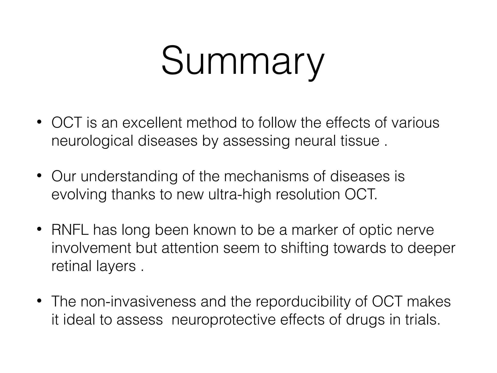
Summary
• OCTis an excellent method to follow the effects of various
neurological diseases by assessing neural tissue .
• Our understanding of the mechanisms of diseases is
evolving thanks to new ultra-high resolution OCT.
• RNFL has long been known to be a marker of optic nerve
involvement but attention seem to shifting towards to deeper
retinal layers .
• The non-invasiveness and the reporducibility of OCT makes
it ideal to assess neuroprotective effects of drugs in trials.
Editor's Notes
#3 We won&apos;t go too much into techinical details of OCT suffice to think that it&apos;s uses slight instead of ultrasound and gives high- resolution images of the retinal layers .
#5 We know that MS is no longer considered a demyelination diseases only and neurodegeneration is hallmark of our most recent understanding of the disease .
OCT allows to monitor the disease from a neurodegeneration.
#6 Now all this is not new ! Almost 40 years ago with only an ophthalmoscope ( underutilized piece of equipment by ophthalmologist and neurologists !) found these slit like defects in patients with ms phenotype but lacked any visual symptoms !
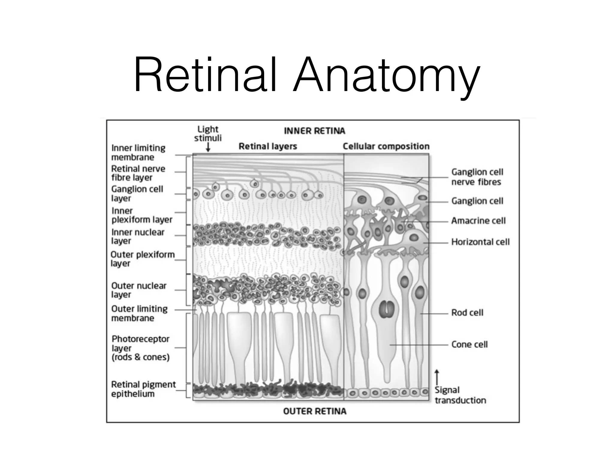
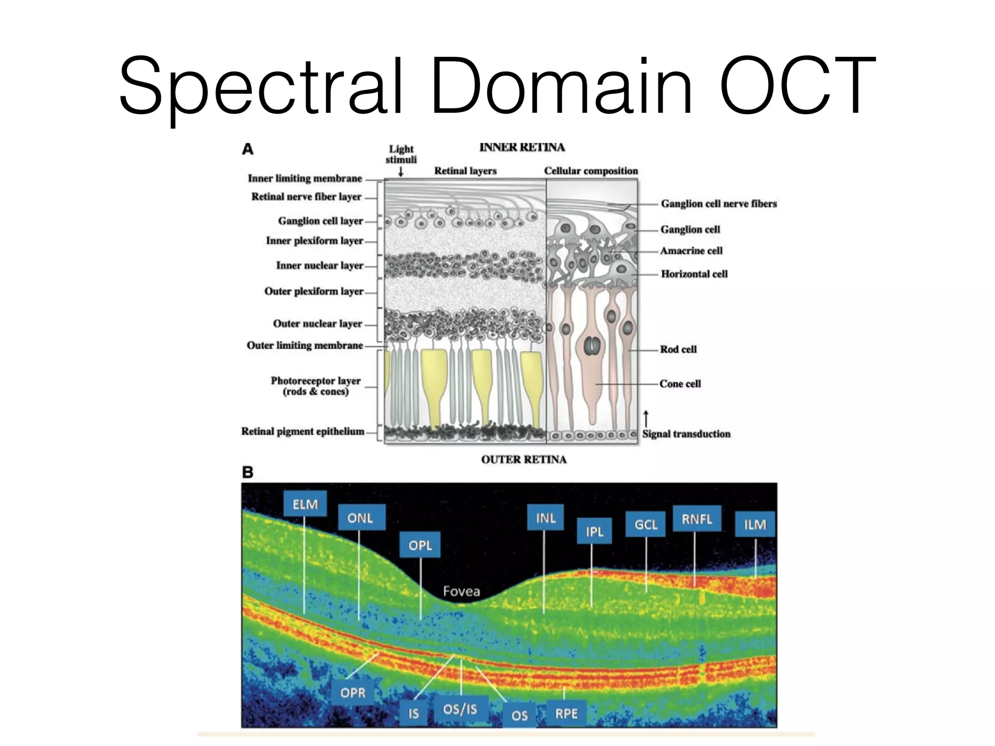
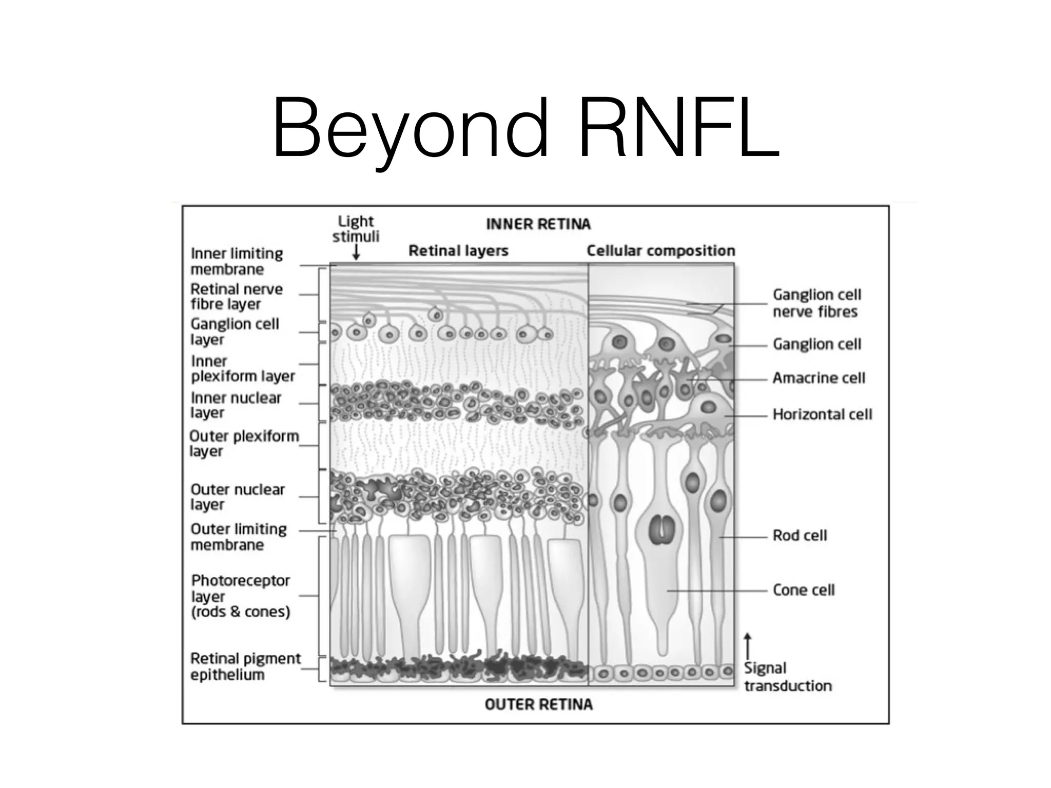
#8 This is just to refamiliarize yourself with the retinal anatomy and as you will see as we talk first bout time domain OCT to spectral domain OCT is that we are going to go deeper and deeper into the retina from inner retina( RNFL) towards outer real a (photoreceptors) and review the impact of MS on all these layers .
#9 This is a comparison between 3rd and 4th generation OCT . Even though one is black and the other is colored you can see the difference in resolution .
Left resolution is 10 microns , right resolution is 3-4 microns . Spectral domain OCT are also 50 times faster .
#11 There arc factors that influence OCT and these are most importantly Age and refractive status. As we age we lose axons , and myopic eyes in general have thinner retinal layers , whereas in hyperopia the eye smaller is smaller and the retinal layers are more compact and crowded and thicker .
#12 Optic neuritis is ideal for OCT since it&apos;s common (most MS patients will develop it either as initial presentation as CIS or in the course of the disease). It&apos;s ideal for studying the effects of MS on the neural tissue and also potentially neuroprotection .
#13 Se the loss is less when compared to the other &quot;Normal&quot;eye and this opens the debate that is opt neuritis a truly unilateral disease or is it always bilateral and asymmetric . Thanks to OCT we know that even the &quot;normal&quot; eye loses axons
#14 So RNFL thinning starts usually around 2-4 weeks following initially. If there is disc edema ( rarely) as optic neuritis is usually retrobulbar , what you see is RNFL thickening followed by thinning.
Even when you don&apos;t you see disc edema by fundoscopy, there is usually mild RNFL thickening . Another concept challenged by OCT , are really most optic neuritis retrobulbar or just the swelling is it subtle for human eyes&apos;
#17 So what happens to the RNFL after optic neuritis you will see thinning of the RNFL for a period of 3-6 months and then it remains stable. It shows that the contrast of what we see clinically when we say patient recovered , as in the vast majority of optic neuritis , while what&apos;s happening in Vivo is axonal loss (neurodegeneration)!
#18 This is another study also by Costello for 2 years follow up of optic neuritis and it sons that RNFL stays stable after 6 months .
#19 This diagram shows the rate of loss and you will see that it is highest between 5-6 months with little change After that ..
#21 What about the RNFL in CIS other than optic neuritis .. It seems that nothing happens much..
#22 Spectral domain thanks to its higher resolution and fast scan time has allowed us to look into greater details.
Focus your attention at the GCL and IPL , as we will talk about this layer in particular and then move deeper into retina.
#23 The GCL was not different between eyes who had CIS and diagnosed CDMS.
#24 And what SDOCT told us is that we have GCL thinning irrespective of whether there initial RNFL swelling or not, which makes it probably a more robust measurement for follow up of optic neuritis .
#25 Macula is very useful for studying effects of neurodegeneration as it is composed of 40% ganglion cells.
#27 So this basically means further drop into RNFL below 70 microns would result is manifest decrease in visual field MD.
#28 And you can see that well in this diagram as the visual filed mean deviation goes down precipitously below 70 microns and as result this value was used threshold for visual recovery, meaning if you have less than 70 microns RNFL , your optic neuritis won&apos;t recover.
#29 Does OCT gives us any predictive information about whethe a patient will develop MS? In the 2 year follow up of optic neuritis by Costello , there was no difference in RNFL between patients who developed MS and those who did not .
#30 Does OCT predict what we see in terms of amount of activity on MRI and lesion load and the answer from one study that no it does not.
#31 There is evidence that there is ongoing axonal loss ,higher than what we see in normal population and perhaps in phases where the disease is seemingly &quot;inactive&quot; .
#32 What makes the macula a good place to study effects of MS is that it is composed of 60% ganglion cells and is not affected by that early phase of swelling in RNFL as you would see in a case of optic neuritis .
#33 What does RNFL tells us about disease severity and subtypes ? It seems that RNFL loss in the temporal region is not a good prognostic sign as it has been Associated with more severity , disability , progression and high relapses .
Also in one study it was found to be more associated with primary progressive ms rather than relapsing remitting type .
#34 This graph shows nicely the relationship between edss and RNFL and as we go higher edss we see corresponding reduction in RNFL .
#36 The newer machines of spectral domain OCT and their higher resolution we have now segmentation algorithms that allows us to examine the layers individually and inspect them closely.
#38 In this study they looked at segmentation alogorithms of patients and they classified them into 3 groups ; normal OCT , abnormal with mostly RNFL thinning , and predominantly macular thinning involvement with involvement of the outer nuclear layers ( horizontal cells , amacrine cells , and photoreceptors ).
#39 Patients with INL and ONL had more progressive disease , and seem to have unique subset of symptoms .