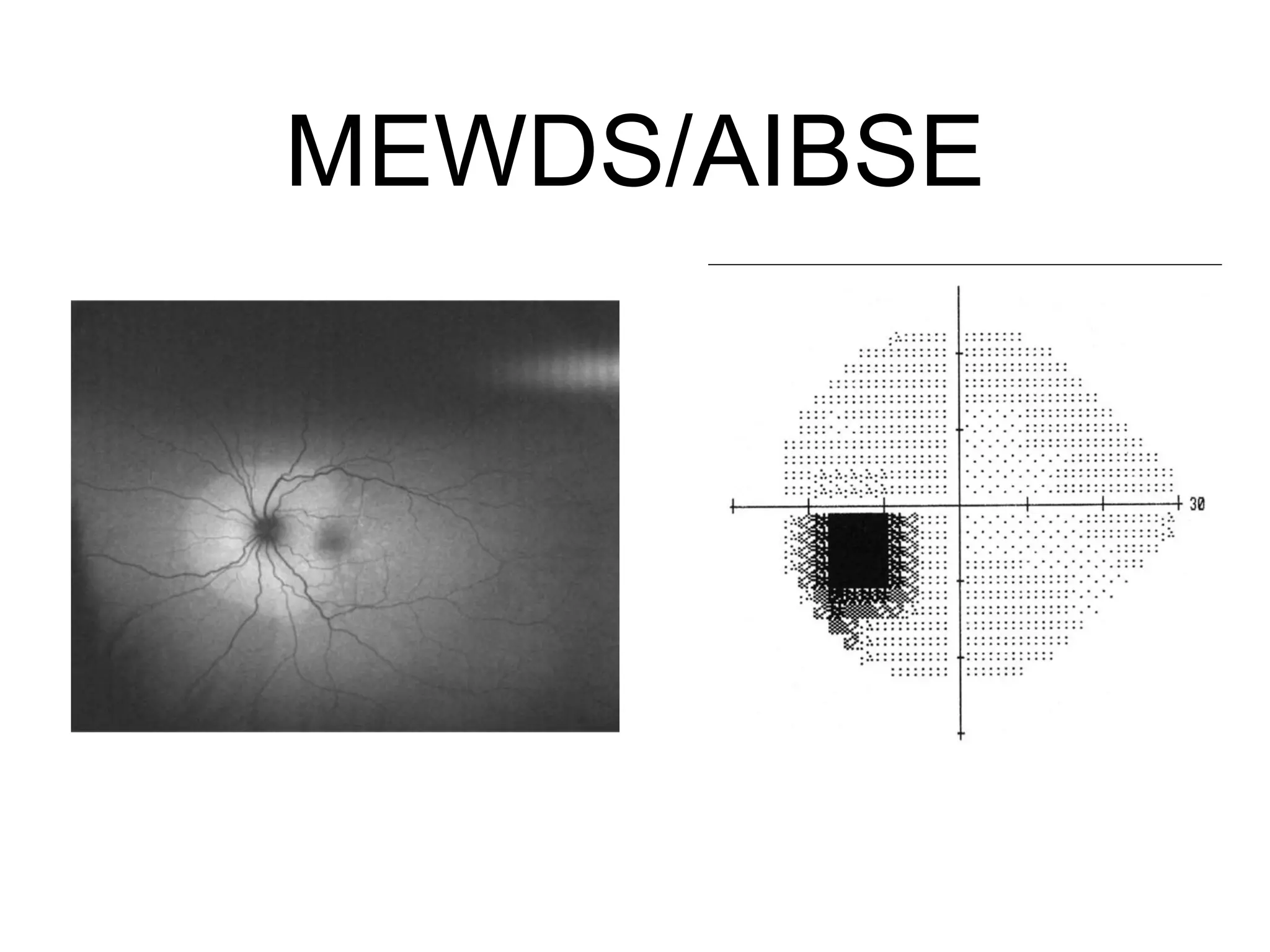
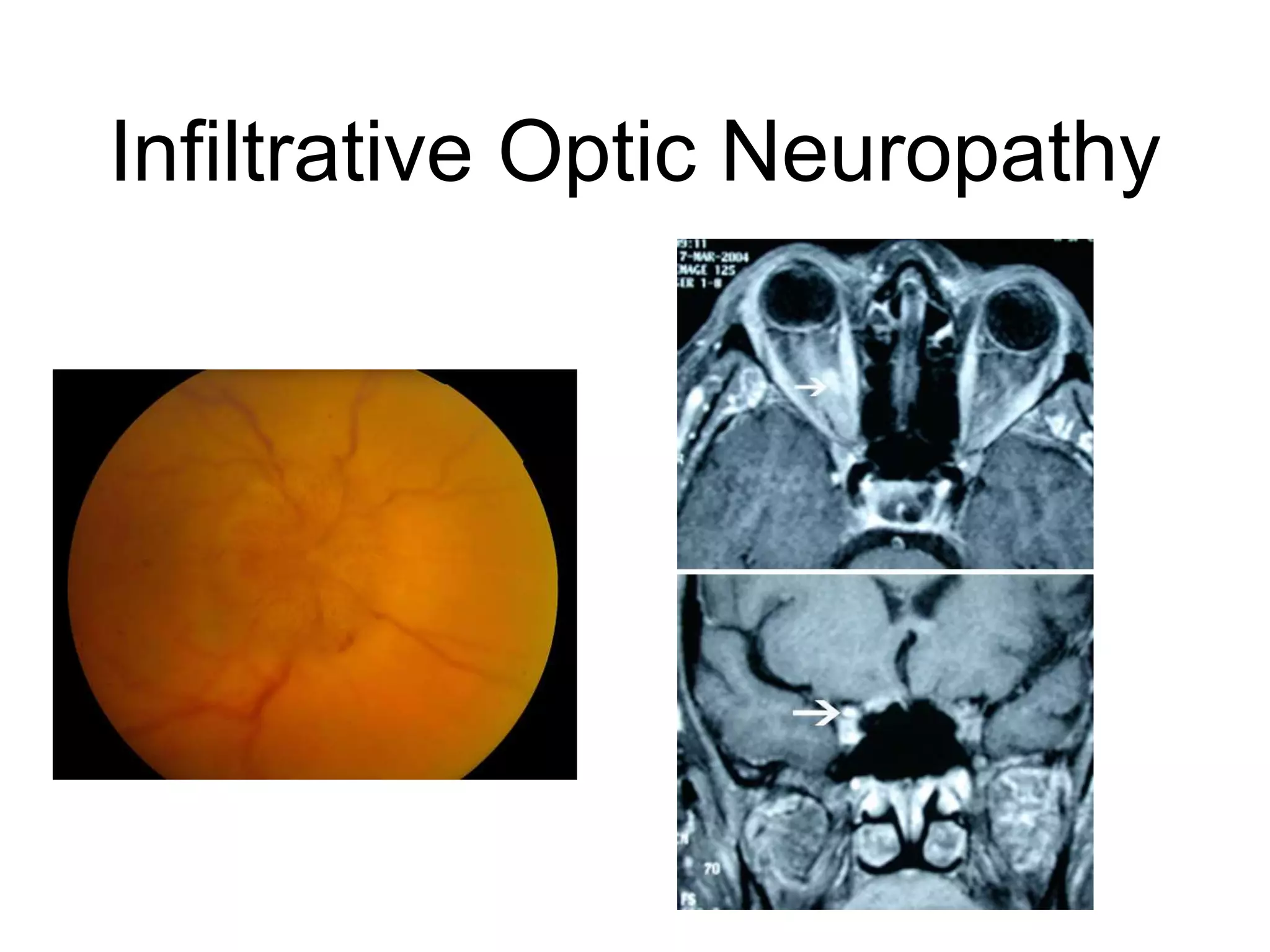
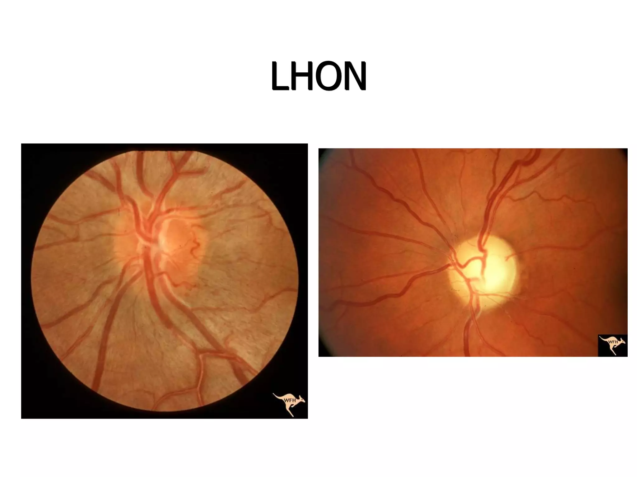
The Approach to A
Patient With Decreased
Vision
Raed Behbehani , MD
Initial Evaluation-History
• Unilateral vs Bilateral
• Onset (acute , sub-acute, chronic)
• Course (constant, progressive,improvement)
• Associated Symptoms
Evaluation-Color Vision
• Optic nerve vs Macular Disease
• Psuedoisochromatic color plates (congenital color
deficiency and acquired optic nerve diseases)
• Other (HRR plates-blue and Yellow , Fansworth-
Munsell D15 Panel)
Evaluation-Pupil
• RAPD is the hall mark of optic neuropathy.
• Swinging flash-light test.
• Reverse RAPD (wrong name) if one pupil is dilated
or does not constrict for mechanical causes.
RAPD
Fundus Evaluation
Visual Field Testing
Ancillary Tests
• Photo-stress Recovery Tests
• IVFA
• OCT
• ERG (Panfiled , Multifocal)
Classification
• Macular Media
• Retinopathy
• Optic Neuropathy
• Chiasmopathy
• Visual Pathway
• Occipital Cortex
MEWDS- AIBSE
• Photopsias
• Temporal Scotoma
• Visual Field-enlarged blind spot
• Mild disc edema, small deep white retinal spots
• Early wrath-like hyperflourescence
• ERG : MFERG (depressed peripapilary regsion) , Full-Field
ERG (depressed a-wave).
• OCT : attenuation of the outer retinal layers
MEWDS/AIBSE
Vitamin A Deficiency
• Bariatric Surgery / Mal-absorption syndromes
• Look for other deficiencies (zinc , copper , Vitamin
B1,B6,B12).
Other Retinopathies
• Cone Dystrophy
• Para-neoplastic Retinopathy
Optic Neuropathy
Non-Arteritic Anterior Ischemic
Optic Neuropathy (NAION)
Non-Arteritic Anterior Ischemic
Optic Nuropathy (NAION)
• Age> 40.
• Unilateral visual acuity/field loss.
• Disc edema ( initially pallid ) , can be sectoral or diffuse.
• Small cup/disc ratio (anamolous disc).
• Vascular risk factors (diabetes,hypertension, smoking,
hypercholesterolemia).
• Usually remains static but can improve in 42.7 % or
progress over several weeks in 25 %.
NAION
Hemorrhage
Anamolous
disc
NAION
Narrowed arterioles
Segmental pallor
Arteritic anterior ischemic
optic neuropathy
• Age >70
• GCA symptoms
• Vision loss is often more severe
• Chalky-white swelling of the disc
• Retinal infarcts/cotton wools spots
• Delayed filling IVFA
• Other disc has normal C/D ratio
Posterior Ischemic Optic
Neuropathy
• After severe blood loss, intra-operative hypotension, renal
dialysis, severe anemia, spinal procedures, and coronary
bypass surgery.
• Disc are usually normal.
• Pupillary light reflexes assessment.
PION
Retinal Artery Occlusion
Optic neuritis
• Young, female
• Pain ( dull-aching , periocular headache , worse with
EOM)
• Visual acuity can be normal.
• RAPD
• Visual field defect
OCT in ON
Acute RNFL thickening
Late RNFL loss
GCL loss in ON
MRI brain
• A 12 year old with history of sudden, sequential, bilateral
vision loss x 4 days.
• First right eye and 1 day later the left eye.
• No photophobia, or redness.
• No fever, headache or neck pain.
• Otherwise healthy.
• No allergies.
Case
Case
• Was admitted to Amiri Hospital .
• CT and MRI brain was normal.
• Received IV steroids x 2 days with mild improvement in
vision.
• CBC , ESR Normal.
• Ophthalmology consult.
Case
• VA : HM , CF 2 meters.
• Color vision : 0/13 OU.
• Pupils : sluggish OU , Right RAPD
• Motility : Full, orthophoric.
• Orbits : Normal RTR.
• TA : 12 mm/Hg OU.
• SLE : Normal anterior segment , no cells or falre.
Neuro-retinitis
• Cat scratch (Bartonella henselae).
• TB
• Syphilis
• Sarcoidosis
• EBV / CMV / HSV / HZV / Mumps
• Lyme
• Toxoplasmosis
Neuro-retinitis
http://medstat.med.utah.edu/NOVEL
Infiltrative Optic Neuropathy
Radiation Optic Neuropathy
 Follows XRT to the head and neck.
 Interval 12-36 months.
 Endothelial cell injury ( ischemia ).
 Associated radiation retinopathy.
 MRI will show optic nerve enhancement with
gadolinium.
 Rule out recurrent tumor !
 ? Hyperbaric oxygen, antiplatelets, and
steroids.
RON
NOVEL online library
RON
Leber’s Hereditary Optic Neuropathy
 Unilateral painless vision loss.
 Usually acute, but some are chronic.
 Sequential bilateral involvement in weeks or
months.
 6-80 year old.
 Central or cecocentral visual field defect.
 disk swelling, thickening of the peripapillary retinal
nerve fiber layer and peripapillary retinal
telangectatic vessels.
LHON
Acute LHON
End stage LHON
LHON
 4 primary mitochondrial genome mutations;
11778, 3460, 14485, 14459.
 males > females ratio = 2.5:1.
 Spontaneous recovery of vision can occur.
 Cardiac conduction defects.
Compressive Optic Neuropathy
 Thyroid eye disease, meningioma, pituitary
tumors, glioma, aneurysms, Hydrocephalus,
and carniopharyngiomas.
 Disc can be pale or normal.
 Visual fields defects help localization.
 Neuro-imaging is essential.
TED
Compressive Optic Neuropathy
• 5%-7% of TED
• Direct compression of the optic nerve at the orbital apex
• Dyschromatopsia , RAPD ( absent if bilateral)
• Disc edema in 40%
• Visual fields
• Often in the active phase of the disease
• Proptosis may be minimal (tight lids)
Thyroid CON
TED
 TED compressive ON: steroids, radiation and
orbital decompression.
Optic Nerve Sheath Meningioma
ONSM
NOVEL online library
Aneurysm
Aneurysm
Orbital Apex Lymphoma
Hereditary Optic Neuropathy
 Dominanat (Kjer’s).
 Recessive.
 X-linked.
 Mitochondrial (Leber’s)
Dominant OA
 Visual acuity 20/20 – CF.
 Blue yellow color vision blindness.
 Central visual field defects.
 Optic disc shows excavation.
 Examine family members.
 OPA1 gene mutation.
Dominant Optic Atrophy
NOVEL online library
Dominant OA
NOVEL online library
Optic atrophy with other
neurological manifestations
 Optic atrophy with spinocerebellar ataxias.
 Wolfram syndrome (DIDMOAD).
 Fredreich's ataxia.
Toxic-nutritional Optic Neuropathy
 Tobacco-alcohol amblyopia in alcohol
abusers, smokers, and in nutritional
deficiency.
 Vitamin deficiency : thiamine (B1), riboflavin
(B2), Folate , B 12 and B6.
 Stop inciting agents and give thiamine.
 Recovery is usually slow.
Toxic ON
Swollen ON in TON
A 35 year old woman underwent a gastric by-pass for weight loss.
Nausea and vomiting, had significant weight loss.
She had no alcohol consumption.
She presented with extra-ocular muscle abnormalities, ptosis, confusion,
ataxia, and responded to IV Thiamine
Methanol Optic Neuropathy
Drug-Induced Optic Neuropathy
 Ethambutol.
 Amiodarone.
 Chloramphenicol.
 Cyclosporine.
 Methotrexate.
Chiasmal Disorders
• Visual fields respecting vertical meridien
• Most common visual field loss - Bitemporal
Hemianopsia
• Junctional scotoma - lesion at junction of the optic
nerve and chiasm
Chiasmal Syndrome
Junctional Scotoma
Band Atrophy
Extrinsic Causes of
Chiasmal Syndrome
• Pituitary adenoma - Apoplexy
• Craniopharyngiona
• Parasellar meningiom
• ICA aneurysm
• Dilated 3d ventricle due obstruction
Delayed Vision Loss Post-
Treatment
•Tumor recurrence
•Delayed radionecrosis of the chiasm or optic
nerves
•Chiasmal distortion due to adhesions or
secondary empty sella syndrome, with descent
and traction on the chiasm
•Chiasmal compression from expansion of
intraoperative overpacking of the sella with fat
Intrinsic Causes of Chiasmal
Disorders
• Infection (TB)
• Inflammation (Sarcoid , NMO, IgG4RD)
• Neoplasm (Glioma)
• Metastatic
• Traumatic
Neuro-sarcoidosis
Retrochiasmal Lesions
• Homonymous Hemianopsia
• Congruity
• Contr-alteral RAPD
Optic Tract Lesions
•Mass lesions (aneurysms)
•Inflammation, demyelination
•Ischemic lesions (rare)
Bow-tie Atrophy
Lateral Geniculate Lesions
•Congruous horizontal sectoranopia
•Posterolateral choroidal artery, a branch of the
PCA
Lateral Geniculate Lesions
Temporal Lobe Lesions
Pie in the Sky
Parietal Lobe Lesions
• “Pie on the floor” homonynous defect.
• Associated neurologic signs and symptoms (e.g.,
hemiplegia, hemisensory loss, visual, or neglect) may be
present.
• Gertsman’s syndrome-dominant lobe (acalculia, agraphia,
finger agnosia, and left right confusion)
• Lesions in the nondominant parietal lobe can produce
contralateral neglect.
Occipital Lobe Lesions
• Congruous homonymous hemianopia, possibly sparing the
fixational region .
• A monocular defect of the temporal crescent involving only the
most anterior portion of the occipital
• lobe
• Homonymous lesion sparing the temporal crescent in the eye
contralateral to the lesion
• Homonymous hemianopia that respects both the vertical and
horizontal meridians
Occipital Lobe Lesions
• Isolated (Stroke)
• Cortical blindness (Bilateral occipital lobe destruction)
• Stroke, severe blood loss, Eclampsia, hypertension, angiography, CO
poisoning, cyclosporine.
Higher Cortical Visual
Dysfunction
“What" Pathway
• Object recognition , color, shape, and pattern.
• Continuation of the parvocellular pathway.
• V1 V2->V4-> inferotemporal cortex-> angular gyrus->
limbic structures.
• Alexeia, anomia, agnosia, amenesia.
“Where” Pathway
• Dorsal stream (occipitoparietal): Spatial orientation ,visual
guidance of movement.
• V1 V3-> V5->Parietal and superotemporal cortex.
• Continuation of magnocellular pathway.
• Simultagnosia, optic ataxia, acquired oculomotor apraxia,
and hemispatial neglect.
Cortical Blindness
• Due to bilateral occipital lobe lesions.
• Often misdiagnosed as functional vision loss.
• Stroke, severe blood loss, Eclampsia,
hypertension, angiography, CO poisoning,
cyclosporine.
Alexia without Agraphia
• Loss of ability to
read but can write.
• Left occipital lobe
and splenium of
corpus callosum.

Clinical approach to acute vision loss

  • 1.
    The Approach toA Patient With Decreased Vision Raed Behbehani , MD
  • 2.
    Initial Evaluation-History • Unilateralvs Bilateral • Onset (acute , sub-acute, chronic) • Course (constant, progressive,improvement) • Associated Symptoms
  • 3.
    Evaluation-Color Vision • Opticnerve vs Macular Disease • Psuedoisochromatic color plates (congenital color deficiency and acquired optic nerve diseases) • Other (HRR plates-blue and Yellow , Fansworth- Munsell D15 Panel)
  • 4.
    Evaluation-Pupil • RAPD isthe hall mark of optic neuropathy. • Swinging flash-light test. • Reverse RAPD (wrong name) if one pupil is dilated or does not constrict for mechanical causes.
  • 5.
  • 6.
  • 7.
  • 8.
    Ancillary Tests • Photo-stressRecovery Tests • IVFA • OCT • ERG (Panfiled , Multifocal)
  • 9.
    Classification • Macular Media •Retinopathy • Optic Neuropathy • Chiasmopathy • Visual Pathway • Occipital Cortex
  • 10.
    MEWDS- AIBSE • Photopsias •Temporal Scotoma • Visual Field-enlarged blind spot • Mild disc edema, small deep white retinal spots • Early wrath-like hyperflourescence • ERG : MFERG (depressed peripapilary regsion) , Full-Field ERG (depressed a-wave). • OCT : attenuation of the outer retinal layers
  • 11.
  • 12.
    Vitamin A Deficiency •Bariatric Surgery / Mal-absorption syndromes • Look for other deficiencies (zinc , copper , Vitamin B1,B6,B12).
  • 13.
    Other Retinopathies • ConeDystrophy • Para-neoplastic Retinopathy
  • 14.
  • 15.
  • 16.
    Non-Arteritic Anterior Ischemic OpticNuropathy (NAION) • Age> 40. • Unilateral visual acuity/field loss. • Disc edema ( initially pallid ) , can be sectoral or diffuse. • Small cup/disc ratio (anamolous disc). • Vascular risk factors (diabetes,hypertension, smoking, hypercholesterolemia). • Usually remains static but can improve in 42.7 % or progress over several weeks in 25 %.
  • 17.
  • 18.
  • 19.
    Arteritic anterior ischemic opticneuropathy • Age >70 • GCA symptoms • Vision loss is often more severe • Chalky-white swelling of the disc • Retinal infarcts/cotton wools spots • Delayed filling IVFA • Other disc has normal C/D ratio
  • 23.
    Posterior Ischemic Optic Neuropathy •After severe blood loss, intra-operative hypotension, renal dialysis, severe anemia, spinal procedures, and coronary bypass surgery. • Disc are usually normal. • Pupillary light reflexes assessment.
  • 24.
  • 25.
  • 27.
    Optic neuritis • Young,female • Pain ( dull-aching , periocular headache , worse with EOM) • Visual acuity can be normal. • RAPD • Visual field defect
  • 28.
    OCT in ON AcuteRNFL thickening Late RNFL loss
  • 29.
  • 30.
  • 31.
    • A 12year old with history of sudden, sequential, bilateral vision loss x 4 days. • First right eye and 1 day later the left eye. • No photophobia, or redness. • No fever, headache or neck pain. • Otherwise healthy. • No allergies. Case
  • 32.
    Case • Was admittedto Amiri Hospital . • CT and MRI brain was normal. • Received IV steroids x 2 days with mild improvement in vision. • CBC , ESR Normal. • Ophthalmology consult.
  • 33.
    Case • VA :HM , CF 2 meters. • Color vision : 0/13 OU. • Pupils : sluggish OU , Right RAPD • Motility : Full, orthophoric. • Orbits : Normal RTR. • TA : 12 mm/Hg OU. • SLE : Normal anterior segment , no cells or falre.
  • 35.
    Neuro-retinitis • Cat scratch(Bartonella henselae). • TB • Syphilis • Sarcoidosis • EBV / CMV / HSV / HZV / Mumps • Lyme • Toxoplasmosis
  • 36.
  • 37.
  • 38.
    Radiation Optic Neuropathy Follows XRT to the head and neck.  Interval 12-36 months.  Endothelial cell injury ( ischemia ).  Associated radiation retinopathy.  MRI will show optic nerve enhancement with gadolinium.  Rule out recurrent tumor !  ? Hyperbaric oxygen, antiplatelets, and steroids.
  • 39.
  • 40.
  • 41.
    Leber’s Hereditary OpticNeuropathy  Unilateral painless vision loss.  Usually acute, but some are chronic.  Sequential bilateral involvement in weeks or months.  6-80 year old.  Central or cecocentral visual field defect.  disk swelling, thickening of the peripapillary retinal nerve fiber layer and peripapillary retinal telangectatic vessels.
  • 42.
  • 43.
    LHON  4 primarymitochondrial genome mutations; 11778, 3460, 14485, 14459.  males > females ratio = 2.5:1.  Spontaneous recovery of vision can occur.  Cardiac conduction defects.
  • 44.
    Compressive Optic Neuropathy Thyroid eye disease, meningioma, pituitary tumors, glioma, aneurysms, Hydrocephalus, and carniopharyngiomas.  Disc can be pale or normal.  Visual fields defects help localization.  Neuro-imaging is essential.
  • 45.
  • 46.
    Compressive Optic Neuropathy •5%-7% of TED • Direct compression of the optic nerve at the orbital apex • Dyschromatopsia , RAPD ( absent if bilateral) • Disc edema in 40% • Visual fields • Often in the active phase of the disease • Proptosis may be minimal (tight lids)
  • 47.
  • 48.
    TED  TED compressiveON: steroids, radiation and orbital decompression.
  • 49.
  • 50.
  • 51.
  • 52.
  • 53.
  • 54.
    Hereditary Optic Neuropathy Dominanat (Kjer’s).  Recessive.  X-linked.  Mitochondrial (Leber’s)
  • 55.
    Dominant OA  Visualacuity 20/20 – CF.  Blue yellow color vision blindness.  Central visual field defects.  Optic disc shows excavation.  Examine family members.  OPA1 gene mutation.
  • 56.
  • 57.
  • 58.
    Optic atrophy withother neurological manifestations  Optic atrophy with spinocerebellar ataxias.  Wolfram syndrome (DIDMOAD).  Fredreich's ataxia.
  • 59.
    Toxic-nutritional Optic Neuropathy Tobacco-alcohol amblyopia in alcohol abusers, smokers, and in nutritional deficiency.  Vitamin deficiency : thiamine (B1), riboflavin (B2), Folate , B 12 and B6.  Stop inciting agents and give thiamine.  Recovery is usually slow.
  • 60.
  • 61.
    Swollen ON inTON A 35 year old woman underwent a gastric by-pass for weight loss. Nausea and vomiting, had significant weight loss. She had no alcohol consumption. She presented with extra-ocular muscle abnormalities, ptosis, confusion, ataxia, and responded to IV Thiamine
  • 62.
  • 63.
    Drug-Induced Optic Neuropathy Ethambutol.  Amiodarone.  Chloramphenicol.  Cyclosporine.  Methotrexate.
  • 64.
    Chiasmal Disorders • Visualfields respecting vertical meridien • Most common visual field loss - Bitemporal Hemianopsia • Junctional scotoma - lesion at junction of the optic nerve and chiasm
  • 71.
  • 72.
  • 73.
  • 74.
    Extrinsic Causes of ChiasmalSyndrome • Pituitary adenoma - Apoplexy • Craniopharyngiona • Parasellar meningiom • ICA aneurysm • Dilated 3d ventricle due obstruction
  • 75.
    Delayed Vision LossPost- Treatment •Tumor recurrence •Delayed radionecrosis of the chiasm or optic nerves •Chiasmal distortion due to adhesions or secondary empty sella syndrome, with descent and traction on the chiasm •Chiasmal compression from expansion of intraoperative overpacking of the sella with fat
  • 76.
    Intrinsic Causes ofChiasmal Disorders • Infection (TB) • Inflammation (Sarcoid , NMO, IgG4RD) • Neoplasm (Glioma) • Metastatic • Traumatic
  • 77.
  • 79.
    Retrochiasmal Lesions • HomonymousHemianopsia • Congruity • Contr-alteral RAPD
  • 80.
    Optic Tract Lesions •Masslesions (aneurysms) •Inflammation, demyelination •Ischemic lesions (rare)
  • 81.
  • 82.
    Lateral Geniculate Lesions •Congruoushorizontal sectoranopia •Posterolateral choroidal artery, a branch of the PCA
  • 83.
  • 84.
  • 85.
  • 86.
    Parietal Lobe Lesions •“Pie on the floor” homonynous defect. • Associated neurologic signs and symptoms (e.g., hemiplegia, hemisensory loss, visual, or neglect) may be present. • Gertsman’s syndrome-dominant lobe (acalculia, agraphia, finger agnosia, and left right confusion) • Lesions in the nondominant parietal lobe can produce contralateral neglect.
  • 87.
  • 88.
    Occipital Lobe Lesions •Congruous homonymous hemianopia, possibly sparing the fixational region . • A monocular defect of the temporal crescent involving only the most anterior portion of the occipital • lobe • Homonymous lesion sparing the temporal crescent in the eye contralateral to the lesion • Homonymous hemianopia that respects both the vertical and horizontal meridians
  • 89.
    Occipital Lobe Lesions •Isolated (Stroke) • Cortical blindness (Bilateral occipital lobe destruction) • Stroke, severe blood loss, Eclampsia, hypertension, angiography, CO poisoning, cyclosporine.
  • 90.
  • 91.
    “What" Pathway • Objectrecognition , color, shape, and pattern. • Continuation of the parvocellular pathway. • V1 V2->V4-> inferotemporal cortex-> angular gyrus-> limbic structures. • Alexeia, anomia, agnosia, amenesia.
  • 92.
    “Where” Pathway • Dorsalstream (occipitoparietal): Spatial orientation ,visual guidance of movement. • V1 V3-> V5->Parietal and superotemporal cortex. • Continuation of magnocellular pathway. • Simultagnosia, optic ataxia, acquired oculomotor apraxia, and hemispatial neglect.
  • 93.
    Cortical Blindness • Dueto bilateral occipital lobe lesions. • Often misdiagnosed as functional vision loss. • Stroke, severe blood loss, Eclampsia, hypertension, angiography, CO poisoning, cyclosporine.
  • 94.
    Alexia without Agraphia •Loss of ability to read but can write. • Left occipital lobe and splenium of corpus callosum.

Editor's Notes

  • #4 Macular disease tend to cause similar decrease in VA and CV. Optic nerve disease can give you 20/20 VA but poor color vision. Certain types of optic neuropathies (like NAION) tend to spare color vision
  • #5 I found that most residents and students DON’t do it it very well. Dim room light , rapid swinging (stun the pupil and catch it in mid-constriction), count 2001 - 2002 Can have the patient look up to prevent accommodation response
  • #9 Photo-stress Recovery Tests: The patient attempts to read the next larger .Snellen visual acuity line above that for BCVA (eg, 20/25 vs 20/20) as soon as possible. Normal photostress recovery time is less than 30 seconds, but patients with maculopathy or severe carotid artery stenosis show prolonged recovery times, frequently 90–180 seconds or more.
  • #72 Early on with chasmal syndromes : the optic disc is often normal (no pallor) Visual fields loss can be advanced with normal optic nerve appearance Cupping ++
  • #80 Generally- the more posterior the more congrous, Recent studies have questioned this rule-of-thumb . In aseries of 538 patients, 59% of optic radiation lesions and 50% of optic tract lesions caused congruent homonymous hemianopia.
  • #82 Ma
  • #84 Upper quadrant -medial aspect of LGN, Lower quadrant- lateral aspect of LGN. Macular fibers- central wedge of LGN (supplied by anterior choroidal artery)
  • #85 Meyer loop : nerve fiber fascicle around the temporal and anterior horn of the lateral ventricle.
  • #86 A patient who left temporal lobectomy for seizure . Damage to temporal lobe anterior to Meyer loop does not cause visual field defect.